. 2023 Jan 19;141(3):260-270.
doi: 10.1182/blood.2022017561.

RUNX1-deficient human megakaryocytes demonstrate thrombopoietic and platelet half-life and functional defects

Affiliations

RUNX1-deficient human megakaryocytes demonstrate thrombopoietic and platelet half-life and functional defects

Kiwon Lee et al. Blood. .

Abstract

Heterozygous defects in runt-related transcription factor 1 (RUNX1) are causative of a familial platelet disorder with associated myeloid malignancy (FPDMM). Because RUNX1-deficient animal models do not mimic bleeding disorder or leukemic risk associated with FPDMM, development of a proper model system is critical to understanding the underlying mechanisms of the observed phenotype and to identifying therapeutic interventions. We previously reported an in vitro megakaryopoiesis system comprising human CD34+ hematopoietic stem and progenitor cells that recapitulated the FPDMM quantitative megakaryocyte defect through a decrease in RUNX1 expression via a lentiviral short hairpin RNA strategy. We now show that shRX-megakaryocytes have a marked reduction in agonist responsiveness. We then infused shRX-megakaryocytes into immunocompromised NOD scid gamma (NSG) mice and demonstrated that these megakaryocytes released fewer platelets than megakaryocytes transfected with a nontargeting shRNA, and these platelets had a diminished half-life. The platelets were also poorly responsive to agonists, unable to correct thrombus formation in NSG mice homozygous for a R1326H mutation in von Willebrand Factor (VWFR1326H), which switches the species-binding specificity of the VWF from mouse to human glycoprotein Ibα. A small-molecule inhibitor RepSox, which blocks the transforming growth factor β1 (TGFβ1) pathway and rescued defective megakaryopoiesis in vitro, corrected the thrombopoietic defect, defects in thrombus formation and platelet half-life, and agonist response in NSG/VWFR1326H mice. Thus, this model recapitulates the defects in FPDMM megakaryocytes and platelets, identifies previously unrecognized defects in thrombopoiesis and platelet half-life, and demonstrates for the first time, reversal of RUNX1 deficiency-induced hemostatic defects by a drug.


Conflict of interest statement

Conflict-of-interest disclosure: The authors declare no competing financial interests.

Figures

None
Graphical abstract
Figure 1.
Figure 1.
Analysis of in vitro–grown megakaryocytes derived from human CD34+cells after RUNX1 suppression. (A) Experimental schema of the studies performed. To mimic FPDMM disease, CD34+ cells were infected with shRX- or shNT-lentiviruses on day 2 of differentiation. Infected cells expressing mCherry (mCherry+) that were sorted on day 4 of differentiation, were the focus of these studies. From day 5 of differentiation, cells were treated with drugs until day 11 or day 13 to 14 of differentiation. These matured megakaryocytes (Mk) were used for either in vitro or in vivo experiments. (B) Representative flow cytometric data on day 11 of differentiation for agonist-induced surface P-selectin exposure. After stimulation of mCherry+ megakaryocytes with indicated doses of thrombin, cells were stained with both anti-hCD41a and hCD62P (P-selectin). (C) The mean ± 1 standard deviation (SD) levels of surface P-selectin were quantified in megakaryocytes stimulated by increasing doses of thrombin as indicated from lighter to darker color. Blue indicates shNT-megakaryocytes; red, shRX-megakaryocytes. In panels D and E, similar studies as in panel C, but shNT- or shRX-megakaryocytes were exposed to TRAP (D) or convulxin (CVX) (E). In panels C-E, N = 3 separate studies, each in duplicate. ∗P ≤ .05, ∗∗P ≤ .01, ∗∗∗P ≤ .001, and ∗∗∗∗P ≤ .0001. P values were calculated by 1-way analysis of variance (ANOVA) comparing shRX-megakaryocyte to each shNT-megakaryocyte sample. Also, see supplemental Figure 5 for all 3 agonists on day 14 megakaryocytes showing that all megakaryocytes that were mCherry for shRX-lentivirus were agonist responsive.
Figure 2.
Figure 2.
Number and half-life of circulating human platelets after donor-derived platelets or megakaryocytes were infused into NSG mice. (A) 4 × 108 donor-derived (dd) human platelets or 3 × 106 CD34+ megakaryocytes were infused into NSG mice. At each time point, peripheral blood was withdrawn to measure circulating human platelet (hPlts) numbers relative to murine platelets after staining with hCD41 and mCD41 antibodies. Mean ± 1 SD is shown. N = 3 per arm. ∗∗∗P ≤ .001 by 1-way ANOVA. (B) Same as in panel A, but for 9 × 106 infused mCherry+ shNT- and shRX-megakaryocytes. N = 3 per arm. ∗P ≤ .05 and ∗∗P ≤ 0.01 by 1-way ANOVA comparing shNT-platelets released vs shRX-platelets released. (C) Similar to panel B, but after 9 × 106 of shNT- or shRX-megakaryocytes were infused and shown to display a drop from peak platelet count, with the 50% and 25% levels indicated. N = 3 per arm. ∗P ≤ .05 by 1-way ANOVA comparing shNT-platelets released vs shRX-platelets released.
Figure 3.
Figure 3.
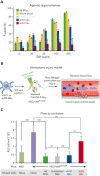
Studies of released human platelets, agonist responsiveness, and hemostatic efficacy. (A) Flow cytometric studies of removed murine blood at 2 hours after infusion of donor-derived platelets or uninfected, shNT-, or shRX-megakaryocytes into NSG mice for P-selectin level analysis after activation with various concentrations of TRAP. Mean ± 1 SD is shown. N = 3 per arm. ∗P ≤ .05 by 1-way ANOVA comparing shNT-platelets released vs shRX-platelets. (B) Schematic of the Rose Bengal photochemical carotid artery thrombotic challenge with NSG/VWFR1326H mice and infused human megakaryocytes or platelets. (C) Same as in panel A except that NSG mice were studied as a hemostatic control because the untreated NSG/VWFR1326H mice had a hemostatic defect. Studies were done 4 hours after infusion of human platelets or megakaryocytes. Mean ± 1 is shown for residual blood flow after carotid artery injury. N = 4 to 6 animals per arm. ∗∗P ≤ .01, ∗∗∗P ≤ .001, and not significant (ns) by 1-way ANOVA. AUC, area under the curve.
Figure 4.
Figure 4.
Drug screening to rescue RUNX1-deficient megakaryocyte yield. Studies of shNT- (A) and shRX-megakaryocytes (B), exposed to the indicated drugs from day 5 to day 14 of differentiation. Megakaryocyte yield was calculated by flow cytometric analysis stained for an anti-hCD42b antibody and for mCherry. The dashed line represents yield of megakaryocytes from uninfected HSPCs not exposed to any drug. Mean ± SD is shown. N = 3 to 5 separate studies, each in duplicate. ∗P ≤ .05, ∗∗∗P ≤ .001, and ∗∗∗∗P ≤ .0001 by 1-way ANOVA compared to each dimethyl sulfoxide control sample.
Figure 5.
Figure 5.
Drug screening to rescue RUNX1-deficient megakaryocyte agonist response. shNT- (A) and shRX (B) megakaryocytes were treated with the indicated drugs from day 5 to day 11 of differentiation. Expression of P-selectin levels was measured by flow cytometry after staining with human CD62P in day 11 megakaryocytes exposed to various concentrations of TRAP as indicated. Mean ± 1 SD is shown. N = 3. ∗P ≤ .05, ∗∗P ≤ .01, and ∗∗∗P ≤ .001 by 1-way ANOVA comparing each shRX data point to its shNT comparative.
Figure 6.
Figure 6.
Drug correction of function and platelet-release defects with RepSox. (A) 3 × 106 shNT-, shRX-, or shRX+RepSox (shRX+RS)–megakaryocytes were infused into NSG mice. At each time point, mouse peripheral blood was withdrawn to monitor human platelet level. The peripheral blood samples were stained with hCD41 and mCD41 antibodies and analyzed by flow cytometry for human vs mouse platelets as in Figure 2. Mean ± 1 SD are shown. N = 3 per arm. ∗P < .05 and ∗∗P < .001 comparing shRX vs shRX+RS studies. (B) Study as in panel A. P-selectin levels on released human platelets in mouse blood were measured by flow cytometry under activation with various concentrations of TRAP. N = 3 per arm. ∗P ≤ .05 and ∗∗P ≤ .001 by 1-way ANOVA comparing shRX vs shRX+RS studies.
Figure 7.
Figure 7.
In vivo hemostatic correction of shRX-platelets by RepSox exposure of developing megakaryocytes. Thrombus formation studies as in Figure 3 with infused shRX-megakaryocytes in a Rose Bengal-photochemical carotid injury model system in NSG/VWFR1326H with NSG mice used as a positive control. To determine in vivo functionalities of infused RepSox-treated shRX-megakaryocytes, we monitored thrombus formation by measuring total blood flow (A) and time to occlusion (B). Mean ± SD are shown. N = 4 to 6 per arm. ∗P ≤ .05, ∗∗P ≤ .01, ∗∗∗P ≤ .001, and ns = not significant by 1-way ANOVA comparing indicated matches.

References

    1. Okuda T, van Deursen J, Hiebert SW, Grosveld G, Downing JR. AML1, the target of multiple chromosomal translocations in human leukemia, is essential for normal fetal liver hematopoiesis. Cell. 1996;84(2):321–330. - PubMed
    1. Wang Q, Stacy T, Miller JD, et al. The CBFbeta subunit is essential for CBFalpha2 (AML1) function in vivo. Cell. 1996;87(4):697–708. - PubMed
    1. Kalev-Zylinska ML, Horsfield JA, Flores MV, et al. Runx1 is required for zebrafish blood and vessel development and expression of a human RUNX1-CBF2T1 transgene advances a model for studies of leukemogenesis. Development. 2002;129(8):2015–2030. - PubMed
    1. Lee TI, Young RA. Transcriptional regulation and its misregulation in disease. Cell. 2013;152(6):1237–1251. - PMC - PubMed
    1. Ichikawa M, Asai T, Saito T, et al. AML-1 is required for megakaryocytic maturation and lymphocytic differentiation, but not for maintenance of hematopoietic stem cells in adult hematopoiesis. Nat Med. 2004;10(3):299–304. - PubMed

Publication types

Substances

Supplementary concepts