. 2022 Dec:66:101611.
doi: 10.1016/j.molmet.2022.101611. Epub 2022 Oct 8.

BDK inhibition acts as a catabolic switch to mimic fasting and improve metabolism in mice

Affiliations

BDK inhibition acts as a catabolic switch to mimic fasting and improve metabolism in mice

Eliza Bollinger et al. Mol Metab. 2022 Dec.

Abstract

Objective: Branched chain amino acid (BCAA) catabolic defects are implicated to be causal determinates of multiple diseases. This work aimed to better understand how enhancing BCAA catabolism affected metabolic homeostasis as well as the mechanisms underlying these improvements.

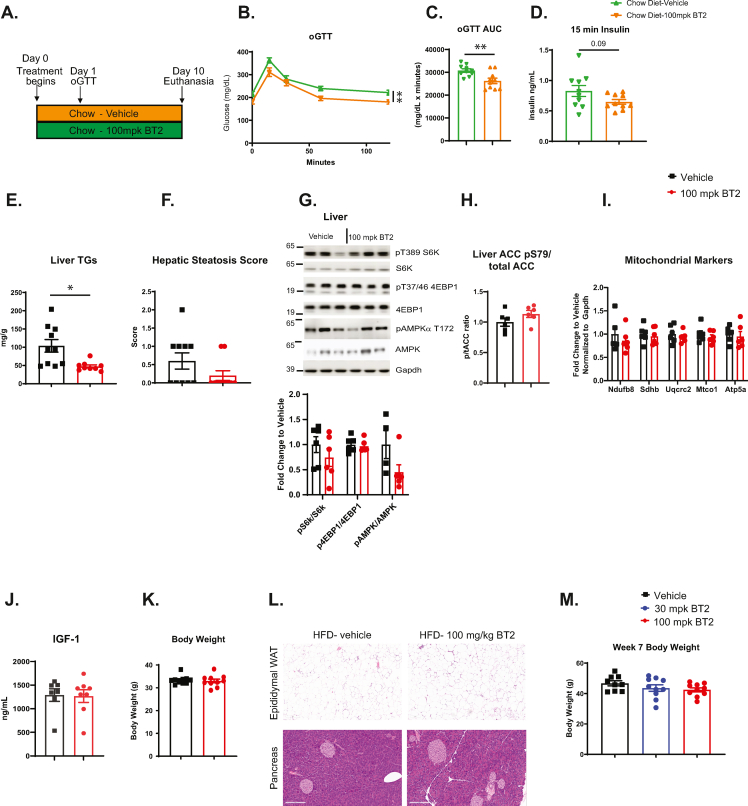
Methods: The rate limiting step of BCAA catabolism is the irreversible decarboxylation by the branched chain ketoacid dehydrogenase (BCKDH) enzyme complex, which is post-translationally controlled through phosphorylation by BCKDH kinase (BDK). This study utilized BT2, a small molecule allosteric inhibitor of BDK, in multiple mouse models of metabolic dysfunction and NAFLD including the high fat diet (HFD) model with acute and chronic treatment paradigms, the choline deficient and methionine minimal high fat diet (CDAHFD) model, and the low-density lipoprotein receptor null mouse model (Ldlr-/-). shRNA was additionally used to knock down BDK in liver to elucidate liver-specific effects of BDK inhibition in HFD-fed mice.

Results: A rapid improvement in insulin sensitivity was observed in HFD-fed and lean mice after BT2 treatment. Resistance to steatosis was assessed in HFD-fed mice, CDAHFD-fed mice, and Ldlr-/- mice. In all cases, BT2 treatment reduced steatosis and/or inflammation. Fasting and refeeding demonstrated a lack of response to feeding-induced changes in plasma metabolites including insulin and beta-hydroxybutyrate and hepatic gene changes in BT2-treated mice. Mechanistically, BT2 treatment acutely altered the expression of genes involved in fatty acid oxidation and lipogenesis in liver, and upstream regulator analysis suggested that BT2 treatment activated PPARα. However, BT2 did not directly activate PPARα in vitro. Conversely, shRNA-AAV-mediated knockdown of BDK specifically in liver in vivo did not demonstrate any effects on glycemia, steatosis, or PPARα-mediated gene expression in mice.

Conclusions: These data suggest that BT2 treatment acutely improves metabolism and liver steatosis in multiple mouse models. While many molecular changes occur in liver in BT2-treated mice, these changes were not observed in mice with AAV-mediated shRNA knockdown of BDK. All together, these data suggest that systemic BDK inhibition is required to improve metabolism and steatosis by prolonging a fasting signature in a paracrine manner. Therefore, BCAA may act as a "fed signal" to promote nutrient storage and reduced systemic BCAA levels as shown in this study via BDK inhibition may act as a "fasting signal" to prolong the catabolic state.

Keywords: BCAA; Diabetes; Metabolic syndrome; Metabolism; NAFLD.


Figures

Figure 1
Figure 1
BT2 treatment reduces BCAA/BCKA and improves glucose tolerance and liver fat in diet-induced obese mice. A-H. Plasma BCAA/BCKA and BT2 levels after a single oral BT2 dose (30 or 100 mpk) in a time course. A. Study Design. B. Leucine, C. Isoleucine, D. Valine, E. Ketoleucine, F. Ketoisoleucine, G. Ketovaline, H. BT2. I–S. Mice were fed 60% HFD for 10 weeks, at which time, mice were dosed with 100 mpk BT2 or vehicle, fasted overnight, and dosed again. One hour later, an oral glucose tolerance test (oGTT) was performed. On day 10, the animals were euthanized. I. Study Design. J. Glucose levels during oGTT. K. Area under the curve (AUC) for glucose during oGTT. L. Insulin levels during oGTT. M-S. Tissues were collected 1 h post the final dose of BT2 or vehicle. M-O. Western blots of pBCKDHE1a, BCKDHE1a or Gapdh (M-O) as a loading control M. Liver, N. Heart, and O. Gastrocnemius muscle. Top, representative Western blot image. Bottom, densitometric analysis. P–S. BCAA/BCKA were quantified by mass spectrometry. P-Q. Heart, R–S. Muscle. T. Mice were dosed with 30 or 100 mpk BT2 or vehicle daily for 8 weeks, at which time liver TG were measured. Data represent the mean ± SEM. N = 3–12 per group; ∗; p < 0.05, ∗∗; p < 0.01, ∗∗∗; p < 0.005, ∗∗∗∗; p < 0.0005, #; p < 0.0001.
Figure 2
Figure 2
Acute BT2 treatment improves insulin sensitivity in diet-induced obese mice. Diet induced obese mice were implanted with a jugular vein catheter, and 13C glucose was infused through one line, and insulin on a second line. Animals were dosed with vehicle or BT2 (30 & 100 mpk) at time 0. Glucose was measured via tail bleed every 5 min 10 min prior to euthanasia, FDG was injected (2 mpk). Animals were euthanized 120 min after BT2 treatment, and 13C glucose ratios as well as skeletal muscle FDG levels were examined by mass spectrometry. A. Study design. B. Glycemia was maintained at 250 mg/dL throughout the study duration for all groups. C-D. Glucose infusion rate over the time course of the study. Arrow denotes time of compound dosing. D. GIR calculation. E. Hepatic glucose production. F. Clamped glucose utilization (Rd). G. FDG-6P in gastrocnemius muscle. Data represent the mean ± SEM. N = 7–11 per group; ∗; p < 0.05, ∗∗∗; p < 0.005.
Figure 3
Figure 3
BT2 treatment improves liver pathology in choline deficient high fat diet. Mice were administered normal chow, or choline deficient, methionine minimal high fat diet (CDAHFD) for 8 weeks. Animals were euthanized at weeks 4 and 8 after diet administration. A. Study design. B. Body weights throughout the study duration. C. Liver weights. D. Liver triglyceride content. E. Plasma ALT. F. Plasma AST. G-K. RNA was extracted from liver, and quantitative RT-PCR was performed for G.Tnfα, H.Ccl2, I.Cd68, J.Ccr2, K.Col1a1. All genes were normalized to Ppia. L-Q. Representative histological sections of liver from mice fed normal chow (control), CDAHFD, or CDAHFD with 100 mpk BT2 for 8 weeks. Sections were stained as indicated. L. Histologic stains and IHC: From Left to Right, H&E, PSR, Iba1, α-SMA. M-N. Histologic qualitative grades for liver steatosis and inflammation across experimental groups. M. Steatosis score. N. Inflammation score. O. Quantification of PSR stain from L. P. Quantification of α-SMA stain from L. Q. Quantification of Iba1 stain from L. Data represent the mean ± SEM. N = 4–12 per group; ∗; p < 0.05, ∗∗; p < 0.01, ∗∗∗; p < 0.005, ∗∗∗∗; p < 0.0005.
Figure 4
Figure 4
BT2 treatment improves liver pathology in Ldlr−/−mice fed Western diet.Ldlr−/− mice were fed Western diet for 8 weeks, at which time treatment with vehicle or BT2 was initiated and continued daily for 8 weeks. A. Study design. B. Body weight. C. Liver weight normalized to body weight. D. Liver triglyceride content. E. H&E-stained liver sections. Left panel (vehicle), grade 4 steatosis; right panel (100 mpk BT2), grade 1 steatosis. F. Qualitative grading of steatosis across experimental groups. G. Plasma ALT. H. Plasma AST. I. RNA was extracted from liver, and quantitative RT-PCR was performed for a number of genes and normalized to Ppia for input control, and vehicle treated control animals for fold change. Data represent the mean ± SEM. N = 4–10 per group; ∗; p < 0.05, ∗∗; p < 0.01, ∗∗∗; p < 0.005.
Figure 5
Figure 5
BT2 treatment prolongs the effects of fasting in diet-induced obese mice. Mice were treated with vehicle or BT2 in the morning, fasted overnight, dosed again with BT2 or vehicle and either remained fasting or were refed for 2 h prior to euthanasia. A. Study design. B-G. Plasma metabolites were measured. B. Glucose, C. Insulin, D. Beta hydroxybutyrate, E. Free fatty acids, F. Triglycerides, G. Glycerol. H-M. Protein was isolated, and Western blots were performed. H. Representative Western blots from skeletal muscle, I. Densitometric analyses of H. J. Representative Western blots from liver, K. Densitometric analysis of J. L. Representative Western blots from white adipose tissue (WAT), M. Densitometric analysis of L. N. RNA was extracted from liver, and RNAseq was performed. Differentially expressed genes in vehicle-treated animals after feeding are shown. This gene signature remains unchanged with BT2 treatment (N = 7–9) O. Plasma FGF-21 levels. Data represent the mean ± SEM. N = 3–14 per group. ∗; p < 0.05, ∗∗; p < 0.01.
Figure 6
Figure 6
Insulin-mediated biochemical events and signaling are not altered in BT2-treated obese mice. Mice were treated with vehicle or BT2 in the morning, fasted overnight, dosed again with BT2 or vehicle, and 1 h later were injected with saline or insulin for 10 min, at which time animals were euthanized, and plasma and tissues were collected. A. Study design. B-D. Plasma metabolites were measured. B. Insulin, C. Glucose, D. Beta hydroxybutyrate. E-H. Protein was isolated, and Western blots were performed. E. Representative Western blots from Liver, F. Densitometric analyses of E. G. Representative Western blots from Quadricep muscle, H. Densitometric analysis of G. I. Representative Western blots from epididymal WAT, J. Densitometric analysis of I. Data represent the mean ± SEM. N = 3–8 per group. ∗; p < 0.05, ∗∗; p < 0.01, ∗∗∗; p < 0.005.
Figure 7
Figure 7
Indirect activation of PPARα in liver of BT2-treated mice. A-D mice were treated with BT2 or vehicle, fasted overnight, and RNAseq was performed in whole liver as described in Figure 5. A. Heat map of differentially expressed genes from the vehicle and BT2 treated, fasted animals. B. Upstream regulator analysis. C. Ingenuity pathway analysis. D. Quantitative RT-PCR was performed for a number of PPARα target genes from livers of vehicle and BT2 treated, fasted animals and normalized to Ppia N = 8–9 per group. E. PPARα coactivator Lanthascreen assay with BT2 or GW7647 as a positive control. Data represent the average of 2 independent experiments. All data represent the mean ± SEM. ∗∗; p < 0.01, ∗∗∗; p < 0.005, ∗∗∗∗; p < 0.0005.
Figure 8
Figure 8
Liver-specific knockdown of BDK does not improve glucose tolerance or NAFLD and does not activate PPARα target genes in HFD-fed mice. BDK shRNA was injected into 12-week-old mice, and they were then put on HFD for 9 weeks. At week 8, an oral glucose tolerance test (oGTT) was performed. A. Study design. B. Body weights over the study duration. C-D. Oral glucose tolerance test. C. Glucose levels during oGTT, D. Insulin levels during oGTT. E. Liver weight as % of body weight. F. Liver triglycerides/mg protein. G-H. mRNA was isolated, and quantitative RT-PCR was performed for G.Bckdk and H.Gfp and normalized to Hprt.I. Protein was isolated, and Western blots were performed for p-Bckdh, Bckdh, Bdk, Gapdh. Top, representative images. Bottom, densitometric analyses. J. BCAA & K. BCKA were measured in terminal plasma samples. L. RNA was isolated from whole liver, and qPCR was performed for PPARα target genes and normalized to Ppia. Data represent the mean ± SEM. N = 4–8 per group. ∗; p < 0.05, ∗∗; p < 0.01, ∗∗∗∗; p < 0.0005. M. Model of the mechanism underlying metabolic improvements by BT2-mediated improvements in BCAA catabolism.
figs1
figs1
figs2
figs2
figs3
figs3
figs4
figs4
figs5
figs5

References

    1. Newgard C.B., An J., Bain J.R., Muehlbauer M.J., Stevens R.D., Lien L.F., et al. A branched-chain amino acid-related metabolic signature that differentiates obese and lean humans and contributes to insulin resistance. Cell Metabolism. 2009;9(4):311–326. - PMC - PubMed
    1. Wang T.J., Larson M.G., Vasan R.S., Cheng S., Rhee E.P., McCabe E., et al. Metabolite profiles and the risk of developing diabetes. Nature Medicine. 2011;17(4):448–453. - PMC - PubMed
    1. Lotta L.A., Scott R.A., Sharp S.J., Burgess S., Luan J., Tillin T., et al. Genetic predisposition to an impaired metabolism of the branched-chain amino acids and risk of type 2 diabetes: a mendelian randomisation analysis. PLoS Medicine. 2016;13(11) - PMC - PubMed
    1. Menni C., Fauman E., Erte I., Perry J.R., Kastenmuller G., Shin S.Y., et al. Biomarkers for type 2 diabetes and impaired fasting glucose using a nontargeted metabolomics approach. Diabetes. 2013;62(12):4270–4276. - PMC - PubMed
    1. Zhou M., Shao J., Wu C.Y., Shu L., Dong W., Liu Y., et al. Targeting BCAA catabolism to treat obesity-associated insulin resistance. Diabetes. 2019;68(9):1730–1746. - PMC - PubMed

Publication types

Substances