. 2022 Oct 11;13(1):5989.
doi: 10.1038/s41467-022-33588-z.

An ancestral interaction module promotes oligomerization in divergent mitochondrial ATP synthases

Affiliations

An ancestral interaction module promotes oligomerization in divergent mitochondrial ATP synthases

Ondřej Gahura et al. Nat Commun. .

Abstract

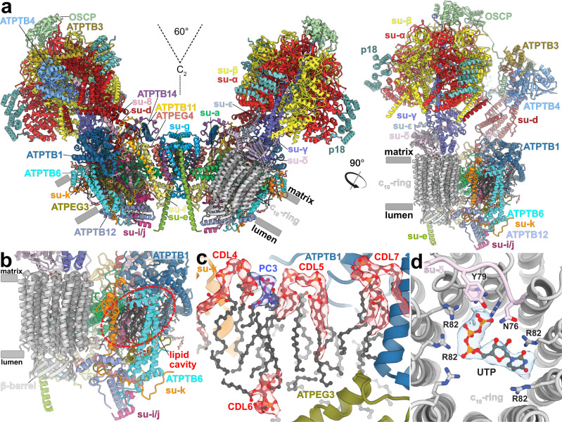
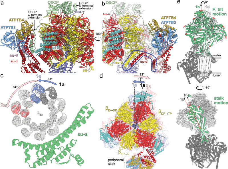
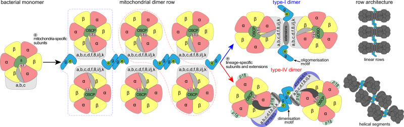
Mitochondrial ATP synthase forms stable dimers arranged into oligomeric assemblies that generate the inner-membrane curvature essential for efficient energy conversion. Here, we report cryo-EM structures of the intact ATP synthase dimer from Trypanosoma brucei in ten different rotational states. The model consists of 25 subunits, including nine lineage-specific, as well as 36 lipids. The rotary mechanism is influenced by the divergent peripheral stalk, conferring a greater conformational flexibility. Proton transfer in the lumenal half-channel occurs via a chain of five ordered water molecules. The dimerization interface is formed by subunit-g that is critical for interactions but not for the catalytic activity. Although overall dimer architecture varies among eukaryotes, we find that subunit-g together with subunit-e form an ancestral oligomerization motif, which is shared between the trypanosomal and mammalian lineages. Therefore, our data defines the subunit-g/e module as a structural component determining ATP synthase oligomeric assemblies.


Conflict of interest statement

The authors declare no competing interests.

Figures

Fig. 1
Fig. 1. The T. brucei ATP synthase structure with lipids and ligands.
a Front and side views of the composite model with both monomers in rotational state 1. The two F1/c10-ring complexes, each augmented by three copies of the phylum-specific p18 subunit, are tied together at a 60°-angle. The membrane-bound Fo region displays a unique architecture and is composed of both conserved and phylum-specific subunits. b Side view of the Fo region showing the lumenal interaction of the ten-stranded β-barrel of the c-ring (grey) with ATPTB12 (pale blue). The lipid-filled peripheral Fo cavity is indicated. c Close-up view of the bound lipids within the peripheral Fo cavity with cryo-EM density shown. d Top view into the decameric c-ring with a bound pyrimidine ribonucleoside triphosphate, assigned as UTP, although not experimentally detected. Map density shown in transparent blue, interacting residues shown.
Fig. 2
Fig. 2. Identification of conserved Fo subunits.
a Top view of the membrane region with T. brucei subunits (coloured) overlaid with S. cerevisiae structure (grey transparent). Close structural superposition and matching topology allowed the assignment of conserved subunits based on matching topology and location. b Superposition of subunits-b, -e and -g with their S. cerevisiae counterparts PDB 6B2Z (S. cerevisiae mitochondrial ATP synthase) confirms their identity. c Schematic representation of transmembrane helices of subunit-b and adjacent subunits in T. brucei, E. gracilis PDB 6TDV (E. gracilis mitochondrial ATP synthase, membrane region) and S. cerevisiae PDB 6B2Z (S. cerevisiae mitochondrial ATP synthase) ATP synthases. PC – phosphatidylcholine.
Fig. 3
Fig. 3. A divergent peripheral stalk allows high flexibility during rotary catalysis.
a N-terminal OSCP extension provides a permanent central stalk attachment, while the C-terminal extension provides a phylum-specific attachment to the divergent peripheral stalk. b The C-terminal helices of subunits -β and -d provide a permanent F1 attachment. c Substeps of the c-ring during transition from rotational state 1 to 2. d F1 motion accommodating steps shown in (c). After advancing along with the rotor to state 1e, the F1 rotates in the opposite direction when transitioning to state 2a. e Tilting motion of F1 and accommodating bending of the peripheral stalk.
Fig. 4
Fig. 4. The lumenal half-channel contains ordered water molecules and is confined by an Fo-bound lipid.
a Subunit-a (green) with the matrix (orange) and lumenal (light blue) channels, and an ordered phosphatidylcholine (PC1; blue). E102 of the c10-ring shown in grey. b Close-up view of the highly conserved R146a and N209a, which coordinate two water molecules between helices H5-6a. c Sideview of the lumenal channel with proton pathway (light blue) and confining phosphatidylcholine (blue). d Chain of ordered water molecules in the lumenal channel. Distances between the W1–W5 (red) are 5.2, 3.9, 7.3, and 4.8 Å, respectively. e The ordered waters extend to H155a, which likely mediates the transfer of protons to D202a.
Fig. 5
Fig. 5. The homotypic dimerization motif of subunit-g generates a conserved oligomerization module.
a Side view with dimerizing subunits coloured. The dimer interface is constituted by b subunit-e’ contacting subunit-a in the membrane and subunit-f in the lumen, c subunits e and g from both monomers forming a subcomplex with bound lipids. d Subunit-g and -e form a dimerization motif in the trypanosomal (type-IV) ATP synthase dimer (this study), the same structural element forms the oligomerization motif in the porcine ATP synthase tetramer. The structural similarity of the pseudo-dimer (i.e., two diagonal monomers from adjacent dimers) in the porcine structure with the trypanosomal dimer suggests that type I and IV ATP synthase dimers have evolved through divergence from a common ancestor. e The dimeric subunit-e/g structures are conserved in Sus scrofa PDB 6ZNA (S. scrofa mitochondrial ATP synthase) and T. brucei (this work) and contain a conserved GXXXG motif (orange) mediating interaction of transmembrane helices. f Models of the ATP synthase dimers fitted into subtomogram averages of short oligomers: matrix view, left; cut-through, middle, lumenal view, right; EMD-3560 (in situ structure of T. brucei mitochondrial ATP synthase).
Fig. 6
Fig. 6. RNAi knockdown of subunit-g results in monomerization of ATP synthase.
a Growth curves of non-induced (solid lines) and tetracycline-induced (dashed lines) RNAi cell lines grown in the presence (black) or absence (brown) of glucose. The insets show relative levels of the respective target mRNA at indicated days post-induction (DPI) normalized to the levels of 18S rRNA (black bars) or β-tubulin (white bars). b Immunoblots of mitochondrial lysates from indicated RNAi cell lines resolved by BN-PAGE probed with antibodies against indicated ATP synthase subunits (n = 2). Positions of molecular weight (MW) marker are shown. c Immunoblots of whole cell lysates from indicated RNAi cell lines probed with indicated antibodies (n = 3). Positions of MW marker are shown. d Quantification of three independent replicates of immunoblots in (c). Values were normalized to the signal of the loading control Hsp70 and to non-induced cells. Plots show individual values, means, and standard deviations (SD; error bars).
Fig. 7
Fig. 7. Monomerization of ATP synthase by subunit-g knockdown results in aberrant mitochondrial ultrastructure but does not abolish catalytic activity.
a Transmission electron micrographs of sections of non-induced or 4 days induced RNAi cell lines. At least 70 micrographs were obtained in each category. Mitochondrial membranes and cristae are marked with blue and red arrowheads, respectively. Top panel shows examples of irregular, elongated and round cross-sections of mitochondria quantified in (b). Scale bars: 500 nm. b Cristae numbers per vesicle from indicated induced (IND) or non-induced (NON) cell lines counted separately in irregular, elongated and round mitochondrial cross-section. Boxes and whiskers show 25th to 75th and 5th to 95th percentiles, respectively. The numbers of analyzed cross-sections are indicated for each data point. Unpaired two-sided t-test, p-values are shown in the graph. c Mitochondrial membrane polarization capacity of non-induced or RNAi-induced cell lines two and four DPI measured by Safranine O. Black and grey arrow indicate addition of ATP and oligomycin, respectively. d ATP production in permeabilized non-induced (0) or RNAi-induced cells 2 and 4 DPI in the presence of indicated substrates and inhibitors. The graphs show individual values of two technical replicates of n = 2 (subunit-8), n = 3 (ATPTB4), or n = 4 (subunit-g) independent experiments and means (bars) and SD (error bars) of the averaged values of the technical replicates. Gly3P DL-glycerol phosphate; KCN potassium cyanide; CATR carboxyatractyloside.
Fig. 8
Fig. 8. The subunit-e/g module is an ancestral oligomerization motif of ATP synthase.
Schematic model of the evolution of type-I and IV ATP synthases. Mitochondrial ATP synthases are derived from a monomeric complex of proteobacterial origin. In a mitochondrial ancestor, acquisition of mitochondria-specific subunits, including the subunit-e/g module resulted in the assembly of ATP synthase double rows, the structural basis for cristae biogenesis. Through divergence, different ATP synthase architectures evolved, with the subunit-e/g module functioning as an oligomerization (type I) or dimerization (type IV) motif, resulting in distinct row assemblies between mitochondrial lineages.

Similar articles

Cited by

References

    1. Paumard P, et al. The ATP synthase is involved in generating mitochondrial cristae morphology. EMBO J. 2002;21:221–230. doi: 10.1093/emboj/21.3.221. - DOI - PMC - PubMed
    1. Davies KM, Anselmi C, Wittig I, Faraldo-Gomez JD, Kuhlbrandt W. Structure of the yeast F1Fo-ATP synthase dimer and its role in shaping the mitochondrial cristae. Proc. Natl Acad. Sci. USA. 2012;109:13602–13607. doi: 10.1073/pnas.1204593109. - DOI - PMC - PubMed
    1. Panek T, Elias M, Vancova M, Lukes J, Hashimi H. Returning to the fold for lessons in mitochondrial crista diversity and evolution. Curr. Biol. 2020;30:R575–R588. doi: 10.1016/j.cub.2020.02.053. - DOI - PubMed
    1. Kuhlbrandt W. Structure and mechanisms of F-Type ATP synthases. Annu. Rev. Biochem. 2019;88:515–549. doi: 10.1146/annurev-biochem-013118-110903. - DOI - PubMed
    1. Spikes TE, Montgomery MG, Walker JE. Structure of the dimeric ATP synthase from bovine mitochondria. Proc. Natl Acad. Sci. USA. 2020;117:23519–23526. doi: 10.1073/pnas.2013998117. - DOI - PMC - PubMed

Publication types