. 2022 Oct 11;13(1):5994.
doi: 10.1038/s41467-022-33714-x.

Sequential action of a tRNA base editor in conversion of cytidine to pseudouridine

Affiliations

Sequential action of a tRNA base editor in conversion of cytidine to pseudouridine

Satoshi Kimura et al. Nat Commun. .

Abstract

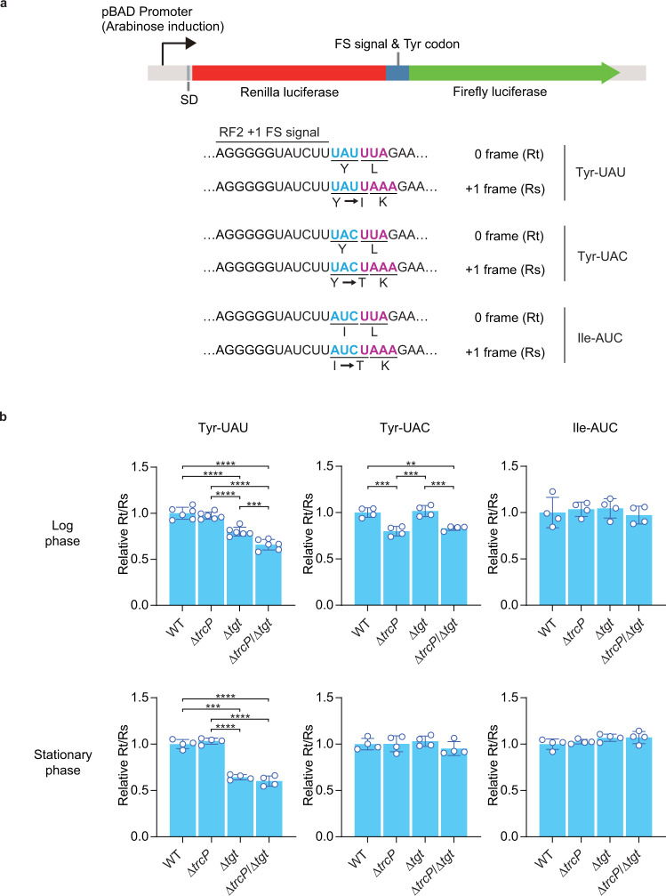
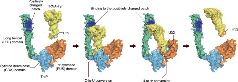
Post-transcriptional RNA editing modulates gene expression in a condition-dependent fashion. We recently discovered C-to-Ψ editing in Vibrio cholerae tRNA. Here, we characterize the biogenesis, regulation, and functions of this previously undescribed RNA editing process. We show that an enzyme, TrcP, mediates the editing of C-to-U followed by the conversion of U to Ψ, consecutively. AlphaFold-2 predicts that TrcP consists of two globular domains (cytidine deaminase and pseudouridylase) and a long helical domain. The latter domain tethers tRNA substrates during both the C-to-U editing and pseudouridylation, likely enabling a substrate channeling mechanism for efficient catalysis all the way to the terminal product. C-to-Ψ editing both requires and suppresses other modifications, creating an interdependent network of modifications in the tRNA anticodon loop that facilitates coupling of tRNA modification states to iron availability. Our findings provide mechanistic insights into an RNA editing process that likely promotes environmental adaptation.


Conflict of interest statement

The authors declare no competing interests.

Figures

Fig. 1
Fig. 1. TrcP is sufficient to catalyze C-to-Ψ editing reaction.
a Secondary structure of V. cholerae tRNA-Tyr. The C at position 32 is edited to Ψ. b Sanger sequencing of amplified cDNA derived from 250 nM tRNAs incubated with recombinant TrcP protein. TrcP concentrations are shown on the right. In the traces, red corresponds to T, and blue corresponds to C. c MALDI-TOF analysis of an oligo-protected portion (positions 10–46) of tRNA-Tyr after incubation with recombinant TrcP protein. The tRNA fragment was treated with acrylonitrile to introduce a cyanoethyl group (53 Da) onto Ψ. RNase A digests of a cyanoethylated RNA fragment were subjected to MS. The m/z values and assigned fragments are shown. Asterisks indicate the fragment corresponding to the tetramer fragment, GAGDp (m/z 1344) derived from positions 13–16. d Multiple sequence alignment of TrcP homologs and V. cholerae RluA, a paralog of the pseudouridine synthetase domain of TrcP. Similar amino acids that are observed in more than 80% of sequences are shaded with colors. Amino acid residues that are mutated in this study are indicated. TrcP NTD and CTD, and domains assigned by structural modeling, i.e., the cytidine deaminase (CDA) domain (1–114 aa and 250–339 aa), the long helical (LHL) domain (115–249 aa), and the pseudouridine synthase (PUS) domain (351–570 aa) are shown.
Fig. 2
Fig. 2. TrcP includes a C-to-U RNA editor and a pseudouridylase.
a Schematic of the C-to-Ψ editing reaction by TrcP. b TrcP domain structure with the position of R507. c MALDI-TOF analysis of an oligo-protected portion (positions 10–46) of tRNA-Tyr after incubation with recombinant TrcP and mutant derivatives. The tRNA fragment was treated with acrylonitrile to introduce a cyanoethyl group (53 Da) to Ψ. RNase A digests of a cyanoethylated RNA fragment were subjected to MS. The m/z values and assigned fragments are shown. Asterisks indicate the fragment corresponding to the tetramer fragment, GAGDp (m/z 1344) derived from positions 13–16.
Fig. 3
Fig. 3. Reaction mechanisms of TrcP catalyzing C-to-U editing.
a A structural model of TrcP protein generate by ColabFold. TrcP is composed of two globular domains, the cytidine deaminase (CDA) domain (light blue) and pseudouridine synthetase (PUS) domain (orange), and a long helical (LHL) domain (green). NTD and CTD are shaded with light blue and light yellow, respectively. b Alignment of the predicted catalytic site in the TrcP CDA domain (light blue) with the Blasticidin-S deaminase (BSD) (yellow). The residues of BSD and its coordinating zinc ion are shown in the yellow background, whereas the residues of TrcP are shown in the light blue background. Oxygen and sulfur residues are also colored red and gold, respectively. c Colorimetric measurement of zinc concentration of TrcP solution. Source data are provided as a Source Data File. d In vivo complementation analysis of TrcP mutants. Sanger sequencing results of tRNA-Tyr cDNA derived from RNA isolated from trcP knockout strains expressing TrcP (WT) or the indicated mutant derivatives. e A schematic of the C-to-Ψ editing reaction by TrcP. TrcP’s NTD uses water as a source of oxygen for C-to-U editing. In U and Ψ, the expected positions of 18O-labeled oxygen from water are shown in red. f Nucleoside analysis of tRNAs incubated with or without TrcP and stable isotope-labeled water (18O-water). The reaction conditions and the detected nucleosides are shown above and right, respectively.
Fig. 4
Fig. 4. The long helical domain facilitates substrate tRNA binding.
a Predicted surface electrostatic potential of the structural model of TrcP. The color key is shown below (unit KbT/ec). b In vivo complementation analysis of the KR mutant. Sanger sequencing results of tRNA-Tyr cDNA derived from RNA isolated from trcP knockout strains expressing TrcP (WT) or mutant derivatives. c Gel mobility shift assay with TrcP (WT and KR mutant) and tRNAs (tRNA-Tyr and tRNA-Asp). Upper and lower arrowheads represent wells and free tRNA signals, respectively. The black line indicates the shifted band, presumably representing the soluble tRNA-TrcP complex. Protein concentrations were 0, 0.1, 1, 2, and 5 μM, and the tRNA concentration was 1 μM. An uncropped blot is provided in Source Data. Representative data from two independent experiments with similar results is shown. d Sanger sequencing of cDNA derived from 250 nM tRNA-Tyr (WT) and the indicated mutants incubated with 750 nM recombinant TrcP protein. The secondary structures of mutant tRNAs are shown in Supplementary Fig. 9. e A schematic of the experiment in f. TrcP-NTD was used for generating 18O-labeled U32 in tRNA-Tyr. Purified tRNA-Tyr was then incubated with indicated enzymes. f Nucleoside analysis of tRNA-Tyr after the reaction with the indicated proteins. Conversion of U into Ψ was assessed by the signals of 18O-labeled U (U + 2). The signals were normalized by m5U. The bar represents the average values of two independent reaction results represented by the points. This experiment was performed once. Source data are provided as a Source Data File.
Fig. 5
Fig. 5. An iron-responsive modification network in V. cholerae tRNA-Tyr.
a MALDI-TOF analysis of the oligo-protected portion (positions 10–46) of in vitro transcribed tRNA-Tyr. tRNA (250 nM) was reacted with 0 nM (top), 25 nM (middle), and 250 nM (bottom) recombinant TrcP. Oligo-protected portions were incubated with acrylonitrile, which specifically cyanoethylates (CE) pseudouridine, increasing its mass by 53 Da. m/z values and assigned fragment sequences are shown. Asterisks indicate the fragment corresponding to the tetramer fragment, GAGDp (m/z 1344) derived from positions 13–16. b Sanger sequence of tRNA-Tyr cDNA from RNA isolated from WT (Top), ΔmiaA (second top), ΔmiaB (middle), ΔmiaE (second bottom), and tgt::Tn (bottom) strains. c Sanger sequence of cDNA of tRNA-Tyr isolated from a ΔtrcP strain (left) or a ΔmiaB strain (right). tRNAs (250 nM) were incubated with 0 nM, 50 nM, and 500 nM TrcP. d Sanger sequence of tRNA-Tyr cDNA from RNA isolated from the WT strain cultured with or without dipyridyl (dip) at the indicated concentrations. e Nucleoside analysis of tRNA-Tyr from WT, ΔtrcP and ΔmiaB strains. The left panel shows log phase results, and the right panel shows stationary phase results. The detected nucleosides are shown on the right. The signal intensity is normalized with m5U signals in each sample. Representative data from two independent experiments with similar results is shown. f Schematic of proposed interdependency network of modifications in the tRNA-Tyr anticodon loop. In WT under iron-replete conditions, ms2io6A37 facilitates C-to-Ψ editing at position 32, which in turn suppresses Q formation at position 34 (Left). In contrast, the absence of miaB or low iron conditions eliminates methyl-thio modification at position 37, suppressing C-to-Ψ editing and thereby relieving inhibition of Q34 biogenesis (Right).
Fig. 6
Fig. 6. C-to-Ψ editing facilitates Tyr decoding.
a Schematic of reporter construct for measuring decoding ability of tRNA-Tyr and tRNA-Ile. b Reporter assays evaluating decoding of Tyr and Ile codons. Relative Rt/Rs value (see methods) is indicated. The top and bottom panels show the results derived from log and stationary phase cultures, respectively. The tested codons are shown above, and the tested strains are labeled below. Ordinary one-way ANOVA was used for a statistical test. Comparisons were made in all combinations among the strains, and Tukey was used for correction for multiple comparisons (**p < 0.01; ***p < 0.001; ****p < 0.0001). The bar represents the average values of biologically independent culture results represented by the points. n = 6 for log phase UAU reporter assay and n = 4 for the other conditions. The standard deviations (SD) were shown as error bars. Source data with exact p values are provided as a Source Data File.
Fig. 7
Fig. 7. Model of TrcP mediated C-to-Ψ editing.
The anticodon loop of tRNA-Tyr locates in the catalytic pocket of the CDA (light blue) domain and undergoes C-to-U conversion. Then the anticodon moves to the PUS domain (orange) and undergoes U-to-Ψ conversion. During both reactions, the positively charged patch (dark blue) in the LHL domain (green) supports the reaction by binding to the upper part of the tRNA. The LHL domain appears to enable the enzyme to adopt a substrate channeling mechanism to carry out its consecutive deamination and pseudouridylation reactions.

Similar articles

Cited by

References

    1. Bjork, G. R. & Hagervall, T. G. Transfer RNA modification: presence, synthesis, and function. EcoSal Plus6, 10.1128/ecosalplus.ESP-0007-2013 (2014). - PubMed
    1. Machnicka MA, et al. MODOMICS: a database of RNA modification pathways-−2013 update. Nucleic Acids Res. 2013;41:D262–D267. doi: 10.1093/nar/gks1007. - DOI - PMC - PubMed
    1. Wiener, D. & Schwartz, S. The epitranscriptome beyond m(6)A. Nat. Rev. Genet., 10.1038/s41576-020-00295-8 (2020). - PubMed
    1. Zaccara S, Ries RJ, Jaffrey SR. Reading, writing and erasing mRNA methylation. Nat. Rev. Mol. Cell Biol. 2019;20:608–624. doi: 10.1038/s41580-019-0168-5. - DOI - PubMed
    1. Ichinose, M. & Sugita, M. RNA Editing and Its Molecular Mechanism in Plant Organelles. Genes (Basel)8, 10.3390/genes8010005 (2016). - PMC - PubMed

Publication types