. 2023 Mar;30(3):716-730.
doi: 10.1038/s41418-022-01074-0. Epub 2022 Oct 11.

C9orf72 functions in the nucleus to regulate DNA damage repair

Affiliations

C9orf72 functions in the nucleus to regulate DNA damage repair

Liying He et al. Cell Death Differ. 2023 Mar.

Abstract

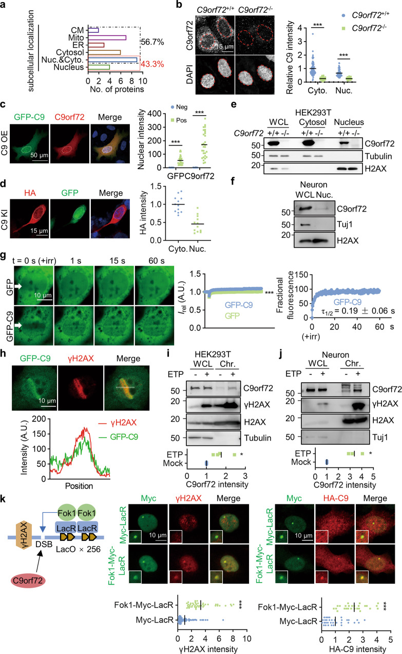
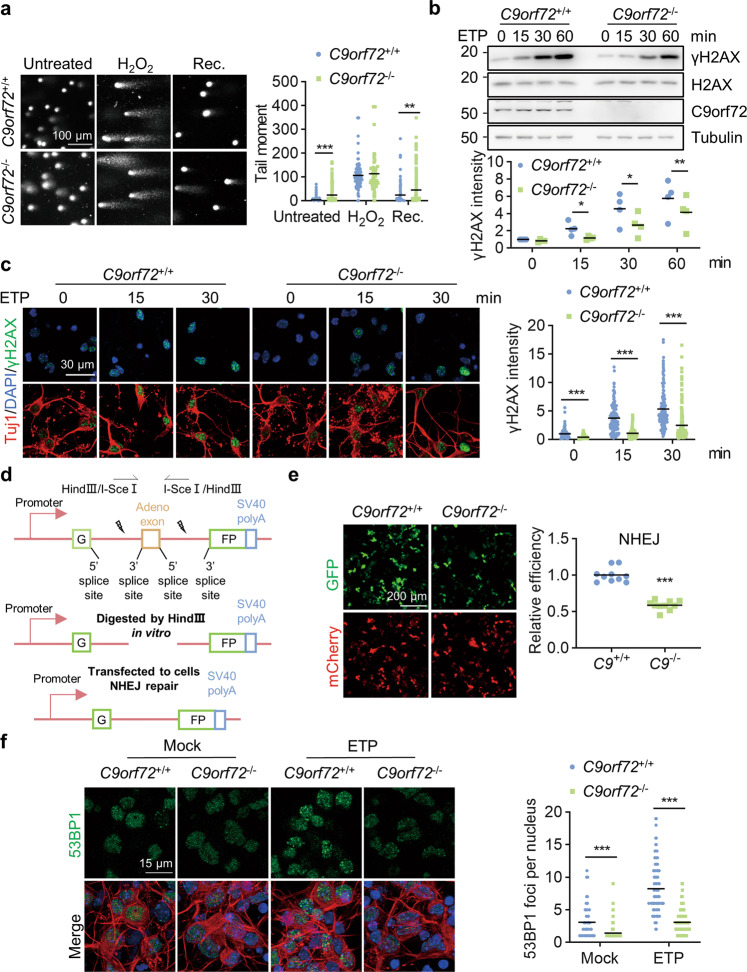
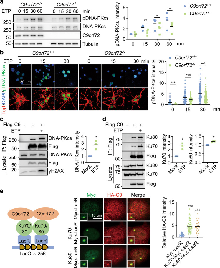
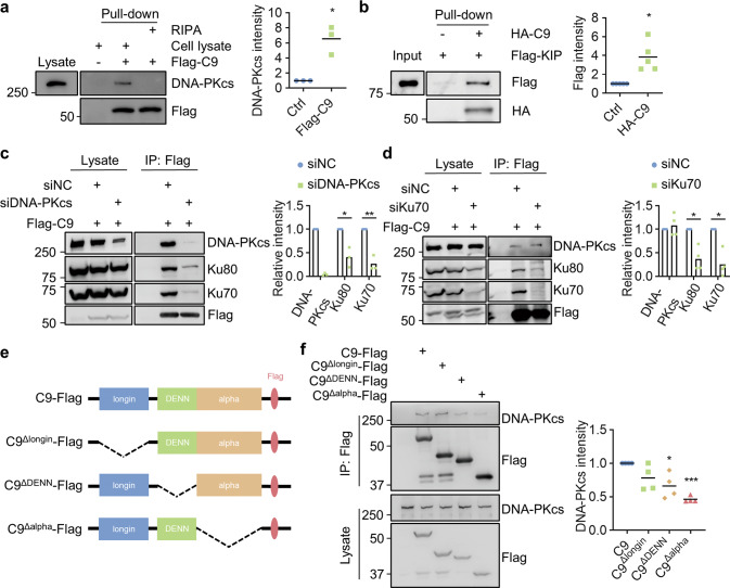
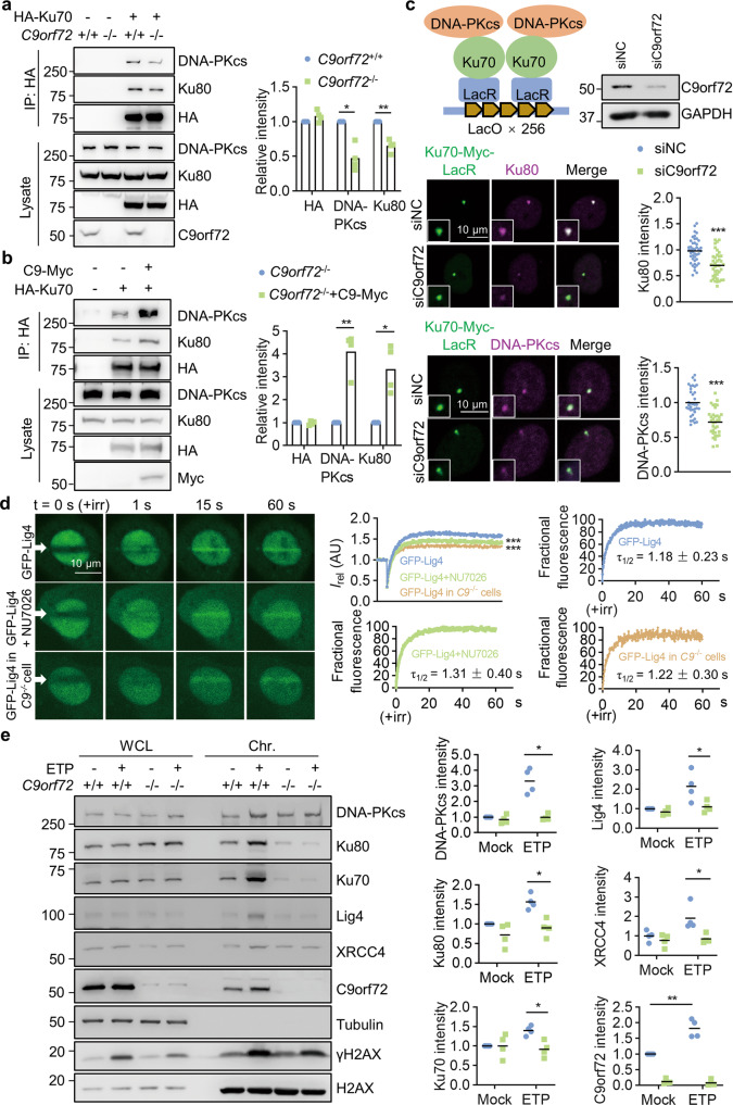
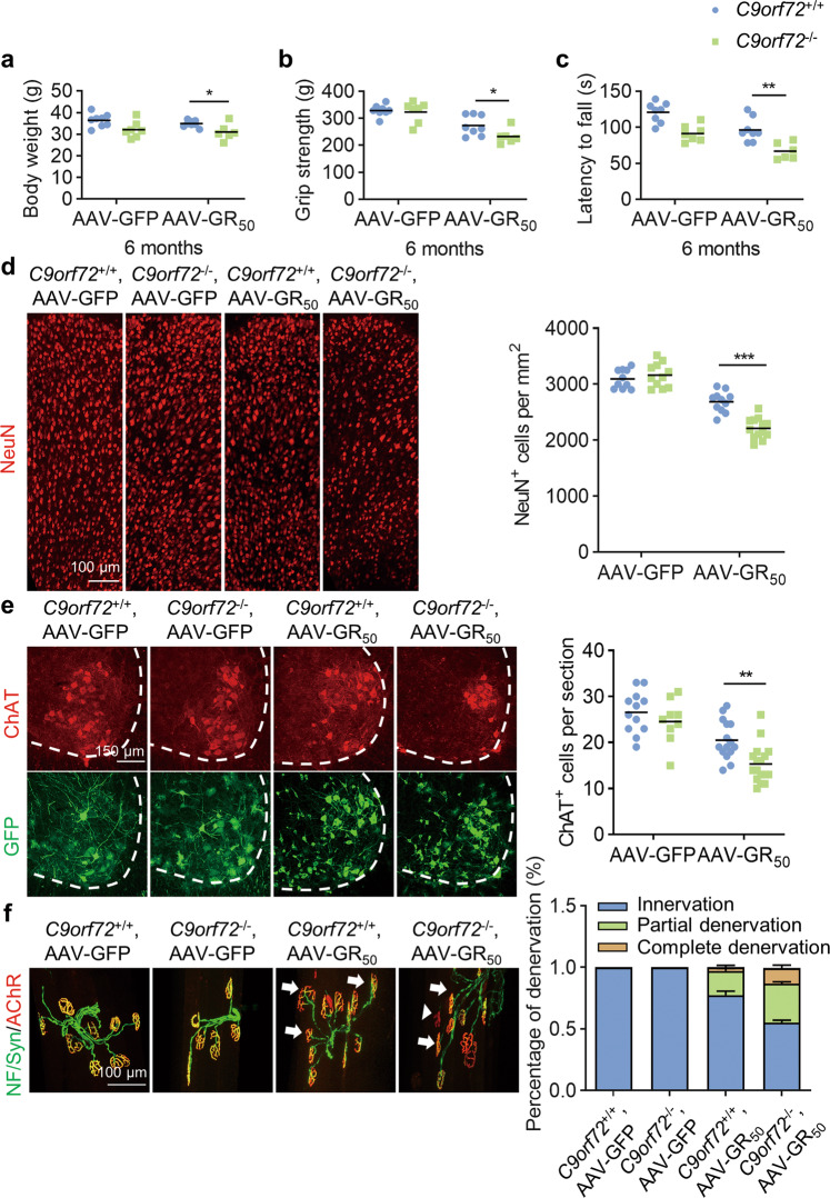
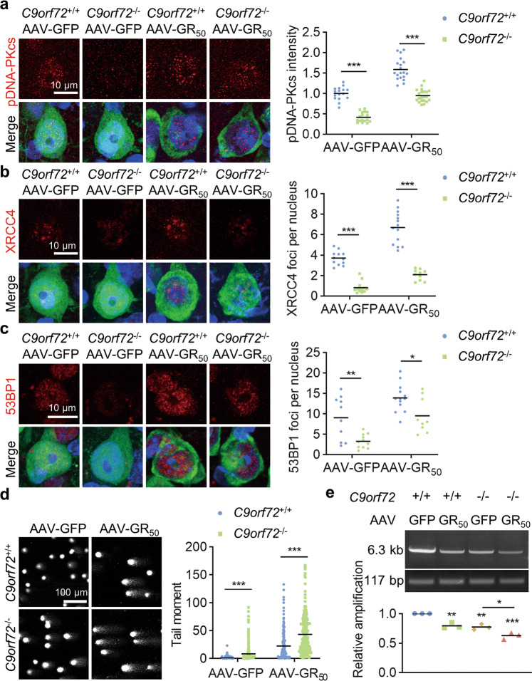
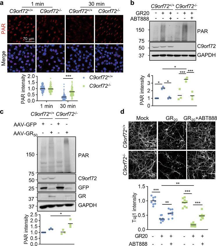
The hexanucleotide GGGGCC repeat expansion in the intronic region of C9orf72 is the most common cause of Amyotrophic lateral sclerosis (ALS) and frontotemporal dementia (FTD). The repeat expansion-generated toxic RNAs and dipeptide repeats (DPRs) including poly-GR, have been extensively studied in neurodegeneration. Moreover, haploinsufficiency has been implicated as a disease mechanism but how C9orf72 deficiency contributes to neurodegeneration remains unclear. Here, we show that C9orf72 deficiency exacerbates poly-GR-induced neurodegeneration by attenuating non-homologous end joining (NHEJ) repair. We demonstrate that C9orf72 localizes to the nucleus and is rapidly recruited to sites of DNA damage. C9orf72 deficiency resulted in impaired NHEJ repair through attenuated DNA-PK complex assembly and DNA damage response (DDR) signaling. In mouse models, we found that C9orf72 deficiency exacerbated poly-GR-induced neuronal loss, glial activation, and neuromuscular deficits. Furthermore, DNA damage accumulated in C9orf72-deficient neurons that expressed poly-GR, resulting in excessive activation of PARP-1. PARP-1 inhibition rescued neuronal death in cultured neurons treated with poly-GR peptides. Together, our results support a pathological mechanism where C9orf72 haploinsufficiency synergizes with poly-GR-induced DNA double-strand breaks to exacerbate the accumulation of DNA damage and PARP-1 overactivation in C9orf72 ALS/FTD patients.


Conflict of interest statement

The authors declare no competing interests.

Figures

Fig. 1
Fig. 1. Accumulation of nuclear C9orf72 at DSB sites.
a Diagram showing subcellular localization of proteins identified as potential C9orf72 binding proteins by IP-MASS analysis. CM, cell membrane; Mito, mitochondria; ER, endoplasmic reticulum. b, c C9orf72 localized to the nucleus and cytosol in cells. b The endogenous C9orf72 was stained with anti-C9orf72 antibody (GTX632041) in WT and C9orf72 KO U2OS cells. Red circles indicate the nuclei (left). Quantitative analysis of C9orf72 intensity in the cytosol and nucleus (right). Data are mean (n = 63-96 cells from 3 independent experiments; unpaired t-test; ***, p < 0.001). c The C9orf72 KO U2OS cells were transfected with GFP-C9orf72. The localization of GFP-C9orf72 was revealed by anti-GFP and anti-C9orf72 (GTX632041) antibodies. White circles indicate the nuclei (left). Quantitative analysis of C9orf72 intensity in the nucleus (right). Data are mean (n = 29 cells from 3 independent experiments; unpaired t-test; ***, p < 0.001). d The HA tag was inserted into endogenous C9orf72 gene in HEK293T cells by CRISPR-Cas9. The localization of C9orf72 was revealed by anti-HA antibody, and cells transfected with CRISPR construct were indicated by GFP. White circles indicate the nuclei (left). Quantitative analysis of HA intensity (right). Data are mean (n = 12 cells from 3 independent experiments). e, f Nuclear localization of endogenous C9orf72 was revealed by subcellular fraction analysis in HEK293T cells (e) and neurons (f) (n = 3 independent experiments). g Accumulation of GFP-C9orf72 protein at sites of DNA DSB. The U2OS cells were transfected with the constructs encoding GFP or GFP-C9orf72. Two days after transfection, cells were stained with Hoechst and subjected to laser-generated DNA lesion by a 405 nm laser (+irr). Time-lapse images of the transfected cells were taken by a confocal microscope. White arrows indicate the damaged regions of interest (ROI) (left). Quantitative analysis of relative fluorescence intensity (Irel) as a function of time at lesioned ROIs for U2OS cells transfected with GFP (green) or GFP-C9orf72 (blue) (middle) (right). Data are mean (n = 12–16 cells from 3 independent experiments; Two-way ANOVA; ***, p < 0.001). h GFP-C9orf72 co-localized with γH2AX in U2OS cells after DNA DSB induction. U2OS cells transfected with GFP-C9orf72 were fixed immediately after laser-induced DNA damage and stained with anti-γH2AX antibody. The fluorescence-intensity profile of GFP-C9orf72 (green) and γH2AX (red) were obtained along the white line. i, j Increased C9orf72 in the chromatin fraction after DNA damage. HEK293T cells (i) and neurons (j) were treated with ETP (10 μM) for 4 h. The whole cell lysate (WCL) and chromatin fraction (Chr.) were isolated for immunoblotting analysis. Quantitative analysis of C9orf72 intensity in the chromatin fraction. Data are mean (n = 3-4 independent experiments; paired t test; *, p < 0.05). k Schematic diagram of the LacO/LacR (Lac operator/Lac repressor) system to analyze recruitment of C9orf72 on chromatin after DNA DSB (left). U2OS LacO cells contain Lac operator array in the genome to tether LacR protein at LacO array. U2OS LacO cells were transfected with Fok1-LacR to tether the nuclease Fok1 at LacO array to generate DNA DSB. U2OS LacO cells expressing Fok1-Myc-LacR or Myc-LacR, together with HA-C9orf72 were stained with anti-γH2AX (middle) (n = 45-88 cells from 3 independent experiments) or anti-HA (right) (n = 26-33 cells from 3 independent experiments) antibodies. Quantitative analysis of γH2AX intensity or HA-C9orf72 intensity. Data are mean (unpaired t-test; ***, p < 0.001).
Fig. 2
Fig. 2. C9orf72 deficiency leads to attenuated NHEJ repair.
a Impaired DNA damage repair in C9orf72−/− neurons. Primary cultured cortical neurons from C9orf72+/+ and C9orf72−/− mice were treated with 200 μM H2O2 for 3 h (H2O2). After withdrawal of H2O2, the neurons were cultured for 24 h to repair DNA damage (Rec.). DNA damage in neurons was assessed by a comet assay. Quantitative analysis of tail moment. Data are mean (n = 67–237 cells from 3 independent experiments; unpaired t-test; **, p < 0.01; ***, p < 0.001). b Reduced γH2AX level in C9orf72 KO cells compared with WT cells after ETP treatment. HEK293T cells with indicated genotypes were treated with 10 μM ETP for the indicated time points. The γH2AX level was revealed by blotting with anti-γH2AX antibody. Quantitative analysis of γH2AX intensity. Data are mean (n = 4 independent experiments; paired t-test; *, p < 0.05; **, p < 0.01). c Decreased γH2AX level in C9orf72−/− neurons compared with WT neurons after ETP treatment. Primary cultured cortical neurons from C9orf72+/+ and C9orf72−/− mice were treated with 10 μM ETP for the indicated time points, fixed and stained with anti-γH2AX antibody (green) and anti-Tuj1 antibody (red). Quantitative analysis of γH2AX intensity. Data are mean (n = 145–200 cells from 3 independent experiments; unpaired t-test; ***, p < 0.001). d Diagram of NHEJ reporter construct. The NHEJ report construct contains a GFP gene with two HindIII sites in the intron region. The DSB was induced by digestion with HindIII in vitro. e Attenuated NHEJ repair capacity in C9orf72 KO cells. DNA DSB was generated by HindIII digestion in the NHEJ reporter construct. Indicated genotypes HEK293T cells were transfected with cleaved reporter construct. The GFP-positive cells were counted as successful NHEJ repair and mCherry was used for transfection control. Quantitative analysis of relative NHEJ efficiency. Data are mean (n = 10 images of 3 independent experiments; unpaired t-test; ***, p < 0.001). f Reduced number of 53BP1 foci in C9orf72−/− neurons. Primary cultured cortical neurons with indicated genotypes were treated with 10 μM ETP for 30 min, fixed, and stained with anti-53BP1 (green) and anti-Tuj1 (red) antibodies. Quantitative analysis of 53BP1 foci in the nucleus. Data are mean (n = 112–150 cells from 3 independent experiments; unpaired t-test; ***, p < 0.001).
Fig. 3
Fig. 3. C9orf72 binds with DNA-PK complex to regulate DNA-PKcs phosphorylation.
a Impaired induction of DNA-PKcs phosphorylation in C9orf72-deficient cells after DNA DSB. HEK293T cells were treated with 10 μM ETP for the indicated time. Cell lysates were blotted for indicated antibodies. Quantitative analysis of pDNA-PKcs intensity. Data are mean (n = 4 independent experiments; paired t-test; *, p < 0.05; **, p < 0.01). b Impaired induction of DNA-PKcs phosphorylation in C9orf72−/− neurons treated with ETP. Primary cultured cortical neurons from C9orf72+/+ and C9orf72−/− mice were treated with 10 μM ETP for the indicated time, fixed, and stained with anti-pDNA-PKcs antibody (green) and anti-Tuj1 antibody (red). Quantitative analysis of pDNA-PKcs intensity. Data are mean (n = 115–150 cells from 3 independent experiments; unpaired t-test; ***, p < 0.001). c, d Increased interaction between C9orf72 and DNA-PK complex after DNA damage. HEK293T cells were transfected with Flag-C9orf72 and treated with ETP. The immunoprecipitated complex was probed with anti-DNA-PKcs antibody (c), and anti-Ku70, and anti-K80 antibodies (d). Quantitative analysis of intensities. Data are mean (n = 3 independent experiments; paired t-test; *, p < 0.05). e Schematic diagram of the LacO/LacR system to analyze C9orf72 interaction with Ku70 and Ku80 on chromatin (left). U2OS LacO cells were transfected with HA-C9orf72 and Ku70-Myc-LacR or Ku80-Myc-LacR, and stained with anti-Myc (green), and anti-HA (red) antibodies. Quantitative analysis of HA-C9orf72 intensity. Data are mean (n = 27–44 cells from 3 independent experiments; one-way ANOVA; ***, p < 0.001).
Fig. 4
Fig. 4. C9orf72 directly interacts with DNA-PKcs.
a, b C9orf72 interacts with DNA-PKcs directly. a The purified Flag-C9orf72 protein pulled down DNA-PKcs in the cell lysate. Quantitative analysis of DNA-PKcs intensity. b HA-C9orf72 protein directly interacted with Flag-tagged KIP domain of DNA-PKcs in in-vitro pulldown assay. Data are mean (n = 3–5 independent experiments; paired t-test; *, p < 0.05). c, d DNA-PKcs deficiency impaired interaction between C9orf72 and Ku70, Ku80. HEK293T cells were transfected with Flag-C9orf72 and siDNA-PKcs (c), or siKu70 (d), and immunoprecipitated with anti-Flag antibody. The immunoprecipitated complex was probed with indicated antibodies. Quantitative analysis of intensities of Ku70, Ku80, and DNA-PKcs. Data are mean (n = 3 independent experiments; paired t-test; *, p < 0.05; **, p < 0.01). e, f The C terminus of C9orf72 is required for interaction with DNA-PKcs. e Schematic diagram of C9orf72 constructs. f HEK293T cells were transfected with C9orf72-Flag or indicated deletions. The immunoprecipitated complex was probed with DNA-PKcs antibody. Quantitative analysis of DNA-PKcs intensity. Data are mean (n = 4 independent experiments; paired t-test; *, p < 0.05; ***, p < 0.001).
Fig. 5
Fig. 5. C9orf72 promotes DNA-PK complex assembly and recruitment of Ligase 4/XCCR4 complex.
a C9orf72 deficiency reduced DNA-PK complex assembly. HEK293T cells with indicated genotypes were transfected with HA-Ku70, and immunoprecipitated with anti-HA antibody. The immunoprecipitated complex was probed with indicated antibodies. Quantitative analysis of intensities of Ku70, Ku80 and DNA-PKcs. Data are mean (n = 4 independent experiments; paired t-test; *, p < 0.05; **, p < 0.01). b C9orf72 overexpression rescues DNA-PK complex assembly in C9orf72 KO cells. C9orf72 KO HEK293T cells were transfected with HA-Ku70 with or without C9orf72-Myc, and immunoprecipitated with anti-HA antibody. The immunoprecipitated complex was probed with indicated antibodies. Quantitative analysis of intensities of Ku70, Ku80, and DNA-PKcs. Data are mean (n = 4 independent experiments; paired t-test; *, p < 0.05; **, p < 0.01). c Schematic diagram and representative images of reduced Ku70 interaction with Ku80 or DNA-PKcs on chromatin in C9orf72-deficient cells. U2OS LacO cells were transfected with control siRNA or C9orf72 siRNA, and followed by transfection with Ku70-Myc-LacR in 12 h. The U2OS cells were stained with indicated antibodies. Quantitative analysis of Ku80 and DNA-PKcs intensities. Data are mean (n = 35–43 cells from 3 independent experiments; unpaired t-test; ***, p < 0.001). d Reduced GFP-Lig4 accumulation at laser-lesioned sites in C9orf72-deficient cells. The U2OS cells with indicated genotypes were transfected with GFP-Lig4. Two days after transfection, cells were stained with Hoechst and subjected to laser-generated DNA lesion by a 405 nm laser (+irr). Time-lapse images of the transfected cells were taken by a confocal microscope. White arrows indicate the damaged regions of interest (ROI). Data are mean (n = 3 independent experiments; Two-way ANOVA; ***, p < 0.001). Quantitative analysis of relative fluorescence intensity (Irel) as a function of time at lesioned ROIs for U2OS cells transfected with GFP-Lig4 (blue, WT cells, n = 11; green, NU7026-treated cells, n = 10; yellow, C9orf72−/− cells, n = 12). Modeling and regression analysis of the quantitative data. e C9orf72 deficiency reduced recruitment of NHEJ repair proteins on the chromatin after DNA damage. HEK293T cells with indicated genotypes were treated with ETP (5 μM) overnight and performed chromatin fraction assay. NHEJ proteins were probed by indicated antibodies in immunoblots. Quantitative analysis of intensities of indicated proteins. Data are mean (n = 4 independent experiments; paired t-test; *, p < 0.05; **, p < 0.01).
Fig. 6
Fig. 6. C9orf72 deficiency exacerbates motor deficits and neurodegeneration in poly-GR expression mice.
a Reduced body weight of C9orf72−/−, AAV-GR50 mice at 6 months of age. Data are mean (n = 6–8 mice; unpaired t-test; *, p < 0.05). b Reduced grip strength of four limbs in C9orf72−/− mice injected with AAV-GR50 at 6 months of age. Data are mean (n = 6–8 mice; unpaired t-test; *, p < 0.05). c Impaired motor performance on an accelerating rotarod test of C9orf72−/−, AAV-GR50 mice at 6 months of age. Data are mean (n = 6–8 mice; unpaired t-test; **, p < 0.01). d Decreased number of NeuN-positive cortical neurons in the motor cortex of C9orf72−/−, AAV-GR50 mice at 6 months of age. The brain slices from mice of indicated genotypes were stained with anti-NeuN antibody to label cortical neurons in the motor cortex. Data are mean (n = 10–13 sections of 3 mice; unpaired t-test; ***, p < 0.001). e Decreased number of ChAT-positive motor neurons in the spinal cord of C9orf72−/−, AAV-GR50 mice at 6 months of age. The lumbar spinal cord slices from mice of indicated genotypes were stained with anti-ChAT antibody to reveal motor neurons in the anterior horn. Data are mean (n = 9–17 sections of 3 mice; unpaired t-test; **, p < 0.01). f Increased neuromuscular junction denervation in C9orf72−/−, AAV-GR50 mice at 6 months of age. The tibialis anterior muscle fibers from mice of indicated genotypes were whole mount stained with CF555-BTX (red) to label AChR clusters and anti-NF/synapsin-1 antibodies (green) to label motor nerve terminals. Arrows, partially denervated NMJ; arrow heads, completely denervated NMJ (n = 9–21 images of 3 mice).
Fig. 7
Fig. 7. C9orf72 deficiency impairs NHEJ repair to induce DNA damage accumulation in poly-GR expression mice.
ac C9orf72 deficiency attenuated NHEJ repair in neurons. The brain slices from mice of indicated genotypes were stained with anti-pDNA-PKcs (a), anti-XRCC4 (b), and anti-53BP1 (c) antibodies, respectively. Data are mean (n = 9–21 sections of 3 mice; unpaired t-test; *, p < 0.05; **, p < 0.01; ***, p < 0.001). d Accumulated DNA damage in C9orf72−/−, AAV-GR50 mice. The cortical neurons were separated from cortex with indicated genotypes. DNA damage in neurons was assessed by the comet assay. Quantitative analysis of tail moment. Data are mean (n = 256 cells from 3 independent experiments; unpaired t-test; ***, p < 0.001). e Reduced genomic integrity in the cortex of C9orf72−/−, AAV-GR50 mice. The genomic DNA was purified from the cortex of indicated genotypes. A 6.3-kb amplicon in Dntt gene was amplified by long amplicon PCR. Quantitative analysis of the long amplicon. Data are mean (n = 3 independent experiments; Two-way ANOVA; *, p < 0.05; **, p < 0.01; ***, p < 0.001).
Fig. 8
Fig. 8. C9orf72 deficiency exacerbates poly-GR-induced neuronal death by PARP-1 overactivation.
a Accumulated PAR polymers at laser-lesioned regions in C9orf72 KO cells. WT and C9orf72 KO U2OS cells were subjected to laser-generated DNA lesion by a 405 nm laser. The cells were fixed at 1 min or 30 min after microirradiation, and stained with anti-PAR antibody (red). Quantitative analysis of PAR intensity. Data are mean (n = 87–99 cells from 3 independent experiments; unpaired t-test; ***, p < 0.001). b Increased PAR in C9orf72 KO neurons after GR20 treatment. Primary neurons were cultured for 24 h and treated with poly-GR peptide (10 μM), with or without 10 μM ABT888 for 24 h. Proteins were analyzed by immunoblot. Data are mean (n = 3 independent experiments; Two-way ANOVA; *, p < 0.05; ***, p < 0.001). c Increased PAR polymers in the cortex of C9orf72−/−, AAV-GR50 mice. Proteins were extracted from the cortex of indicated genotypes and PAR polymers were analyzed by immunoblot. Quantitative analysis of PAR intensity. Data are mean (n = 3 independent experiments; paired t-test; *, p < 0.05). d PARP inhibition rescues poly-GR-induced neurodegeneration. The cortical neurons were cultured for 48 h and treated with poly-GR peptide (10 μM), with or without 10 μM ABT888 for 48 h. The neurons were fixed and stained with anti-Tuj1 antibody to reveal neurites. Quantitative analysis of Tuj1 intensity. Data are mean (n = 3 independent experiments; Two-way ANOVA; **, p < 0.01; ***, p < 0.001).

References

    1. DeJesus-Hernandez M, Mackenzie IR, Boeve BF, Boxer AL, Baker M, Rutherford NJ, et al. Expanded GGGGCC hexanucleotide repeat in noncoding region of C9ORF72 causes chromosome 9p-linked FTD and ALS. Neuron. 2011;72:245–56. doi: 10.1016/j.neuron.2011.09.011. - DOI - PMC - PubMed
    1. Renton AE, Majounie E, Waite A, Simon-Sanchez J, Rollinson S, Gibbs JR, et al. A hexanucleotide repeat expansion in C9ORF72 is the cause of chromosome 9p21-linked ALS-FTD. Neuron. 2011;72:257–68. doi: 10.1016/j.neuron.2011.09.010. - DOI - PMC - PubMed
    1. Balendra R, Isaacs AM. C9orf72-mediated ALS and FTD: multiple pathways to disease. Nat Rev Neurol. 2018;14:544–58. doi: 10.1038/s41582-018-0047-2. - DOI - PMC - PubMed
    1. Donnelly CJ, Zhang PW, Pham JT, Haeusler AR, Mistry NA, Vidensky S, et al. RNA toxicity from the ALS/FTD C9ORF72 expansion is mitigated by antisense intervention. Neuron. 2013;80:415–28. doi: 10.1016/j.neuron.2013.10.015. - DOI - PMC - PubMed
    1. Walker C, Herranz-Martin S, Karyka E, Liao C, Lewis K, Elsayed W, et al. C9orf72 expansion disrupts ATM-mediated chromosomal break repair. Nat Neurosci. 2017;20:1225–35. doi: 10.1038/nn.4604. - DOI - PMC - PubMed

Publication types