. 2022 Oct 11;21(1):205.
doi: 10.1186/s12933-022-01643-0.

Neuregulin-4 attenuates diabetic cardiomyopathy by regulating autophagy via the AMPK/mTOR signalling pathway

Affiliations

Neuregulin-4 attenuates diabetic cardiomyopathy by regulating autophagy via the AMPK/mTOR signalling pathway

Hongchao Wang et al. Cardiovasc Diabetol. .

Abstract

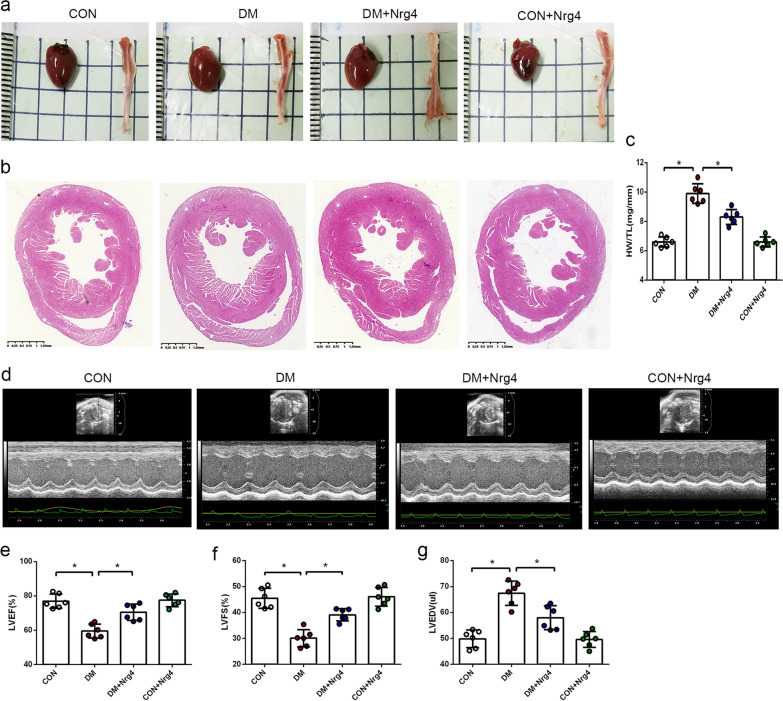
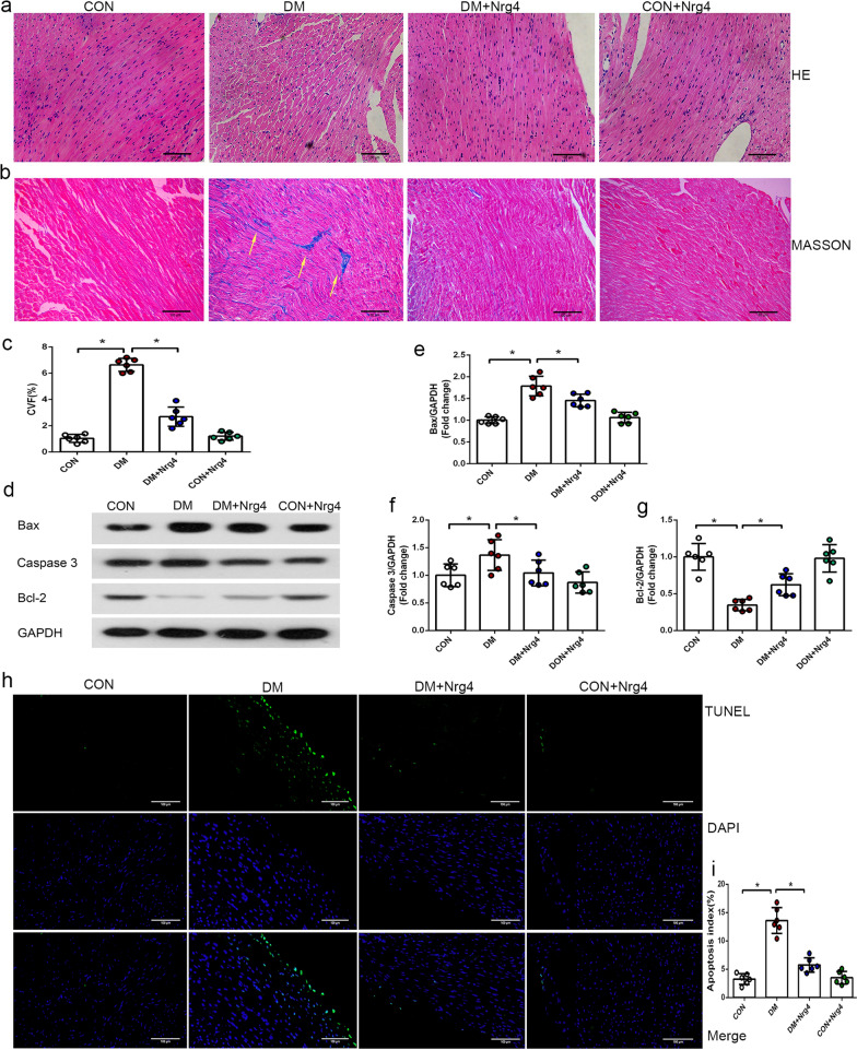
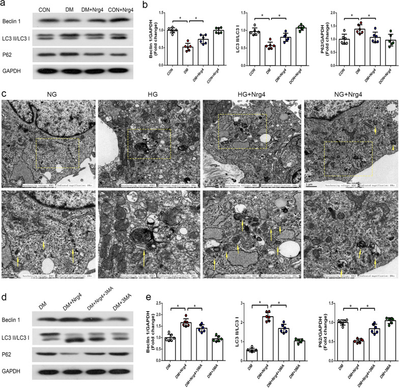
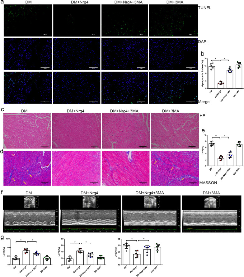
Background: Diabetic cardiomyopathy is characterized by left ventricle dysfunction, cardiomyocyte apoptosis, and interstitial fibrosis and is a serious complication of diabetes mellitus (DM). Autophagy is a mechanism that is essential for maintaining normal heart morphology and function, and its dysregulation can produce pathological effects on diabetic hearts. Neuregulin-4 (Nrg4) is an adipokine that exerts protective effects against metabolic disorders and insulin resistance. The aim of this study was to explore whether Nrg4 could ameliorate DM-induced myocardial injury by regulating autophagy.

Methods: Four weeks after the establishment of a model of type 1 diabetes in mice, the mice received Nrg4 treatment (with or without an autophagy inhibitor) for another 4 weeks. The cardiac functions, histological structures and cardiomyocyte apoptosis were investigated. Autophagy-related protein levels along with related signalling pathways that regulate autophagy were evaluated. In addition, the effects of Nrg4 on autophagy were also determined in cultured primary cardiomyocytes.

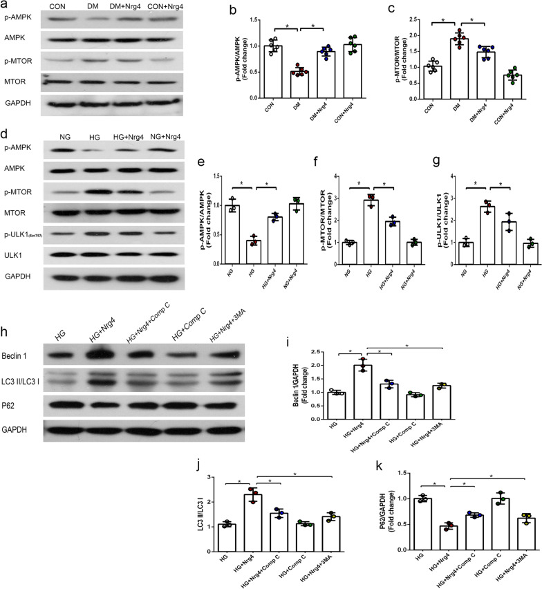
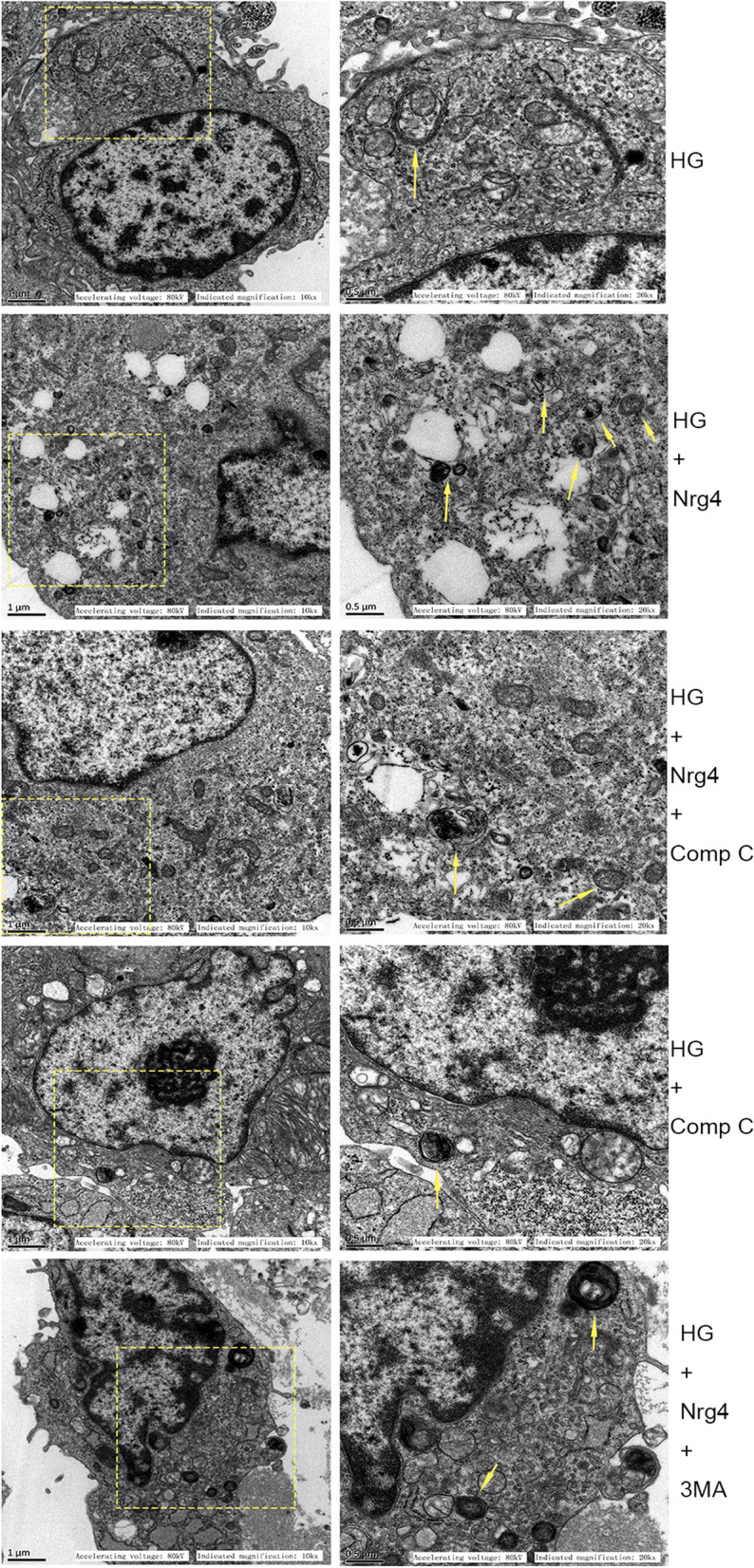
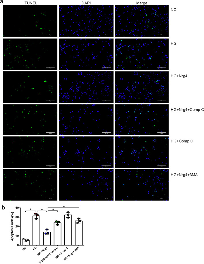
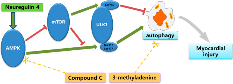
Results: Nrg4 alleviated myocardial injury both in vivo and in vitro. The autophagy level was decreased in type 1 diabetic mice, and Nrg4 intervention reactivated autophagy. Furthermore, Nrg4 intervention was found to activate autophagy via the AMPK/mTOR signalling pathway. Moreover, when autophagy was suppressed or the AMPK/mTOR pathway was inhibited, the beneficial effects of Nrg4 were diminished.

Conclusion: Nrg4 intervention attenuated diabetic cardiomyopathy by promoting autophagy in type 1 diabetic mice. Additionally, Nrg4 induced autophagy via the AMPK/mTOR signalling pathway.

Keywords: Autophagy; Diabetic cardiomyopathy; Neuregulin-4; Signalling pathway.


Conflict of interest statement

The authors declare that they have no competing interests.

Figures

Fig. 1
Fig. 1
Nrg4 alleviates cardiac hypertrophy and cardiac dysfunction. a Representative cardiac and tibial specimens of each group. b Cross-sectional view of cardiac specimens from each group. c Ratio of heart weight to tibia length (HW/TL). d Representative echocardiographic images. e Left ventricular ejection fraction (LVEF). f Left ventricular fraction shortening (LVFS). g Left ventricular end diastolic volume (LVEDV). Data are presented as the mean ± SD (n = 6 per group). Significant differences are depicted with *P < 0.05
Fig. 2
Fig. 2
Nrg4 alleviates cardiac fibrosis and cardiomyocyte apoptosis. a Left ventricle stained with HE (original magnification ✕ 200). b Interstitial fibrosis of the left ventricles by Masson staining. The collagen fibres are stained blue (with arrows, original magnification ✕ 200). c Comparison of the collagen volume fraction (CVF) in each group. dg Western blot and quantitative analysis of apoptosis-related proteins (Bax, Caspase-3 and Bcl-2). h Representative immunofluorescent images with TUNEL (green) and DAPI (blue) staining (original magnification ✕ 200). i Comparison of the apoptosis index in each group. Data are presented as the mean ± SD (n = 6 per group). Significant differences are depicted with *P < 0.05
Fig. 3
Fig. 3
Nrg4 upregulates the suppressed autophagy in type 1 DM, and 3-MA attenuates Nrg4-upregulated autophagy. a, b Western blot and quantitative analysis of autophagy-related proteins (Beclin1, LC3-II/I and P62) in vivo. c Representative TEM images depicting autophagosomes in vitro, with arrows indicating autophagosomes. d, e Western blot and quantitative analysis of autophagy-related proteins in step 2 of the animal experiment. Data are presented as the mean ± SD (n = 6 per group). Significant differences are depicted with *P < 0.05
Fig. 4
Fig. 4
3-MA attenuates the protective effect of Nrg4 on diabetic cardiomyopathy. a Representative immunofluorescent images with TUNEL (green) and DAPI (blue) staining (original magnification ✕ 200). b Comparison of the apoptosis index in each group. c Stained with HE (original magnification ✕ 200). d Stained with Masson staining (original magnification ✕ 200). e Comparison of CVF in each group. f Representative echocardiographic images. g Quantitative analysis of LVEF, LVFS and LVEDV. Data are presented as the mean ± SD (n = 6 per group). Significant differences are depicted with *P < 0.05
Fig. 5
Fig. 5
Nrg4 activates autophagy via the AMPK/mTOR signalling pathway. a–c Western blot and quantitative analysis of the expression of p-AMPK and p-mTOR in vivo. (n = 6 per group). d–g Western blot and quantitative analysis of p-AMPK, p-mTOR and p-ULK1 in vitro (n = 3 independent experiments). h–k Western blot and quantitative analysis of autophagy-related proteins in vitro (n = 3 independent experiments). All data are presented as the mean ± SD. Significant differences are depicted with *P < 0.05
Fig. 6
Fig. 6
Representative TEM images depicting autophagosomes in the vitro experiment (with arrows indicating autophagosomes)
Fig. 7
Fig. 7
Compound C attenuates the protective effect of Nrg4 on cardiomyocyte apoptosis. a Representative immunofluorescent images with TUNEL in vitro (original magnification × 200). b Apoptosis indexes of different groups in vitro. Data are presented as the mean ± SD (n = 3 independent experiments). Significant differences are depicted with *P < 0.05
Fig. 8
Fig. 8
Schematic diagram of the mechanisms of autophagy induced by Nrg4 intervention

Similar articles

Cited by

References

    1. Tate M, Grieve DJ, Ritchie RH. Are targeted therapies for diabetic cardiomyopathy on the horizon. Clin Sci (Lond) 2017;131(10):897–915. doi: 10.1042/CS20160491. - DOI - PubMed
    1. Kenny HC, Abel ED. Heart failure in type 2 diabetes mellitus. Circ Res. 2019;124(1):121–41. doi: 10.1161/CIRCRESAHA.118.311371. - DOI - PMC - PubMed
    1. Jia G, Hill MA, Sowers JR. Diabetic cardiomyopathy: an update of mechanisms contributing to this clinical entity. Circ Res. 2018;122(4):624–38. doi: 10.1161/CIRCRESAHA.117.311586. - DOI - PMC - PubMed
    1. Seferović PM, Paulus WJ. Clinical diabetic cardiomyopathy: a two-faced disease with restrictive and dilated phenotypes. Eur Heart J. 2015;36(27):1718–27. doi: 10.1093/eurheartj/ehv134. - DOI - PubMed
    1. Wang L, Cai Y, Jian L, Cheung CW, Zhang L, Xia Z. Impact of peroxisome proliferator-activated receptor-α on diabetic cardiomyopathy. Cardiovasc Diabetol. 2021;20(1):2. doi: 10.1186/s12933-020-01188-0. - DOI - PMC - PubMed

Publication types

MeSH terms