Review
. 2022 Oct 7;101(40):e31062.
doi: 10.1097/MD.0000000000031062.

Primary bronchial schwannoma: A case report

Affiliations
Review

Primary bronchial schwannoma: A case report

Yosuke Aoyama et al. Medicine (Baltimore). .

Abstract

Rationale: Bronchial schwannomas are extremely rare among the benign tracheobronchial tumors and little are known about its epidemiology and optimal clinical management. Here, we report a case of bronchial schwannoma in a young Japanese man and clinical implications about epidemiology, symptom, diagnosis, and treatment of bronchial schwannoma.

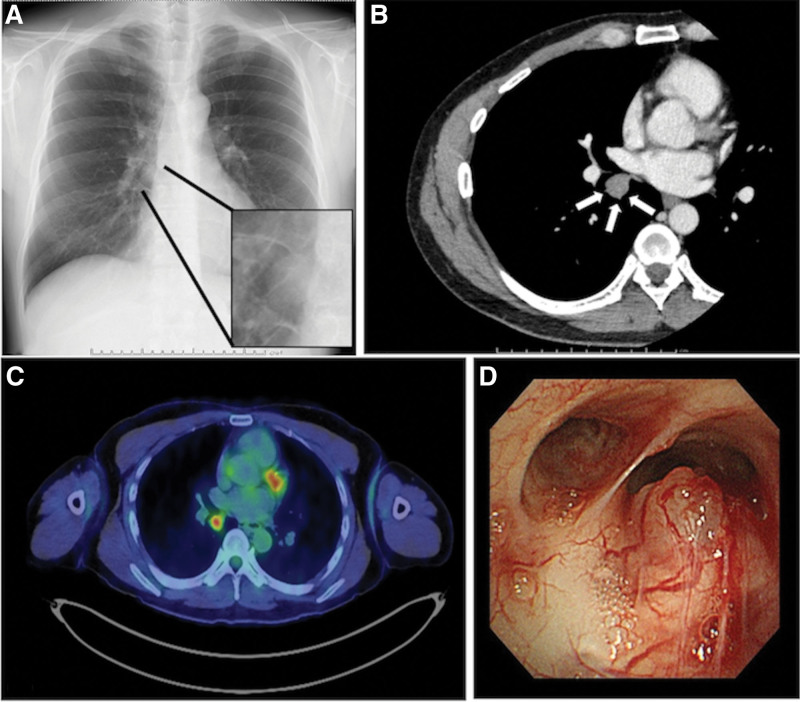
Patients concern: A 37-year-old man visited our department with a nodule incidentally found on his chest radiograph during a routine medical checkup.

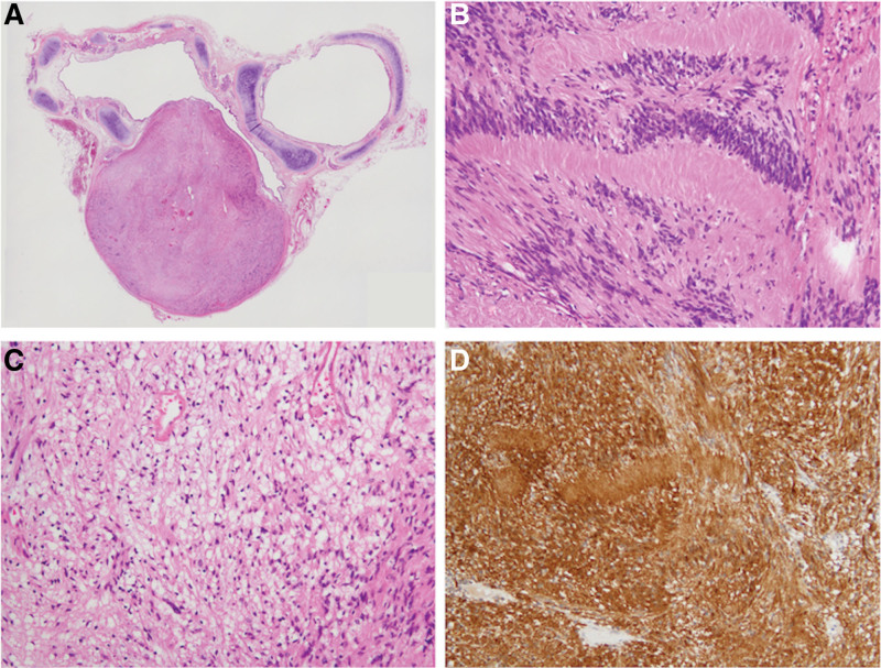
Diagnosis: The tumor was diagnosed as a bronchial schwannoma after pathological evaluation. Microscopically, the tumor consisted of spindle cell proliferation characterized by an alternating highly ordered cellular Antoni A component with occasional nuclear palisading and a loose myxoid Antoni B component. Tumor cells were immunoreactive for S100 but not for smooth muscle actin or KIT.

Interventions: A video-assisted right middle and lower bilobectomy was performed.

Outcome: He remains under observation without recurrence.

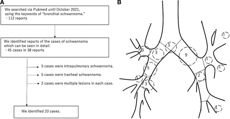
Lessons: In our review, many reports have come from Asian countries. Bronchial schwannoma can occur within a wide range of age groups and in both men and women. No difference in incidence was observed between right and left bronchial tree. Bronchial schwannoma is sometimes difficult to differentiate from malignant diseases. We should include bronchial schwannoma as one of the differential diagnoses of primary bronchial tumors.


Conflict of interest statement

The authors have no funding and conflicts of interest to disclose.

Figures

Figure 1.
Figure 1.
(a) A nodule is noted on the chest radiograph (black arrows). (b) A 17-mm-sized nodule, with a mild contrast effect, is noted on contrast-enhanced CT (white arrows). (c) 18F-fluorodeoxyglucose (FDG) positron emission tomography/computed tomography (PET-CT) reveals abnormal FDG accumulation in the nodule, with a maximum standardized uptake value of 4.07. (d) Bronchoscopy reveals a non-pulsating submucosal nodule, with proliferating capillary vessels, on the mucosal surface of the proximal end of the right intermediate bronchus.
Figure 2.
Figure 2.
(a) Loupe view. A dumbbell-shaped nodule, measuring 20 × 15 × 15 mm in dimension, appears to be localized within the bronchial wall, limited by the bronchial adventitia, and protruding between the cricoid cartilages into the lumen. (b–d) Histological and immunohistochemical findings. (b, c) hematoxylin and eosin stains. The tumor consists of spindle cell proliferation characterized by an alternating highly ordered cellular Antoni A component (b) with occasional nuclear palisading and a loose myxoid Antoni B component (c). Tumor cells were immunoreactive to S100 (d). The nodule was histologically diagnosed as a schwannoma.
Figure 3.
Figure 3.
(a) The review process of previously reported cases of bronchial schwannomas. (b) The number of schwannomas encountered per bronchial location is tallied from the previously reported cases. Three schwannomas were in the carina, three in the right main bronchus, three in the right upper bronchus, five in the intermediate bronchus, one in the right middle bronchus, three in the right lower bronchus, nine in the left main bronchus, three in left upper bronchus area, and three in left lower bronchus area.

References

    1. Shah H, Garbe L, Nussbaum E, et al. . Benign tumors of the tracheobronchial tree. Endoscopic characteristics and role of laser resection. Chest. 1995;107:1744–51. - PubMed
    1. Hilton DA, Hanemann CO. Schwannomas and their pathogenesis. Brain Pathol. 2014;24:205–20. - PMC - PubMed
    1. Komatsu M, Hachiya T, Takahashi H, et al. . Left main bronchial stenosis due to schwannoma. Intern Med. 2018;57:1947–8. - PMC - PubMed
    1. Zhou D, Xing X, Fan J, et al. . PD-1/PD-L1 negative schwannoma mimicking obstructive bronchial malignancy: a case report. Thorac Cancer. 2020;11:2335–8. - PMC - PubMed
    1. Guerreiro C, Dionísio J, da Costa JD. Endobronchial schwannoma involving the carina. Arch Bronconeumol. 2017;53:452. - PubMed