Effect of sedatives or anesthetics on the measurement of resting brain function in common marmosets
- PMID: 36222604
- PMCID: PMC10151911
- DOI: 10.1093/cercor/bhac406
Effect of sedatives or anesthetics on the measurement of resting brain function in common marmosets
Abstract
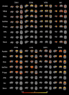
Common marmosets are promising laboratory animals for the study of higher brain functions. Although there are many opportunities to use sedatives and anesthetics in resting brain function measurements in marmosets, their effects on the resting-state network remain unclear. In this study, the effects of sedatives or anesthetics such as midazolam, dexmedetomidine, co-administration of isoflurane and dexmedetomidine, propofol, alfaxalone, isoflurane, and sevoflurane on the resting brain function in common marmosets were evaluated using independent component analysis, dual regression analysis, and graph-theoretic analysis; and the sedatives or anesthetics suitable for the evaluation of resting brain function were investigated. The results show that network preservation tendency under light sedative with midazolam and dexmedetomidine is similar regardless of the type of target receptor. Moreover, alfaxalone, isoflurane, and sevoflurane have similar effects on resting state brain function, but only propofol exhibits different tendencies, as resting brain function is more preserved than it is following the administration of the other anesthetics. Co-administration of isoflurane and dexmedetomidine shows middle effect between sedatives and anesthetics.
Keywords: anesthetic effects; common marmoset; resting state network; resting-state functional magnetic resonance imaging.
© The Author(s) 2022. Published by Oxford University Press.
Figures


Similar articles
-
Effect of prolonged sedation with dexmedetomidine, midazolam, propofol, and sevoflurane on sleep homeostasis in rats.Br J Anaesth. 2024 Jun;132(6):1248-1259. doi: 10.1016/j.bja.2023.11.014. Epub 2023 Dec 8. Br J Anaesth. 2024. PMID: 38071152 Free PMC article.
-
Cerebrospinal fluid metabolic profiling reveals divergent modulation of pentose phosphate pathway by midazolam, propofol and dexmedetomidine in patients with subarachnoid hemorrhage: a cohort study.BMC Anesthesiol. 2022 Jan 27;22(1):34. doi: 10.1186/s12871-022-01574-z. BMC Anesthesiol. 2022. PMID: 35086470 Free PMC article.
-
Bispectral analysis measures sedation and memory effects of propofol, midazolam, isoflurane, and alfentanil in healthy volunteers.Anesthesiology. 1997 Apr;86(4):836-47. doi: 10.1097/00000542-199704000-00014. Anesthesiology. 1997. PMID: 9105228
-
Anesthetic Agents Commonly Used by Oral and Maxillofacial Surgeons.Oral Maxillofac Surg Clin North Am. 2018 May;30(2):155-164. doi: 10.1016/j.coms.2018.01.003. Oral Maxillofac Surg Clin North Am. 2018. PMID: 29622309 Review.
-
Sedative choice in drug-induced sleep endoscopy: A neuropharmacology-based review.Laryngoscope. 2017 Jan;127(1):273-279. doi: 10.1002/lary.26132. Epub 2016 Jul 1. Laryngoscope. 2017. PMID: 27363604 Review.
Cited by
-
Comprehensive profiling of anaesthetised brain dynamics across phylogeny.bioRxiv [Preprint]. 2025 Mar 24:2025.03.22.644729. doi: 10.1101/2025.03.22.644729. bioRxiv. 2025. PMID: 40196621 Free PMC article. Preprint.
-
A Novel Directed Seed-Based Connectivity Analysis Toolbox Applied to Human and Marmoset Resting-State FMRI.J Neurosci. 2024 Nov 6;44(45):e0389242024. doi: 10.1523/JNEUROSCI.0389-24.2024. J Neurosci. 2024. PMID: 39299799 Free PMC article.
-
Projection-Specific Intersectional Optogenetics for Precise Excitation and Inhibition in the Marmoset Brain.bioRxiv [Preprint]. 2025 Aug 15:2025.06.18.660378. doi: 10.1101/2025.06.18.660378. bioRxiv. 2025. PMID: 40667064 Free PMC article. Preprint.
-
A reappraisal of the default mode and frontoparietal networks in the common marmoset brain.Front Neuroimaging. 2024 Jan 9;2:1345643. doi: 10.3389/fnimg.2023.1345643. eCollection 2023. Front Neuroimaging. 2024. PMID: 38264540 Free PMC article.
-
Commonality and variance of resting-state networks in common marmoset brains.Sci Rep. 2024 Apr 9;14(1):8316. doi: 10.1038/s41598-024-58799-w. Sci Rep. 2024. PMID: 38594386 Free PMC article.
References
Publication types
MeSH terms
Substances
LinkOut - more resources
Full Text Sources

