. 2022 Dec 15;28(24):5383-5395.
doi: 10.1158/1078-0432.CCR-22-1206.

Gene-Expression Profiling of Mucinous Ovarian Tumors and Comparison with Upper and Lower Gastrointestinal Tumors Identifies Markers Associated with Adverse Outcomes

Nicola S Meagher  1   2 Kylie L Gorringe  3   4 Matthew Wakefield  5   6   7 Adelyn Bolithon  1   2 Chi Nam Ignatius Pang  8   9 Derek S Chiu  10 Michael S Anglesio  10   11 Kylie-Ann Mallitt  1   12 Jennifer A Doherty  13 Holly R Harris  14   15 Joellen M Schildkraut  16 Andrew Berchuck  17 Kara L Cushing-Haugen  14 Ksenia Chezar  18 Angela Chou  19   20   21 Adeline Tan  22   23 Jennifer Alsop  24 Ellen Barlow  25 Matthias W Beckmann  26 Jessica Boros  21   27   28 David D L Bowtell  3   4 AOCS GroupAlison H Brand  21   28 James D Brenton  29 Ian Campbell  3   4 Dane Cheasley  3   4 Joshua Cohen  30 Cezary Cybulski  31 Esther Elishaev  32 Ramona Erber  33 Rhonda Farrell  21   34 Anna Fischer  35 Zhuxuan Fu  36 Blake Gilks  37 Anthony J Gill  19   20   21 Australian Pancreatic Genome InitiativeCharlie Gourley  38 Marcel Grube  39 Paul R Harnett  21   40 Arndt Hartmann  35 Anusha Hettiaratchi  41 Claus K Høgdall  42 Tomasz Huzarski  31   43 Anna Jakubowska  31   44 Mercedes Jimenez-Linan  45 Catherine J Kennedy  21   27   28 Byoung-Gie Kim  46 Jae-Weon Kim  47 Jae-Hoon Kim  48 Kayla Klett  49 Jennifer M Koziak  50 Tiffany Lai  30 Angela Laslavic  51 Jenny Lester  30 Yee Leung  22   52   53 Na Li  3   54 Winston Liauw  1   55 Belle W X Lim  3 Anna Linder  56 Jan Lubiński  31 Sakshi Mahale  3 Constantina Mateoiu  57 Simone McInerny  3   54 Janusz Menkiszak  58 Parham Minoo  18 Suzana Mittelstadt  39 David Morris  59 Sandra Orsulic  30 Sang-Yoon Park  60 Celeste Leigh Pearce  61   62 John V Pearson  63 Malcolm C Pike  62   64 Carmel M Quinn  41 Ganendra Raj Mohan  52   65   66 Jianyu Rao  67 Marjorie J Riggan  17 Matthias Ruebner  26 Stuart Salfinger  65 Clare L Scott  4   5   6   7 Mitul Shah  24 Helen Steed  68   69 Colin J R Stewart  70 Deepak Subramanian  3 Soseul Sung  71   72   73 Katrina Tang  74 Paul Timpson  19 Robyn L Ward  21 Rebekka Wiedenhoefer  35 Heather Thorne  3 kConFab InvestigatorsPaul A Cohen  22   65 Philip Crowe  1   75 Peter A Fasching  26 Jacek Gronwald  31 Nicholas J Hawkins  1 Estrid Høgdall  76 David G Huntsman  11   77 Paul A James  3   54 Beth Y Karlan  30 Linda E Kelemen  78 Stefan Kommoss  39 Gottfried E Konecny  30 Francesmary Modugno  36   49   51 Sue K Park  72   73   79 Annette Staebler  35 Karin Sundfeldt  56 Anna H Wu  62 Aline Talhouk  10   11   37 Paul D P Pharoah  24   80 Lyndal Anderson  21   81 Anna DeFazio  21   27   28   82 Martin Köbel  18 Michael L Friedlander  1   25   83 Susan J Ramus  1   2
Affiliations

Gene-Expression Profiling of Mucinous Ovarian Tumors and Comparison with Upper and Lower Gastrointestinal Tumors Identifies Markers Associated with Adverse Outcomes

Nicola S Meagher et al. Clin Cancer Res. .

Abstract

Purpose: Advanced-stage mucinous ovarian carcinoma (MOC) has poor chemotherapy response and prognosis and lacks biomarkers to aid stage I adjuvant treatment. Differentiating primary MOC from gastrointestinal (GI) metastases to the ovary is also challenging due to phenotypic similarities. Clinicopathologic and gene-expression data were analyzed to identify prognostic and diagnostic features.

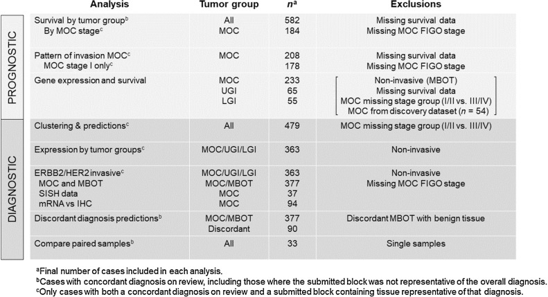
Experimental design: Discovery analyses selected 19 genes with prognostic/diagnostic potential. Validation was performed through the Ovarian Tumor Tissue Analysis consortium and GI cancer biobanks comprising 604 patients with MOC (n = 333), mucinous borderline ovarian tumors (MBOT, n = 151), and upper GI (n = 65) and lower GI tumors (n = 55).

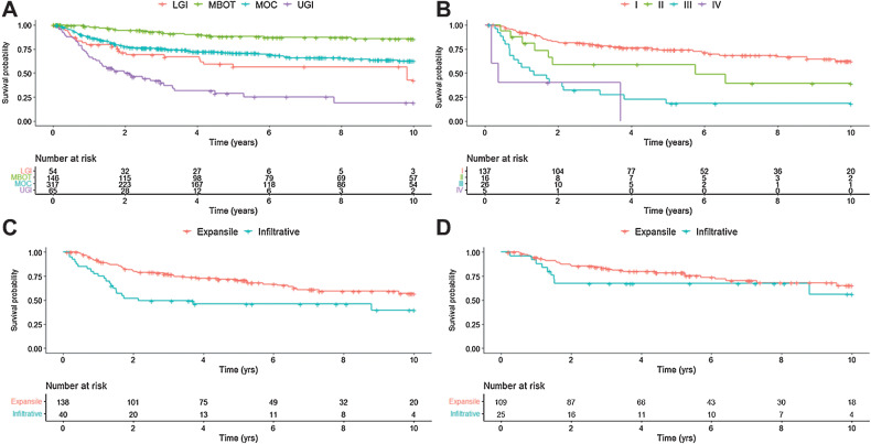
Results: Infiltrative pattern of invasion was associated with decreased overall survival (OS) within 2 years from diagnosis, compared with expansile pattern in stage I MOC [hazard ratio (HR), 2.77; 95% confidence interval (CI), 1.04-7.41, P = 0.042]. Increased expression of THBS2 and TAGLN was associated with shorter OS in MOC patients (HR, 1.25; 95% CI, 1.04-1.51, P = 0.016) and (HR, 1.21; 95% CI, 1.01-1.45, P = 0.043), respectively. ERBB2 (HER2) amplification or high mRNA expression was evident in 64 of 243 (26%) of MOCs, but only 8 of 243 (3%) were also infiltrative (4/39, 10%) or stage III/IV (4/31, 13%).

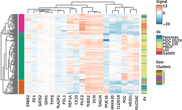
Conclusions: An infiltrative growth pattern infers poor prognosis within 2 years from diagnosis and may help select stage I patients for adjuvant therapy. High expression of THBS2 and TAGLN in MOC confers an adverse prognosis and is upregulated in the infiltrative subtype, which warrants further investigation. Anti-HER2 therapy should be investigated in a subset of patients. MOC samples clustered with upper GI, yet markers to differentiate these entities remain elusive, suggesting similar underlying biology and shared treatment strategies.


Figures

Figure 1. Schema of study numbers for each analysis to describe different cohort numbers due to pathology review and missing data. MOC, mucinous ovarian carcinoma; MBOT, mucinous borderline ovarian tumor; LGI, lower gastrointestinal; UGI, upper gastrointestinal; SISH, silver in situ hybridization.
Figure 1.
Schema of study numbers for each analysis to describe different cohort numbers due to pathology review and missing data. MOC, mucinous ovarian carcinoma; MBOT, mucinous borderline ovarian tumor; LGI, lower gastrointestinal; UGI, upper gastrointestinal; SISH, silver in situ hybridization.
Figure 2. Kaplan–Meier curves of OS in (A) main tumor groups (n = 582)—MBOT, MOC, LGI, and UGI—B, patients with MOC by FIGO stage (n = 184); C, patients with MOC by pattern of invasion in all stages (n = 178); D, patients with stage I MOC (n = 134) by pattern of invasion.
Figure 2.
Kaplan–Meier curves of OS in (A) main tumor groups (n = 582)—MBOT, MOC, LGI, and UGI; (B) patients with MOC by FIGO stage (n = 184); (C) patients with MOC by pattern of invasion in all stages (n = 178); and (D) patients with stage I MOC (n = 134), by pattern of invasion.
Figure 3. Heat map of unsupervised clustering analysis. Contains all samples with a concordant pathology diagnosis (n = 497), with MOC grouped by FIGO stage. Labels show main clusters and diagnoses. Gene-expression values are normalized and logarithm base 2 transformed. Dx, diagnosis.
Figure 3.
Heat map of unsupervised clustering analysis. Contains all samples with a concordant pathology diagnosis (n = 497), with MOC grouped by FIGO stage. Labels show main clusters and diagnoses. Gene-expression values are normalized and logarithm base 2 transformed. dx, diagnosis.

References

    1. Kelemen LE, Kobel M. Mucinous carcinomas of the ovary and colorectum: different organ, same dilemma. Lancet Oncol 2011;12:1071–80. - PubMed
    1. Zaino RJ, Brady MF, Lele SM, Michael H, Greer B, Bookman MA. Advanced stage mucinous adenocarcinoma of the ovary is both rare and highly lethal: a gynecologic oncology group study. Cancer 2011;117:554–62. - PMC - PubMed
    1. Seidman JD, Kurman RJ, Ronnett BM. Primary and metastatic mucinous adenocarcinomas in the ovaries: incidence in routine practice with a new approach to improve intraoperative diagnosis. Am J Surg Pathol 2003;27:985–93. - PubMed
    1. Meagher NS, Wang L, Rambau PF, Intermaggio MP, Huntsman DG, Wilkens LR, et al. A combination of the immunohistochemical markers CK7 and SATB2 is highly sensitive and specific for distinguishing primary ovarian mucinous tumors from colorectal and appendiceal metastases. Mod Pathol 2019;32:1834–46. - PMC - PubMed
    1. Ackroyd SA, Goetsch L, Brown J, Houck K, Wang C, Hernandez E. Pancreaticobiliary metastasis presenting as primary mucinous ovarian neoplasm: a systematic literature review. Gynecol Oncol Rep 2019;28:109–15. - PMC - PubMed

Publication types