

Clinical bracket failure rates between different bonding techniques: a systematic review and meta-analysis
- PMID: 36222731
- PMCID: PMC10065138
- DOI: 10.1093/ejo/cjac050
Clinical bracket failure rates between different bonding techniques: a systematic review and meta-analysis
Abstract
Background: Bracket failure increases the treatment time of orthodontic therapy and burdens patients with unnecessary costs, increased chair time, and possible new appointments.
Objective: To compare the bond failures of different orthodontic materials based on the results of available clinical studies.
Search methods: A systematic search of clinical trials was performed in the Cochrane, Embase, and Pubmed databases with no limitations. The list of investigated techniques contained conventional acid-etch primer (CM-AEP), self-etch primer (SEP), self-cure resin (SCR), and simple or resin-modified glass ionomer (RM-GIC) materials and procedures.
Selection criteria: Clinical studies reporting the failure rate of bonded brackets after using direct adhesive techniques on buccal sites of healthy teeth were included.
Data collection and analysis: Bracket failure rates from eligible studies were extracted by two authors independently. Risk ratios (RRs) were pooled using the random-effects model with DerSimonian-Laird estimation.
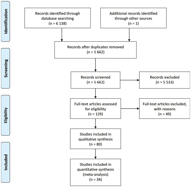
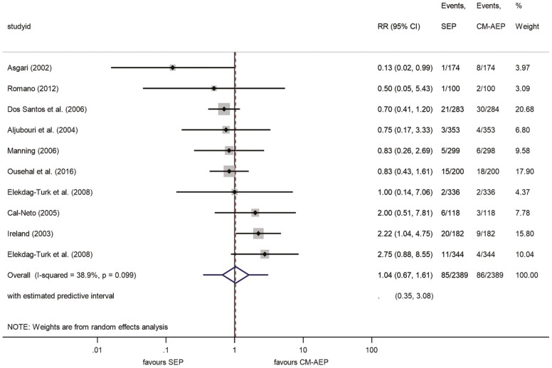
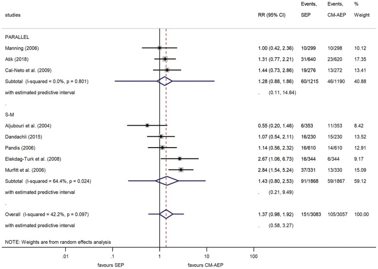
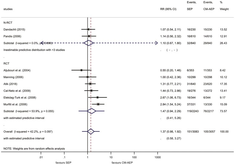
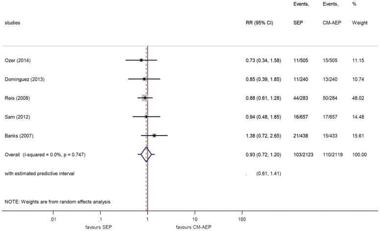
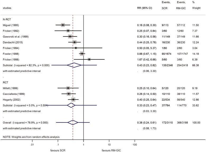
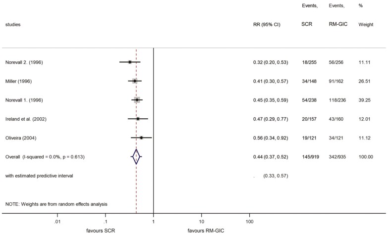
Results: Thirty-four publications, involving 1221 patients, were included. Our meta-analysis revealed no significant difference in the risk of bracket failures between SEP and CM-AEP. After 6, 12, and 18 months of bonding, the values of RR were 1.04 [95% confidence interval (CI), 0.67-1.61], 1.37 (95% CI, 0.98-1.92), and 0.93 (95% CI, 0.72-1.20), respectively. At 18 months, bracket failure was 4.9 and 5.2% for SEP and CM-AEP, respectively. Heterogeneity was good or moderate (I2 < 42.2%). The results of RM-GIC at 12 months indicated a 57% lower risk of bracket failure using SCR as compared with RM-GIC (RR: 0.38; 95% CI, 0.24-0.61). At 18 months, bracket failures for SCR and RM-GIC were 15.8 and 36.6%, respectively (RR: 0.44; 95% CI, 0.37-0.52, I2 = 78.9%), demonstrating three to six times higher failure rate than in the case of etching primer applications.
Limitations: A major limitation of the present work is that the included clinical trials, with no exceptions, showed variable levels of risk of bias. Another possible problem affecting the outcome is the difference between the clustering effects of the split mouth and the parallel group bracket allocation methods.
Conclusions and implications: The results revealed no significant difference between SEP and CM-AEP up to 18 months after application. RM-GIC had much worse failure rates than acid-etching methods; additionally, the superiority of SCR over RM-GIC was evident, indicating strong clinical relevance.
Registration: Prospero with CRD42020163362.
© The Author(s) 2022. Published by Oxford University Press on behalf of the European Orthodontic Society.
Conflict of interest statement
None to declare.
Figures
Similar articles
-
Comparison of self-etch primers with conventional acid-etch technique for bonding brackets in orthodontics: a systematic review and meta-analysis.Eur J Orthod. 2022 Aug 16;44(4):385-395. doi: 10.1093/ejo/cjab076. Eur J Orthod. 2022. PMID: 35022707
-
Comparison of the failure rate, bonding time and ARI score of two orthodontic bonding systems: Self-Etch Primer and Conventional Etching Primer: A systematic review and meta-analysis.Int Orthod. 2021 Dec;19(4):566-579. doi: 10.1016/j.ortho.2021.09.001. Epub 2021 Oct 8. Int Orthod. 2021. PMID: 34629307
-
Clinical bond failure rates of adhesive precoated self-ligating brackets using a self-etching primer.Angle Orthod. 2014 Jan;84(1):155-60. doi: 10.2319/022013-149.1. Epub 2013 Jul 2. Angle Orthod. 2014. PMID: 23819593 Free PMC article. Clinical Trial.
-
Six-month bracket failure rate evaluation of a self-etching primer.Eur J Orthod. 2008 Apr;30(2):211-6. doi: 10.1093/ejo/cjm119. Epub 2008 Jan 23. Eur J Orthod. 2008. PMID: 18216373 Clinical Trial.
-
A clinical evaluation of a glass ionomer cement as an orthodontic bonding adhesive compared with an acrylic resin.Eur J Orthod. 1996 Aug;18(4):373-84. doi: 10.1093/ejo/18.4.373. Eur J Orthod. 1996. PMID: 8921659 Clinical Trial.
Cited by
-
Bracket Bond Failures: Incidence and Association with Different Risk Factors-A Retrospective Study.Int J Environ Res Public Health. 2023 Mar 2;20(5):4452. doi: 10.3390/ijerph20054452. Int J Environ Res Public Health. 2023. PMID: 36901461 Free PMC article.
-
Does the use of universal adhesive systems improve the durability of the bond strength of orthodontic brackets to enamel?J Clin Exp Dent. 2024 Feb 1;16(2):e178-e185. doi: 10.4317/jced.61247. eCollection 2024 Feb. J Clin Exp Dent. 2024. PMID: 38496803 Free PMC article.
-
Shear bond strength of a RMGIC for orthodontic bracket bonding to enamel.BDJ Open. 2024 Jan 2;10(1):1. doi: 10.1038/s41405-023-00181-5. BDJ Open. 2024. PMID: 38167700 Free PMC article.
-
Metallic vs Ceramic Bracket Failures After 12 Months of Treatment: A Prospective Clinical Trial.Int Dent J. 2024 Dec;74(6):1371-1377. doi: 10.1016/j.identj.2024.04.023. Epub 2024 May 13. Int Dent J. 2024. PMID: 38744578 Free PMC article. Clinical Trial.
-
Evaluation of the Conventional Acid-Etching System and the Self-Etching Primer in Bonding Metallic Orthodontic Brackets: An In-Vitro and In-Vivo Study.Cureus. 2024 Oct 23;16(10):e72226. doi: 10.7759/cureus.72226. eCollection 2024 Oct. Cureus. 2024. PMID: 39445044 Free PMC article.
References
-
- Gange, P. (2015) The evolution of bonding in orthodontics. American Journal of Orthodontics and Dentofacial Orthopedics, 147, S56–S63. - PubMed
-
- Skidmore, K.J., Brook, K.J., Thomson, W.M. and Harding, W.J. (2006) Factors influencing treatment time in orthodontic patients. American Journal of Orthodontics and Dentofacial Orthopedics, 129, 230–238. - PubMed
-
- Brown, K. (2009) The impact of bonding material on bracket failure rate. Vital, 6, 28–30.
-
- Chinvipas, N. and Hasegawa, Y. (2014) Repeated bonding of fixed retainer increases the risk of enamel fracture. Odontology, 102, 89–97. - PubMed
-
- Chaudhari, P.K., Goyal, L., Rana, S.S., Dhingra, K. and Kshetrimayum, N. (2018). Nanocomposites and nanoionomers for orthodontic bracket bonding. In Asiri A.M., Mohammad A. (eds.), Applications of Nanocomposite Materials in Dentistry. Woodhead Publishing Series in Biomaterials, Elsevier, pp. 171–180.
