Review
. 2022 Oct 1;28(5):1435-1475.
doi: 10.1212/CON.0000000000001159.

The Dystonias

Review

The Dystonias

Christopher D Stephen. Continuum (Minneap Minn). .

Abstract

Purpose of review: This article discusses the most recent findings regarding the diagnosis, classification, and management of genetic and idiopathic dystonia.

Recent findings: A new approach to classifying dystonia has been created with the aim to increase the recognition and diagnosis of dystonia. Molecular biology and genetic studies have identified several genes and biological pathways involved in dystonia.

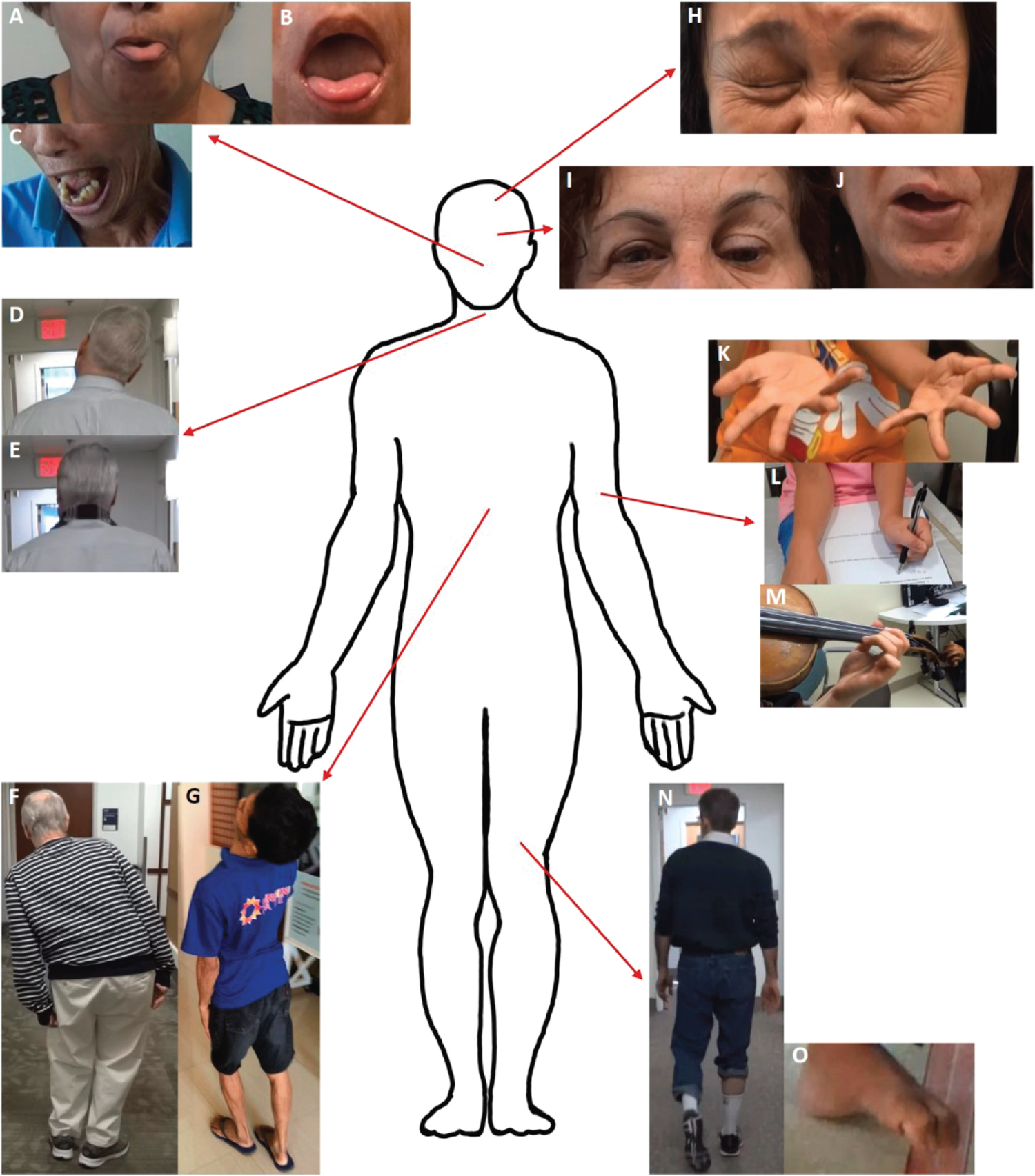
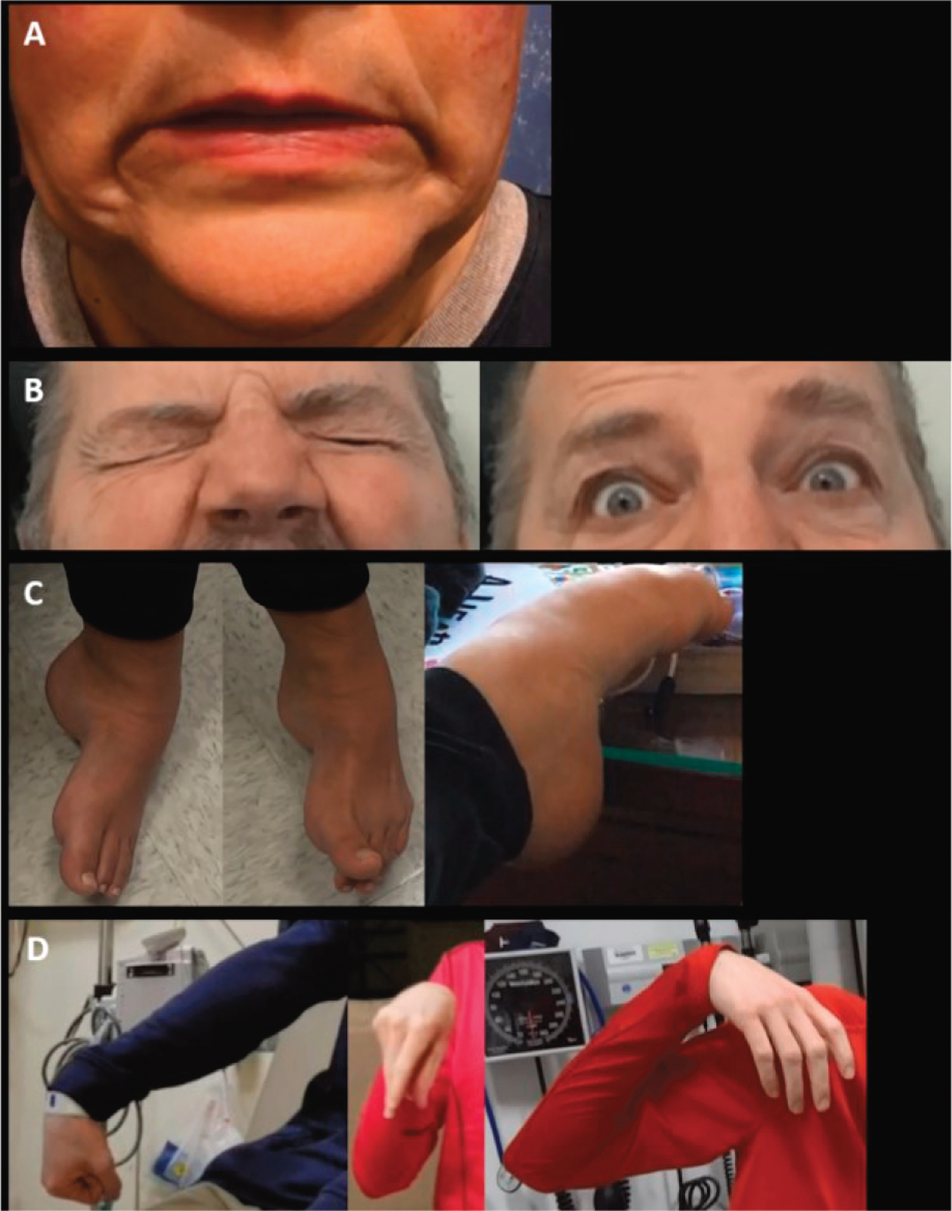
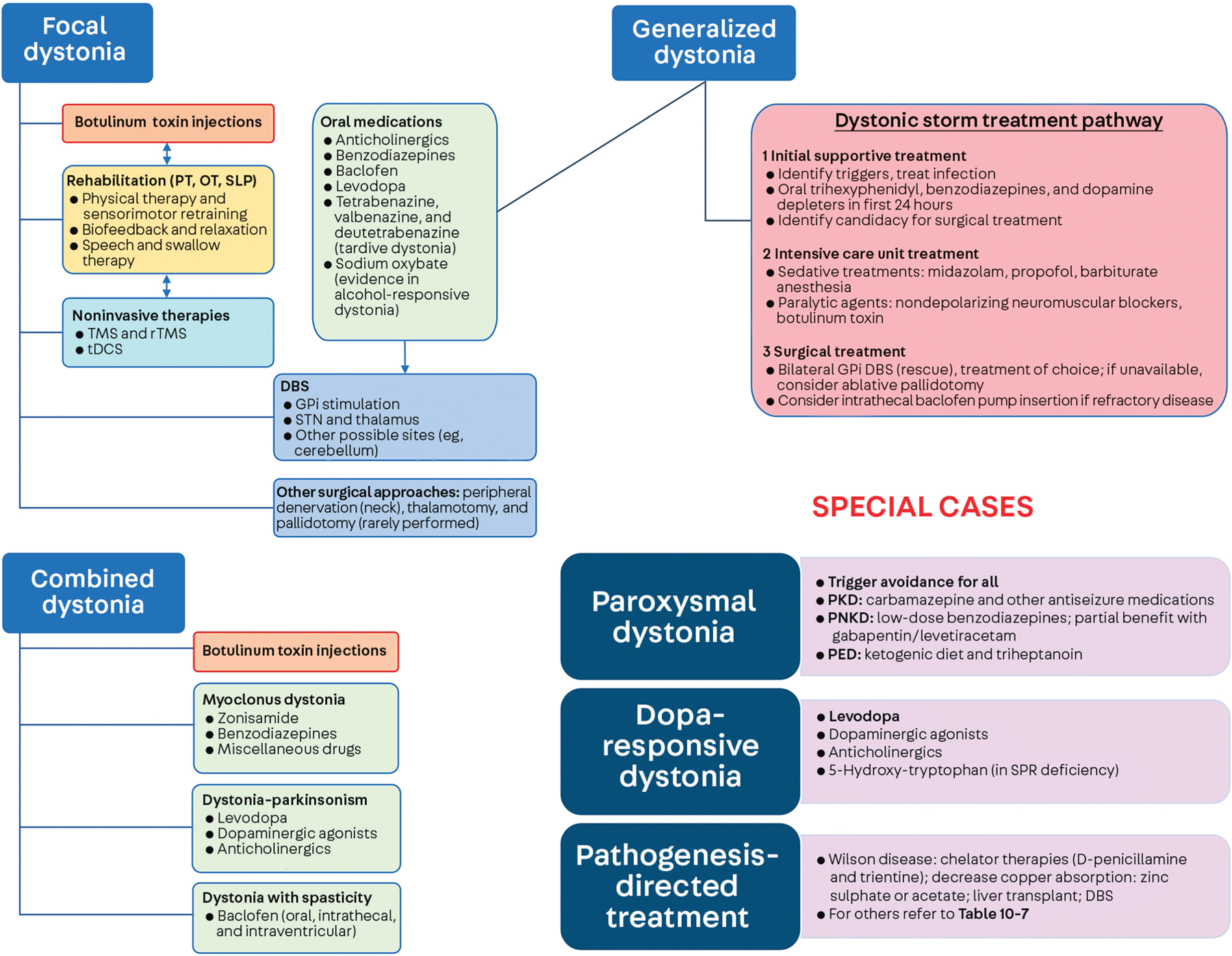
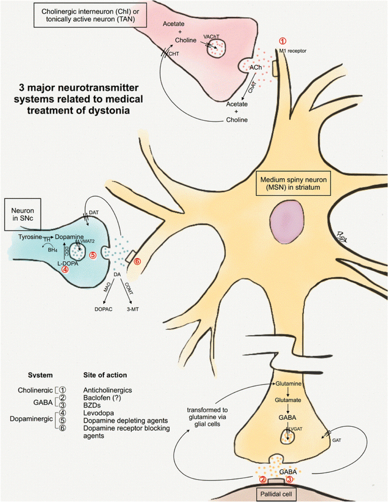
Summary: Dystonia is a common movement disorder involving abnormal, often twisting, postures and is a challenging condition to diagnose. The pathophysiology of dystonia involves abnormalities in brain motor networks in the context of genetic factors. Dystonia has genetic, idiopathic, and acquired forms, with a wide phenotypic spectrum, and is a common feature in complex neurologic disorders. Dystonia can be isolated or combined with another movement disorder and may be focal, segmental, multifocal, or generalized in distribution, with some forms only occurring during the performance of specific tasks (task-specific dystonia). Dystonia is classified by clinical characteristics and presumed etiology. The management of dystonia involves accurate diagnosis, followed by treatment with botulinum toxin injections, oral medications, and surgical therapies (mainly deep brain stimulation), as well as pathogenesis-directed treatments, including the prospect of disease-modifying or gene therapies.


Figures

FIGURE 10–1
FIGURE 10–1
Clinical phenomenology of dystonia. A, Oromandibular dystonia with tongue protrusion posturing is shown in a patient with tardive dystonia; B, in the same patient, the abnormal tongue posture, with curling in the mouth, is highlighted. C, In a patient with X-linked dystonia parkinsonism (DYT/PARK-TAF1, DYT3), severe jaw opening dystonia is shown as a part of multifocal dystonia, predominantly involving head and neck segmental dystonia. D, Image shows focal idiopathic cervical dystonia with laterocollis (lateral flexion) to the right, (E) with normalization of neck posturing in the same patient by using a sensory trick of wearing a scarf and applying tension to the posterior neck. F, Focal truncal dystonia with Pisa syndrome is shown in a patient with Parkinson disease. G, Image shows severe segmental dystonia involving truncal extension and neck extension (retrocollis). H, Segmental dystonia with idiopathic Meige syndrome is shown, highlighting bilateral eyelid spasm and lower facial spasm. I, Idiopathic left hemifacial spasm is shown, highlighting the left eyebrow elevation in keeping with the “other Babinski sign” (co-contraction of the orbicularis and frontalis muscles, causing ipsilateral eyebrow elevation during unilateral eye closure, which cannot be reproduced voluntarily) and (J) abnormal left lower facial posturing related to lower facial spasms in the same patient with a tortuous vertebrobasilar system, which may be an etiologic factor. K, Multifocal bilateral hand dystonia is shown in a patient with rapid-onset dystonia parkinsonism (DYT/PARK-ATP1A3). L, Multifocal bilateral arm dystonia is shown with notable abnormal wrist extensor posturing and right shoulder adduction in a patient with DYT-THAP1 (DYT6) dystonia. M, Task-specific focal hand dystonia is shown in a violinist who exhibits flexion dystonia of the left little greater than ring fingers. N, Focal left foot dystonia involves excessive plantarflexion in a patient with task-specific walking/running dystonia. O, Left foot dystonia with toe curling is shown in a patient with generalized dystonia in the setting of X-linked dystonia parkinsonism (DYT/PARK-TAF1, DYT3). Reprinted with permission from Stephen CD, et al, Elsevier. © 2022 Elsevier.
FIGURE 10–2
FIGURE 10–2
Examples of functional dystonia phenomenology, which is distinct from that seen in “organic” dystonia. A, Functional cranial dystonia is shown in a patient with bilateral lip pulling (pulling of this kind is typically unilateral in “organic” dystonia other than in the setting of risus sardonicus, which is phenomenologically distinct). B, Patient exhibits functional blepharospasm with eyes tightly shut (left) and forcefully open when concentrating (right) (distinct from “organic” dystonia, as the eye closure was sporadic and very severe; in this case, provocative maneuvers, such as tight eye closure, did not trigger spasms, which is highly atypical and highlights the inconsistency). C, Three examples of functional foot dystonia illustrating the typical posturing involving fixed dystonia with plantarflexion and inversion (left); in this same patient, extension of the great toe with flexion of the others toes is shown (middle); in a different patient, paroxysmal dystonia involving plantarflexion and toe curling is shown (right); this is distinct from organic dystonia, as fixed dystonia at onset is highly unusual and tends to occur in more advanced disease, and the symptomatology is inconsistent. D, Three examples are shown of varying dystonic upper extremity posturing in a patient with paroxysmal functional dystonia: the right arm extended with wrist flexion and fisting (left); elbow flexion with wrist flexion akin to carpopedal spasm (middle); and shoulder abduction, elbow flexion, and wrist flexion with a limp hand (right) (distinct from “organic” dystonia where the phenomenology of episodes in paroxysmal dystonia/dyskinesia is typically stereotyped). Reprinted with permission from Frucht L, et al, Front Neurol. © 2020 Frontiers Media S. A.
FIGURE 10–3
FIGURE 10–3
Treatment modalities in dystonia. Treatment approaches to focal, generalized, and combined dystonia, as well as for special cases involving paroxysmal dystonia/dyskinesia, dopa-responsive disorders, and specific pathogenesis-directed treatments. DBS = deep brain stimulation; GPi = globus pallidus internus; OT = occupational therapy; PED = paroxysmal exercise-induced dyskinesia/dystonia; PKD = paroxysmal kinesigenic dyskinesia/dystonia; PNKD = paroxysmal nonkinesigenic dyskinesia/dystonia; PT = physical therapy; rTMS = repetitive transcranial magnetic stimulation; SLP = speech and language pathology; SPR: sepiapterin reductase; STN = subthalamic nucleus; tDCS = transcranial direct current stimulation; TMS = transcranial magnetic stimulation. Modified with permission from Balint B, et al, Nat Rev Dis Primers. © 2018 Springer Nature Limited.
FIGURE 10–4
FIGURE 10–4
Neurochemical pathways and targets for oral pharmacologic treatment in dystonia. The figure illustrates the three striatal neurotransmitters (cholinergic [pink], γ-aminobutyric acid–mediated (GABA-ergic) [yellow, brown], and dopaminergic [blue]), and targets relevant to oral dystonia medications. 1) Cholinergic system: Acetylcholine (ACh) is synthesized in presynaptic terminals, catalyzed by choline acetyltransferase (ChAT), and transported into vesicles. ACh then binds to muscarinic and/or nicotinic receptors for cellular effects. The remaining ACh is metabolized by acetylcholinesterase (AChE), with uptake into the presynaptic terminal by choline transporter [CHT]. 2) GABA-ergic system: GABA is synthesized from glutamate presynaptically and transported to vesicles by vesicular GABA transporter (VGAT), released into synaptic clefts, and binds to postsynaptic receptors. The remaining GABA at the synaptic clefts is transported to presynaptic terminals by direct reuptake and indirect transport. 3) Dopaminergic system: The medium spiny neurons receive input from substantia nigra pars compacta (SNc) neurons. Dopamine is presynaptically synthesized from tyrosine by tyrosine hydroxylase (TH), transported into vesicles by vesicular monoamine transporter 2 (VMAT2), and released into the synaptic cleft, binding to postsynaptic dopamine receptors. Dopamine is degraded by monoamine oxidase (MAO) and catechol-O-methyl transferase (COMT), with remaining dopamine transported to presynaptic terminals by dopamine transporters (DAT). Anticholinergics are postsynaptic muscarinic antagonists. Baclofen is a GABAB agonist acting presynaptically and postsynaptically. Benzodiazepines (BZDs) bind to GABAA receptors, causing increased chloride channel opening and inhibitory signals. Levodopa is postsynaptically converted to dopamine for direct effects. Dopamine-depleting agents (eg, tetrabenazine [TBZ]), are VMAT2 inhibitors, impairing dopamine transport into vesicles, while dopamine receptor blocking agents block postsynaptic dopamine receptors. BH4 = tetrahydrobiopterin; ChI = cholinergic interneurons; DA = dopamine; DDC = dopa decarboxylase; DOPAC = 3, 4-dihydroxyphenylacetic acid; GAT = GABA transporter; L-DOPA = levodopa; MSN = medium spiny neuron; 3-MT = 3-methoxytyramine;TAN = tonically active neuron; VAChT = vesicular ACh transporter. Reprinted with permission from Termsarasab P, et al, J Clin Mov Disord. © 2016 The Authors.

References

    1. Albanese A, Bhatia K, Bressman SB, et al..Phenomenology and classification of dystonia: a consensus update. Mov Disord 2013;28(7): 863–873. doi:10.1002/mds.25475 - DOI - PMC - PubMed
    1. Balint B, Mencacci NE, Valente EM, et al. Dystonia. Nat Rev Dis Primers 2018;4(1):25. doi:10.1038/s41572-018-0023-6 - DOI - PubMed
    1. Battistella G, Termsarasab P, Ramdhani RA, Fuertinger S, Simonyan K. Isolated focal dystonia as a disorder of large-scale functional networks. Cereb Cortex 2017;27(2):1203–1215. doi:10.1093/cercor/bhv313 - DOI - PMC - PubMed
    1. Lalli S, Albanese A. The diagnostic challenge of primary dystonia: evidence from misdiagnosis. Mov Disord 2010;25(11):1619–1626. doi:10.1002/mds.23137 - DOI - PubMed
    1. Evatt ML, Freeman A, Factor S. Adult-onset dystonia. Handb Clin Neurol 2011;100:481–511. doi:10.1016/b978-0-444-52014-2.00037-9 - DOI - PubMed

Substances