Review
. 2022 Oct 12;26(3):31.
doi: 10.1007/s00792-022-01279-8.

Electron transfer of extremophiles in bioelectrochemical systems

Affiliations
Review

Electron transfer of extremophiles in bioelectrochemical systems

Miriam Edel et al. Extremophiles. .

Abstract

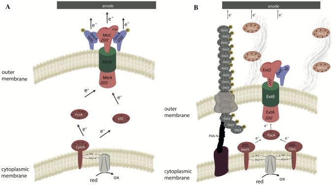
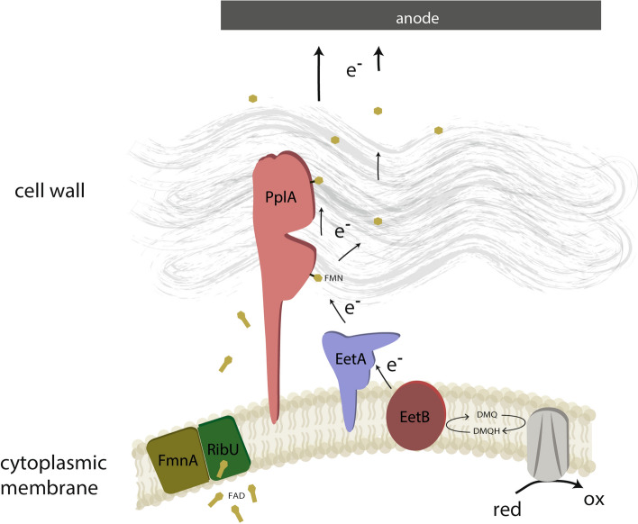
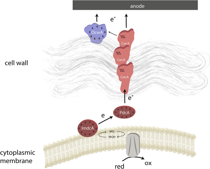
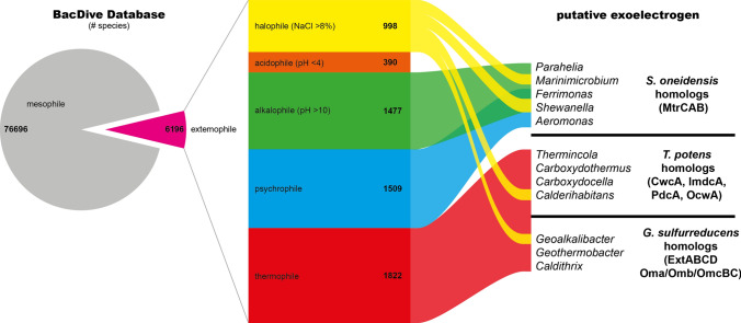
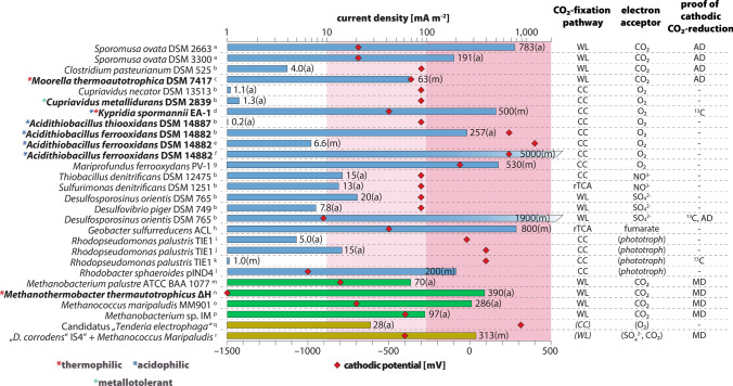
The interaction of bacteria and archaea with electrodes is a relatively new research field which spans from fundamental to applied research and influences interdisciplinary research in the fields of microbiology, biochemistry, biotechnology as well as process engineering. Although a substantial understanding of electron transfer processes between microbes and anodes and between microbes and cathodes has been achieved in mesophilic organisms, the mechanisms used by microbes under extremophilic conditions are still in the early stages of discovery. Here, we review our current knowledge on the biochemical solutions that evolved for the interaction of extremophilic organisms with electrodes. To this end, the available knowledge on pure cultures of extremophilic microorganisms has been compiled and the study has been extended with the help of bioinformatic analyses on the potential distribution of different electron transfer mechanisms in extremophilic microorganisms.

Keywords: Anode interaction; Bioelectrochemical systems; Cathode interaction; Exoelectrogens; Extremophiles; Microbiology; c-type cytochromes.


Figures

Fig. 1
Fig. 1
Scheme of the extracellular electron transfer pathways in S. oneidensis (A) and G. sulfurreducens (B). Diamonds indicate the number of heme cofactors in the respective c-type cytochromes. Scheme modified from Philipp et al. (2020)
Fig. 2
Fig. 2
Model of the extracellular electron transfer in L. monocytogenes. Scheme modified from Light et al. (2018, 2019)
Fig. 3
Fig. 3
Scheme of the extracellular electron transfer pathway in T. ferriacetica. Diamonds indicate the number of heme cofactors in the respective c-type cytochromes. Scheme modified from Faustino et al. (2021)
Fig. 4
Fig. 4
Extremophiles in the BacDive database and genera carrying genes for c-type cytochrome-based EET genes. 7.5% of species listed in the BacDive database are considered as extremophilic. Within these, species of 12 genera carry genes for EET, which are grouped on the righthand side
Fig. 5
Fig. 5
Cathodic current consumption of electroactive, autotrophic organisms, the corresponding CO2-fixation pathway and electron acceptor, and the applied method of cathodic CO2-fixation evidence. Care was taken in the selection of studies to ensure that the cathode served as the sole electron source for CO2-reduction. Extended and modified from Logan (Logan et al. 2019). Bar length corresponds to average (a) or maximum (m) current density. Cathodic potential is indicated by the position of the red diamond. Blue bars are associated with Bacteria, green bars were chosen for Archaea. Yellow bars refer to mixed cultures. WL Wood-Ljungdahl pathway, CC Calvin cycle, rTCA reductive citrate cycle, AD acetate detection, MD methane detection, 13C proof of CO2 fixation by isotope analyses. a: Aryal et al. (2017); b: de Campos Rodrigues and Rosenbaum (2014); c: Yu et al. (2017); d: Reiner et al. (2020); e: Ishii et al. (2015); f: Carbajosa et al. (2010); g: Summers et al. (2013); h: Ueki et al. (2018); i: Doud and Angenent (2014); j: Bose et al. (2014); k: Guzman et al. (2019); l: Schmid et al. (2021); m: Cheng et al. ; n: Sato et al. (2013); o: Lohner et al. (2014); p: Beese-Vasbender, Grote, et al. (2015); q: Wang et al. (2015); r: Deutzmann and Spormann (2016)

References

    1. Aryal N, Tremblay PL, Lizak DM, Zhang T. Performance of different Sporomusa species for the microbial electrosynthesis of acetate from carbon dioxide. Bioresour Technol. 2017;233:184–190. doi: 10.1016/j.biortech.2017.02.128. - DOI - PubMed
    1. Beblawy S, Bursac T, Paquete C, Louro R, Clarke TA, Gescher J. Extracellular reduction of solid electron acceptors by Shewanella oneidensis. Mol Microbiol. 2018;109(5):571–583. doi: 10.1111/MMI.14067. - DOI - PubMed
    1. Beese-Vasbender PF, Nayak S, Erbe A, Stratmann M, Mayrhofer KJJ. Electrochemical characterization of direct electron uptake in electrical microbially influenced corrosion of iron by the lithoautotrophic SRB Desulfopila corrodens strain IS4. Electrochim Acta. 2015;167:321–329. doi: 10.1016/J.ELECTACTA.2015.03.184. - DOI
    1. Bose A, Gardel EJ, Vidoudez C, Parra EA, Girgui PR. Electron uptake by iron-oxidizing phototrophic bacteria. Nat Commun. 2014;5:3391. doi: 10.1038/ncomms4391. - DOI - PubMed
    1. Bratsch SG. Standard electrode potentials and temperature coefficients in water at 298.15 K. J Phys Chem Ref Data. 1989;18:1–21. doi: 10.1063/1.555839. - DOI