Review
. 2024 Mar;291(6):1049-1079.
doi: 10.1111/febs.16650. Epub 2022 Oct 31.

Neuromodulation and the toolkit for behavioural evolution: can ecdysis shed light on an old problem?

Affiliations
Review

Neuromodulation and the toolkit for behavioural evolution: can ecdysis shed light on an old problem?

Luis F Sullivan et al. FEBS J. 2024 Mar.

Abstract

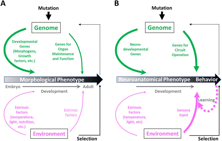
The geneticist Thomas Dobzhansky famously declared: 'Nothing in biology makes sense except in the light of evolution'. A key evolutionary adaptation of Metazoa is directed movement, which has been elaborated into a spectacularly varied number of behaviours in animal clades. The mechanisms by which animal behaviours have evolved, however, remain unresolved. This is due, in part, to the indirect control of behaviour by the genome, which provides the components for both building and operating the brain circuits that generate behaviour. These brain circuits are adapted to respond flexibly to environmental contingencies and physiological needs and can change as a function of experience. The resulting plasticity of behavioural expression makes it difficult to characterize homologous elements of behaviour and to track their evolution. Here, we evaluate progress in identifying the genetic substrates of behavioural evolution and suggest that examining adaptive changes in neuromodulatory signalling may be a particularly productive focus for future studies. We propose that the behavioural sequences used by ecdysozoans to moult are an attractive model for studying the role of neuromodulation in behavioural evolution.

Keywords: Ecdysozoa; Evo-Devo; GPCR; motor programme; neuropeptide.


Conflict of interest statement

Conflicts of Interest: the authors cite no conflicts of interest

Figures

Figure 1:
Figure 1:. Evolution of morphological and behavioral traits.
A) Animal bodies are built during development by genetic programs, which are governed by developmental genes (thick green arrow). Mutations that alter these genes—or where and when they are expressed—give rise to changes in morphological phenotype. Environmental selection of adaptive phenotypes (solid black arrow) fixes the corresponding mutations in the genome (dotted black arrow). Environmental factors (in magenta) can also influence morphological phenotypes both during development and adulthood (e.g. seasonal changes in coat color), but play a secondary role to genetic factors, as indicated by the thinner magenta arrows. Genes for maintaining the function of morphological features, such as organs, are also required post-developmentally. B) The organ most critical for behavior, the nervous system, is composed of neural circuits, which are anatomical features built during development. They are therefore subject to alterations by mutational change in neurodevelopmental genes. These alterations can cause evolutionary changes in neuroanatomical phenotypes and thus animal behavior, which is the target of selection. However, behavior will also be strongly affected by mutations affecting genes responsible for neural circuit operation. This is because behavior is a product of both the structure and function of neural circuits. Importantly, the behavior of an individual animal can also change in response to its interactions with the environment (i.e. sensory input) by learning (thick dotted arrow). Learning alters neural circuit operation and is ultimately also dependent on the genes expressed by neurons in these circuits. Line thicknesses, in general, represent the relative importance of contributions to phenotype, with learning emphasized for behavior in higher animals.
Figure 2:
Figure 2:. Emerging methods for identifying genetic and neural substrates of behavioral evolution
New techniques for characterizing and manipulating both genomes and nervous systems are revolutionizing studies of behavioral evolution. On the side of genomics: whole genome sequencing has enabled genome-wide association studies and quantitative trait loci (QTL) mapping, while transcriptomics has enabled the temporal tracking of gene expression in the brain. On the side of the brain: a range of new genetically encoded tools for monitoring and manipulating neuronal function have enabled the study of brain circuits. Genetic methods now allow many of these methods to be combined so that gene expression, electrical activity, and neuronal function can be selectively characterized or manipulated in targeted neurons. Sophisticated electrophysiological methods, such as the dynamic clamp, also permit manipulation of function. These methods are complemented by a range of new tools for behavioral phenotyping, including computational methods for automating the acquisition and analysis of behavioral.
Figure 3:
Figure 3:. Neuromodulators and their cellular and behavioral actions
A) Neuromodulators represent a diverse group of molecules. These include simple biogenic amines such as dopamine, serotonin, and adrenaline, a broad range of neuropeptides and small peptide hormones, and several gaseous molecules, such as nitric oxide. Most of these molecules act by binding to GPCRs as indicated by the highlight. B) At the cellular level, neuromodulator binding to GPCRs leads to the release of heterotrimeric G-protein subunits, which typically induce the generation of “second messengers,” such as cAMP, Ca++, and IP3. These, in turn, alter the activity of ion channels, enzymes, or GPCRs to change the electrical and synaptic properties of neurons. C) Neuromodulators often act on many cells and cell types within neural circuits to induce changes in cellular and synaptic activity. These circuit-level changes can alter the function of sensorimotor processing and decision-making to alter behavioral output. D) While most molecular components of GPCR signaling are quite conserved in opisthokont lineages (i.e. eukaryotes most closely related to animals), the number of GPCRs themselves has dramatically expanded specifically in animals. Shown is the median number of signaling components of three types (heterotrimeric G-proteins, regulators of G-protein signaling, and GPCRs), calculated for taxa of each clade shown (Figure adapted from de Mendoza et al. [196]; Creative Commons License: http://creativecommons.org/licenses/by-nc/3.0/)
Figure 4:
Figure 4:. Phylogenetic classification of metazoan and ecdysozoan lineages.
A) The Ecdysozoa represent a major clade of Metazoans. They are distinguished by an exoskeleton composed at least partly of chitin that is periodically shed. Together with Lophotrochazoans, the Ecdysozoa make up the Protostomes, one of the two major subdivisions of bilaterian animals. Deuterostomes, which includes vertebrate animals, make up the second major subdivision. (Adapted from Semmens et al. [197]; Creative Commons License: http://creativecommons.org/licenses/by/4.0/. Vertebrate and urochordate images obtained directly from http://phylopic.org.) B) The Ecdysozoa comprise two major groups when classified based on morphology: the Panarthropods and the Cycloneuralians [71]. The former group comprises such segmented animals as insects, crustaceans, myriapods (centipedes, millipedes) and chelicerates (spiders, scorpions, ticks), while the latter group includes nematodes and a variety of other animals distinguished by a central nerve ring. Red circles indicate those taxa for which the endocrinological basis of molting has been at least partially characterized (see text). Descriptions of ecdysis across Ecdysozoan species reveal common recurring themes—behaviors and related features that may represent conserved characteristics within certain groups. Several such features (represented by colored dots) are indicated, together with the groups in which they have been observed and their possible origins in Ecdysozoan evolution. These assignments are necessarily speculative and are intended as suggestive rather than definitive. For many taxa, the number of species studied is small and the behavioral descriptions fragmentary. The absence of a particular feature in the table may thus simply reflect a gap in the published record. Future efforts to categorize ecdysis behaviors for comparative purposes will benefit not only from more extensive and careful observations, but from development of a controlled vocabulary for characterizing ecdysial events. (Phylogenetic tree adapted from Schumann et al. [154]; Creative Commons license: http://creativecommons.org/licenses/by/4.0/.)
Figure 5:
Figure 5:. Endocrinology of insect molting behavior
A) Timeline of Drosophila development showing the fluctuating titers of the steroid hormone 20-hydroxyecdysone (20E), which regulates molting [198]. Peaks in 20E are followed by key developmental transitions, four of them molting events at which the animal sheds its old cuticle and secretes a new one (red lines, “molt”). Molting behaviors (i.e. ecdysis) at each developmental stage are governed by the peptide hormone ETH, which is secreted by Inka cells located on the trachea. Non-molting transitions (hatching, “h,” and puparium formation, “pf”) are also indicated. B) The working model for the endocrinological control of ecdysis in Manduca (see [85]). Low-level of ETH secretion drives “pre-ecdysis” motor patterns by activating abdominal neurons (ABLK) expressing the neuropeptides Leucokinin (Lk) and DH44. ETH also initiates release of the Eclosion Hormone (EH) from ventromedial (Vm) neurons. EH feeds back on the Inka cells to further promote ETH release and surging ETH and EH levels evoke release of Crustacean Cardioactive Peptide (CCAP), which drives the motor programs responsible for “ecdysis” proper and the actual removal of the cuticle. By analogy to its action in flies, bursicon has been proposed to mediate the “postecdysis” motor patterns. In Drosophila, ETH production in the Inka cells has also been shown to be controlled by ecdysone, which initiates a transcriptional cascade in which early-, mid-, and late-acting transcription factors are sequentially activated. Expression of the ETH gene is under the direct control of EcR at the top of the cascade, but ETH release is dependent upon the action of a late-acting transcription factor known as FTZ-f1—a “competence factor” for secretion. In Manduca, the neuropeptide corazonin (Crz) acts as a proximal signal for the release of ETH. C) The principal hormones implicated in the control of insect ecdysis are listed together with the types of receptor they act on. Most receptors of ecdysis hormones are GPCRs.
Figure 6:
Figure 6:. Insect evolution, metamorphosis, and ecdysis
A) The evolution of insects gave rise to three major phylogenetic subdivisions, each characterized by a distinct developmental strategy. The basal Ametabola (blue, a group that also includes non-insect hexapods, the Entognatha) do not undergo metamorphosis and molt throughout their lives; the Hemimetabola (green), undergo partial metamorphosis, typically deploying wings at the final, adult molt; and the most derived orders comprising the Holometabola (yellow) undergo complete metamorphosis, with molts frequently punctuating changes in body plan and habitat.
Figure 7:
Figure 7:. Possible neuromodulatory mechanisms of behavioral evolution
A) Cell types (“Source Neurons”) that express a neuromodulator (NM1) that promotes a particular behavior at an early point in evolution may come to express additional neuromodulators (NM2, NM3) that synergistically promote the same behavior as evolution proceeds. The additional neuromodulators may act on the same or different target cells to refine or augment the original behavior to make it more effective. A’) A neuromodulator (NM) that originally targets a particular set of target cells or circuits (T1) may accrue additional targets (T2, T3) over the course of evolution if the pattern of expression of its receptor broadens. This may happen if it results in the recruitment of new targets that selectively improve behavioral performance by adding to or altering the original behavior(s) evoked by NM. B) A model for the sequential execution of ecdysis motor programs in response to ETH. Neuronal targets of ETH express one of two receptor isoforms: ETHRA or ETHRB. ETHRB has substantially higher affinity for ETH than ETHRA and neurons that express it are proposed to prime the ecdysis network, activating that part governing the first behavioral phase as well as inhibitory nodes that suppress neurons that express ETHRA. The latter neurons release neuromodulators (NM) that act on downstream targets to activate motor networks that generate specific behavioral phases. ETHRB thus regulates delay circuits that determine when the nodes regulated by ETHRA become active. Sequential inactivation of ETHRB-mediated delay circuits (perhaps by sensory feedback signals reporting the successful conclusion of a step in the sequence) serially activates the networks driving the phasic motor patterns of the ecdysis sequence, which are critically modulated by neurons expressing ETHRA. Mutations that alter the patterns of expression of ETHRB, ETHRA, or the receptors of neuromodulators such as CCAP and bursicon are predicted to alter the character and progression of the ecdysis sequence. For example: 1) ETHRA expression patterns identify which neuromodulatory neurons are important for ecdysis behavior and changes in these patterns should change behavioral output. 2) ETHRB expression patterns identify nodes in inhibitory circuits and changes in these patterns should change the order in which distinct motor patterns occur in the ecdysis sequence. 3) Receptor expression patterns of hormones acting downstream of ETH (e.g. CCAP, MIPs, etc) identify neurons that generate distinct motor outputs and changes in these patterns may change the character of these outputs. 4) Changing the pattern of neuromodulator expression at any given ETHRA node (e.g. by adding expression of a new neuromodulator) will alter (e.g. facilitate) the motor output associated with that node.
Figure 8:
Figure 8:. Molecular signaling in ecdysis and the mammalian hypothalamic-pituitary-thyroid axis.
The mammalian H-P-T axis regulates growth and metabolic homeostasis via a canonical signaling pathway. Thryotropin Releasing Hormone (TRH) from hypothalamic neurons acts via its receptor (TRHR) on pituitary thyrotropes to cause the release of Thyroid Stimulating Hormone (TSH). TSH (a heterodimeric glycoprotein with α and β subunits) binds to receptors (TSHR) in the thyroid gland to stimulate thyroid hormone (TH) production. TH acts via a heterodimeric nuclear receptor consisting of TR and RXR subunits, which are expressed in many tissues of the body including the in the hypothalamic neurons that make TRH. TH thus provides negative feedback to maintain metabolic homeostasis. In insects, such as Drosophila, ecdysis also involves the coordination of metabolism and growth. While not a homeostatic process, it involves alternating cycles of ecdysone production (by the prothoracic gland) corresponding to periods of growth, which are briefly interrupted by shedding of the exoskeleton and renewed growth. Shedding is mediated by ETH and bursicon and the organization of signaling in the ecdysis pathway is similar to that of the H-P-T axis. Evolutionarily related signaling molecules act at analogous points in the two pathways. For example, the subunits of bursicon are paralogs of ancestral α and β subunits that gave rise to the TSH subunits; the closest homolog of Drosophila ETHR is human TRHR; DLGR2 (the bursicon receptor) belongs to the same family of long-leucine-rich repeat GPCRs as TSHR; and EcR and TR belong to subfamily 1 of the nuclear receptors, while their binding partners (USP and RXR) are orthologous. Whether this similarity in organization reflects variations on the organization of an ancestral signaling pathway involved in coordinating growth and/or metabolism remains to be determined. (Images were obtained from http://phylopic.org.)
Figure 9:
Figure 9:. Possible mechanisms of ecdysis evolution based on hierarchies of neuromodulator action and behavior.
A) Neuromodulation of synaptically-connected circuits has been divided into three types based on whether the source of neuromodulator (red cell) lies within the circuit (“Intrinsic neuromodulation”), outside the circuit (“Extrinsic neuromodulation”), or upstream of another neuromodulatory cell (blue circle) that acts on the circuit (“metamodulation). Circles; synaptically connected neurons. Sticks with flat and ball endings; inhibitory and excitatory synapses, respectively. B) Left column: Hierarchy of motor components in behavior, increasing in complexity from muscle contractions to goal-directed sequences. At each level, the motor components are composites of components found in levels above. Middle column: examples of ecdysis motor components and their neuromodulatory control, showing that neuromodulators shape ecdysis behavior at all levels. Right column: examples of how behavior at each level might change with evolutionary changes in neuromodulator action of different types.

References

    1. Darwin C (1859) On the origin of species by means of natural selection, or, The preservation of favoured races in the struggle for life, Murray J, London. - PMC - PubMed
    1. Maynard Smith J (1982) Evolution and the theory of games, Cambridge University Press, Cambridge ; New York.
    1. Tinbergen N (1951) The Study of Instinct, Oxford University Press, London.
    1. Arguello JR & Benton R (2017) Open questions: Tackling Darwin's "instincts": the genetic basis of behavioral evolution, Bmc Biology. 15. - PMC - PubMed
    1. Shubin N, Tabin C & Carroll S (2009) Deep homology and the origins of evolutionary novelty, Nature. 457, 818–823. - PubMed