Syncope in a Patient With Giant Left Main Coronary Aneurysm: Is There a Link With Ventricular Arrhythmias?
- PMID: 36223218
- PMCID: PMC9632393
- DOI: 10.14503/THIJ-21-7557
Syncope in a Patient With Giant Left Main Coronary Aneurysm: Is There a Link With Ventricular Arrhythmias?
Abstract
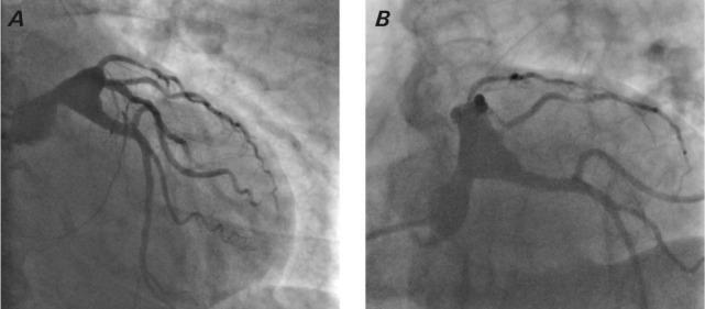
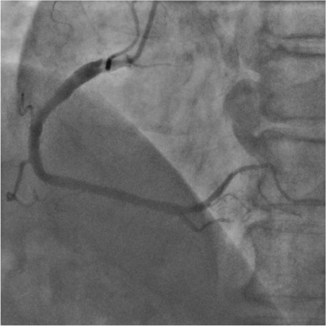
Giant aneurysm of the left main coronary artery is exceedingly rare and accounts for less than 2% of patients undergoing coronary angiography. The etiology varies depending on the patient's age and geographic area, but half are of atherosclerotic origin. In most cases, coronary aneurysms are asymptomatic, however, symptomatic patients present with symptoms characteristic of coronary artery disease such as chest pain (angina pectoris), myocardial infarction, congestive heart failure, and even sudden death. Coronary angiography is considered the gold standard tool to determine the presence or absence of coronary artery disease, and if present, its size and location. Herein, we report a case of giant aneurysm of the left main coronary artery presenting as syncope and documented nonsustained ventricular tachycardia.
Keywords: Coronary aneurysm; cardiogenic syncope; coronary angiography; tachycardia, ventricular.
© 2022 by the Texas Heart® Institute, Houston.
Conflict of interest statement
Figures
Similar articles
-
Giant coronary artery aneurysm of the proximal left anterior descending artery in a patient with two vessel coronary artery disease presented with angina pectoris.Acta Cardiol. 2023 Feb;78(1):158-159. doi: 10.1080/00015385.2022.2059859. Epub 2022 Apr 1. Acta Cardiol. 2023. PMID: 35363109 No abstract available.
-
Giant right coronary artery aneurysm complicated by acute myocardial infarction.Gen Thorac Cardiovasc Surg. 2010 Apr;58(4):186-9. doi: 10.1007/s11748-009-0478-1. Epub 2010 Apr 18. Gen Thorac Cardiovasc Surg. 2010. PMID: 20401712
-
Bifurcating aneurysm of the left main coronary artery involving left anterior descending and left circumflex arteries--a case report.Angiology. 1999 May;50(5):417-20. doi: 10.1177/000331979905000508. Angiology. 1999. PMID: 10348430
-
Giant right coronary artery aneurysm in an adult male patient with non-ST myocardial infarction.Hellenic J Cardiol. 2013 Jan-Feb;54(1):69-76. Hellenic J Cardiol. 2013. PMID: 23340133 Review.
-
Giant coronary artery aneurysms: review and update.Tex Heart Inst J. 2014 Dec 1;41(6):603-8. doi: 10.14503/THIJ-13-3896. eCollection 2014 Dec. Tex Heart Inst J. 2014. PMID: 25593524 Free PMC article. Review.
Cited by
-
Giant right coronary artery aneurysm presenting with recurrent syncope: A diagnostic challenge.Radiol Case Rep. 2024 Nov 27;20(2):1053-1057. doi: 10.1016/j.radcr.2024.10.118. eCollection 2025 Feb. Radiol Case Rep. 2024. PMID: 39659694 Free PMC article.
References
-
- Huang CY, Huang CH, Chen JW, Liu TM. Atherosclerotic left main coronary aneurysm. Int J Angiol . 2004;13:89–91. doi: 10.1007/s00547-004-1066-y. - DOI
Publication types
MeSH terms
LinkOut - more resources
Full Text Sources
Medical