

Proteomics coupled with in vitro model to study the early crosstalk occurring between newly excysted juveniles of Fasciola hepatica and host intestinal cells
- PMID: 36223411
- PMCID: PMC9555655
- DOI: 10.1371/journal.pntd.0010811
Proteomics coupled with in vitro model to study the early crosstalk occurring between newly excysted juveniles of Fasciola hepatica and host intestinal cells
Abstract
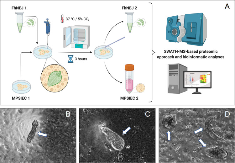
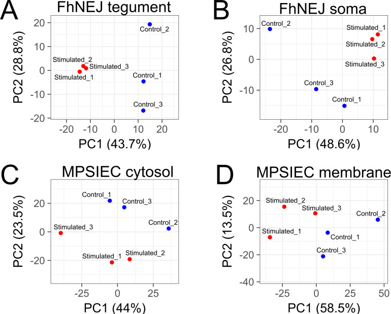
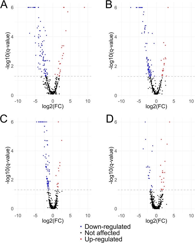
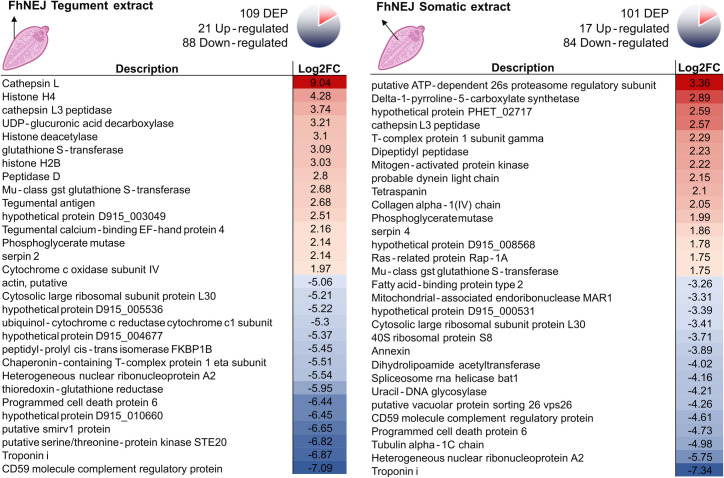
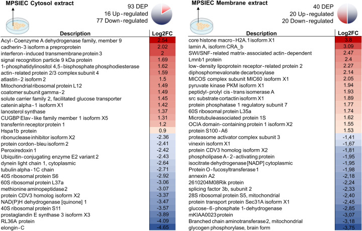
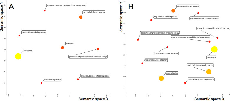
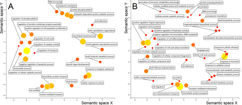
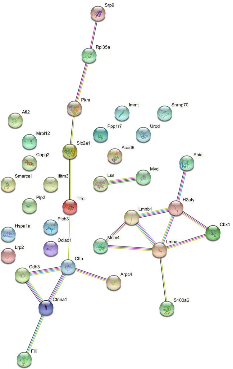
Fasciolosis caused by the trematode Fasciola hepatica is a zoonotic neglected disease affecting animals and humans worldwide. Infection occurs upon ingestion of aquatic plants or water contaminated with metacercariae. These release the newly excysted juveniles (FhNEJ) in the host duodenum, where they establish contact with the epithelium and cross the intestinal barrier to reach the peritoneum within 2-3 h after infection. Juveniles crawl up the peritoneum towards the liver, and migrate through the hepatic tissue before reaching their definitive location inside the major biliary ducts, where they mature into adult worms. Fasciolosis is treated with triclabendazole, although resistant isolates of the parasite are increasingly being reported. This, together with the limited efficacy of the assayed vaccines against this infection, poses fasciolosis as a veterinary and human health problem of growing concern. In this context, the study of early host-parasite interactions is of paramount importance for the definition of new targets for the treatment and prevention of fasciolosis. Here, we develop a new in vitro model that replicates the first interaction between FhNEJ and mouse primary small intestinal epithelial cells (MPSIEC). FhNEJ and MPSIEC were co-incubated for 3 h and protein extracts (tegument and soma of FhNEJ and membrane and cytosol of MPSIEC) were subjected to quantitative SWATH-MS proteomics and compared to respective controls (MPSIEC and FhNEJ left alone for 3h in culture medium) to evaluate protein expression changes in both the parasite and the host. Results show that the interaction between FhNEJ and MPSIEC triggers a rapid protein expression change of FhNEJ in response to the host epithelial barrier, including cathepsins L3 and L4 and several immunoregulatory proteins. Regarding MPSIEC, stimulation with FhNEJ results in alterations in the protein profile related to immunomodulation and cell-cell interactions, together with a drastic reduction in the expression of proteins linked with ribosome function. The molecules identified in this model of early host-parasite interactions could help define new tools against fasciolosis.
Conflict of interest statement
The authors have declared that no competing interests exist.
Figures
Similar articles
-
Fasciola hepatica juveniles interact with the host fibrinolytic system as a potential early-stage invasion mechanism.PLoS Negl Trop Dis. 2023 Apr 21;17(4):e0010936. doi: 10.1371/journal.pntd.0010936. eCollection 2023 Apr. PLoS Negl Trop Dis. 2023. PMID: 37083884 Free PMC article.
-
Study of the migration of Fasciola hepatica juveniles across the intestinal barrier of the host by quantitative proteomics in an ex vivo model.PLoS Negl Trop Dis. 2022 Sep 16;16(9):e0010766. doi: 10.1371/journal.pntd.0010766. eCollection 2022 Sep. PLoS Negl Trop Dis. 2022. PMID: 36112664 Free PMC article.
-
Set up of an in vitro model to study early host-parasite interactions between newly excysted juveniles of Fasciola hepatica and host intestinal cells using a quantitative proteomics approach.Vet Parasitol. 2020 Feb;278:109028. doi: 10.1016/j.vetpar.2020.109028. Epub 2020 Jan 15. Vet Parasitol. 2020. PMID: 31986420
-
Insights into Fasciola hepatica Juveniles: Crossing the Fasciolosis Rubicon.Trends Parasitol. 2021 Jan;37(1):35-47. doi: 10.1016/j.pt.2020.09.007. Epub 2020 Oct 13. Trends Parasitol. 2021. PMID: 33067132 Review.
-
Unraveling the microRNAs Involved in Fasciolosis: Master Regulators of the Host-Parasite Crosstalk.Int J Mol Sci. 2024 Dec 29;26(1):204. doi: 10.3390/ijms26010204. Int J Mol Sci. 2024. PMID: 39796061 Free PMC article. Review.
Cited by
-
Fasciola hepatica juveniles interact with the host fibrinolytic system as a potential early-stage invasion mechanism.PLoS Negl Trop Dis. 2023 Apr 21;17(4):e0010936. doi: 10.1371/journal.pntd.0010936. eCollection 2023 Apr. PLoS Negl Trop Dis. 2023. PMID: 37083884 Free PMC article.
-
Molecular Characterization of the Interplay between Fasciola hepatica Juveniles and Laminin as a Mechanism to Adhere to and Break through the Host Intestinal Wall.Int J Mol Sci. 2023 May 3;24(9):8165. doi: 10.3390/ijms24098165. Int J Mol Sci. 2023. PMID: 37175870 Free PMC article.
-
Antigens from the Helminth Fasciola hepatica Exert Antiviral Effects against SARS-CoV-2 In Vitro.Int J Mol Sci. 2023 Jul 18;24(14):11597. doi: 10.3390/ijms241411597. Int J Mol Sci. 2023. PMID: 37511355 Free PMC article.
-
The role of helminths and their antigens in cancer therapy: insights from cell line models.Infect Agent Cancer. 2024 Oct 9;19(1):52. doi: 10.1186/s13027-024-00613-3. Infect Agent Cancer. 2024. PMID: 39385244 Free PMC article. Review.
-
In vitro co-culture of Fasciola hepatica newly excysted juveniles (NEJs) with 3D HepG2 spheroids permits novel investigation of host-parasite interactions.Virulence. 2025 Dec;16(1):2482159. doi: 10.1080/21505594.2025.2482159. Epub 2025 Mar 25. Virulence. 2025. PMID: 40132201 Free PMC article.
References
Publication types
MeSH terms
Substances
LinkOut - more resources
Full Text Sources
