. 2022 Oct 12;18(10):e1010907.
doi: 10.1371/journal.ppat.1010907. eCollection 2022 Oct.

SERINC5 restricts influenza virus infectivity

Affiliations

SERINC5 restricts influenza virus infectivity

Fei Zhao et al. PLoS Pathog. .

Abstract

SERINC5 is a multi-span transmembrane protein that is incorporated into HIV-1 particles in producing cells and inhibits HIV-1 entry. Multiple retroviruses like HIV-1, equine infectious anemia virus and murine leukemia virus are subject to SERINC5 inhibition, while HIV-1 pseudotyped with envelope glycoproteins of vesicular stomatitis virus and Ebola virus are resistant to SERINC5. The antiviral spectrum and the underlying mechanisms of SERINC5 restriction are not completely understood. Here we show that SERINC5 inhibits influenza A virus infection by targeting virus-cell membrane fusion at an early step of infection. Further results show that different influenza hemagglutinin (HA) subtypes exhibit diverse sensitivities to SERINC5 restriction. Analysis of the amino acid sequences of influenza HA1 strains indicates that HA glycosylation sites correlate with the sensitivity of influenza HA to SERINC5, and the inhibitory effect of SERINC5 was lost when certain HA glycosylation sites were mutated. Our study not only expands the antiviral spectrum of SERINC5, but also reveals the role of viral envelope glycosylation in resisting SERINC5 restriction.


Conflict of interest statement

The authors have declared that no competing interests exist.

Figures
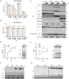
Fig 1
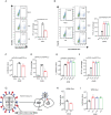
Fig 1. Exogenous SERINC5 inhibits IAV infection.
A-C: Pesudoviruses were produced by transfecting HEK293T cells using plasmid DNA ΔEnv/NefG2A HIV-1NL4-3 (containing mutated Nef) together with Env (HXB2), Env (YU-2), VSV-G, or HA/NA, and the indicated amounts of SERINC5 plasmid or empty vector. (A) Infectivity of the pseudoviruses was measured by infection of the TZM-bl indicator cells with equal p24Gag amounts of viruses. (B) Viral particle production was quantified by p24Gag ELISA. (C) Expression of SERINC5, and p24 in the cell lysates and supernatants were examined by Western blotting. HIV-1 pseudovirus particles were purified by ultracentrifugation through a 20% sucrose cushion. YU-2pp, pseudovirus with YU-2 Env. HXB2pp, pseudovirus with HXB2 Env. VSVGpp, pseudovirus with VSV-G protein. WSNpp, pseudovirus with WSN HA/NA proteins. D-E: Effects of exogenous SERINC5 on influenza A virus long-term infection. A549 cells, either stably overexpressing exogenous SERINC5 (MOI = 0.001) (D) or knocked out of SERINC5 (MOI = 0.0001) (E), were infected with virus A/WSN/33. Virus titers in the culture supernatants were determined by plaque assays. 48 h post infection, cells were harvested to measure the expression of SERINC5, HA and NP proteins. F-G: Effects of exogenous SERINC5 on influenza A virus short-term infection. SERINC5-overexpressing A549 cells (MOI = 5) and SERINC5-knockout cells (MOI = 1) were incubated with WSN. 2h, 4h, 6h and 8h post infection cells were collected and protein expression were determined as described above. Results shown are the averages of three independent experiments. Statistical significance was calculated with unpaired t-test. ns: not significant; *: P<0.05; **: P<0.01.
Fig 2
Fig 2. SERINC5 inhibits IAV infection when expressed in virus target cells.
A-B: Pseudoviruses ΔEnv/NefG2A-GFP HIV-1NL4-3 bearing IAV HA/NA proteins were used to infect A549 SERINC5-overexpressing cells (20ng p24 antigen) or SERINC5-knockout cells (10ng p24 antigen). Virus infection was examined by scoring GFP-positive cells with flow cytometry. C-F: Pseudovirions psPAX2 with influenza HA/NA proteins infected A549 SERINC5-overexpressing cell (20ng p24 antigen) (C, D) and SERINC5-knockout cell (10ng p24 antigen) (E, F). Half of the A549 cells were lysed to measure luciferase activity (C, E), the other half of A549 cells were fixed to measure pseudovirus infectivity by flow cytometry. Percentages of FITC-positive cells are shown in the column (D, F). G: Illustration of IAV for single-round infection. The HA ORF was replaced by Gluc. HA protein was expressed from the plasmid DNA. The expression plasmid of HA and 7 plasmids encoding IAV genomes were co-transfected to obtain the single-round IAV which can infect cells and produce secreted Gluc, but does not express HA protein and does not produce infectious virus particles for the next round of infection. This IAV is competent for a single-round infection. H-I: The single-round IAV was used to infected A549 SERINC5-overexpressing cells (MOI = 0.05) (H) or SERINC5-knockout cells (MOI = 0.01) (I). Levels of Gluc in the culture supernatants were measured 8 h.p.i. Results shown are the averages of three independent experiments. Statistical significance was calculated by unpaired t-test. ns: not significant; *: P<0.05; **: P<0.01.
Fig 3
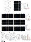
Fig 3. SERINC5 inhibits the fusion of IAV with cellular membrane.
A-B: Levels of IAV NP vRNA in infected A549 SERINC5-overexpressing cells (A) or SERINC5-knockout cells (B). C: A549 cells with SERINC5 overexpression were incubated with IAV WSN (MOI = 50) for 60 min, or 90 min, then fixed and stained for nuclei (blue), PB1 vRNA (green). Scale bar, 10 μm. Ten views were selected and vRNA numbers per cell are summarized in the graph. D: A549 SERINC5-overexpressing cells were infected with DiOC18 and R18 labled WSN (MOI = 20), cells were fixed and stained for nuclei (blue). Virus were shown in red and virus fusion were shown in green. Scale bar, 5 μm. Seven views were selected and number of fusion sites per cell are summarized in the graph. E-F: BlaM assay to measure IAV fusion with target cells. A549 cells with SERINC5 overexpression (20ng p24 antigen) (E) or SERINC5 knockout (10ng p24 antigen) (F) were incubated with IAV WSNpp that contain BlaM-Vpr for 2 h. Cells with cleaved CCF2-AM were scored with flow cytometry. Percentages of cells with CCF2-AM cleavage are summarized in the graph. G: 293T cells expressing envelope and HIV-1 Tat-flag were co-cultured with TZM-bl cells transfected with SERINC5. HA/NA cell fusion was triggered with acidic medium. After 40 hours, cells were lysed to measure luciferase activity. Results shown are the averages of three independent experiments. NC: Negative control, 293T cells transfect with empty vector and incubated with TZM-bl cells. Statistical significance was calculated by unpaired t-test. ns: not significant; *: P<0.05; **: P<0.01.
Fig 4
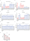
Fig 4. SERINC5 inhibits multiple IAV strains.
A-B: Pseudoviruses were produced by transfecting HEK293T cells with plasmid DNA psPAX2, LentiGFP/Luc, pCAGGS-HA (expressing H1 to H16), pCAGGS-NA(WSN), or VSV G. The pseudoviruses were then used to infect A549 cells that either overexpress SERINC5 (A) or have SERINC5 knocked out (B). Luciferase activity was measured to report viral infection. C-F: ΔEnv/NefG2A HIV-1NL4-3 (C, D) or ΔEnv/NefWT HIV-1NL4-3 (E, F) was used to produce pseudoviruses carrying IAV HA protein (HA1 to HA16) in the presence of SERINC5. Viral particle production (C, E) and viral infectivity (D, F) were measured, and the results of three independent experiments are shown. G: A549 cells with SERINC5 overexpression were infected with A/WSN/33, A/PR/8, A/H1N1pdm09 or A/H3N2 (MOI = 0.05). Titers of IAV in the supernatants were determined by viral plaque assays. Statistical significance was analyzed by unpaired t-test. ns: not significant; *: P<0.05; **: P<0.01.
Fig 5
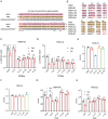
Fig 5. HA glycosylation sites modulate the sensitivity to SERINC5.
A: The glycosylation sites in the HA head domain of HA1-WSN, HA1-PR8 and HA1-A/Brisbane/59/2007. Different amino acids are marked with asterisks. N-glycosylation site were highlighted with red. B: Schematic representation of IAV WSN and PR8 HA mutations. C-D: HA mutants were used to pseudotype lentivirus particles that were produced by transfecting HEK293T cells with plasmid psPAX2, LentiGFP/Luc and pCAGGS-NA. Viruses of equal viral p24Gag amounts were used to infect A549 cells with SERINC5 overexpression (C) or SERINC5 knockout (D). Luciferase activity was measured to report viral infection. E-H: IAV was produced by transfecting HEK293T cells with the mutated WSN-HA (E, G) or PR8-HA (F, H) and the rest 7 viral DNA segments. Titers of viruses were determined by viral plaque assays (E, F). After infecting A549 cells that stably overexpress SERINC5 at MOI = 0.01 for 24 h, titers of IAV in the supernatant were determined by viral plaque assay (G, H). Results shown are the averages of three independent experiments. Statistical significance was calculated with unpaired t-test. ns: not significant; *: P<0.05; **: P<0.01.

Similar articles

Cited by

References

    1. Inuzuka M, Hayakawa M, Ingi T. Serinc, an activity-regulated protein family, incorporates serine into membrane lipid synthesis. The Journal of biological chemistry. 2005;280(42):35776–83. Epub 2005/08/27. doi: 10.1074/jbc.M505712200 . - DOI - PubMed
    1. Usami Y, Wu Y, Gottlinger HG. SERINC3 and SERINC5 restrict HIV-1 infectivity and are counteracted by Nef. Nature. 2015;526(7572):218–23. Epub 2015/09/30. doi: 10.1038/nature15400 . - DOI - PMC - PubMed
    1. Rosa A, Chande A, Ziglio S, De Sanctis V, Bertorelli R, Goh SL, et al.. HIV-1 Nef promotes infection by excluding SERINC5 from virion incorporation. Nature. 2015;526(7572):212–7. Epub 2015/09/30. doi: 10.1038/nature15399 . - DOI - PMC - PubMed
    1. Staudt RP, Smithgall TE. Nef homodimers down-regulate SERINC5 by AP-2-mediated endocytosis to promote HIV-1 infectivity. J Biol Chem. 2020;295(46):15540–52. Epub 2020/09/03. doi: 10.1074/jbc.RA120.014668 . - DOI - PMC - PubMed
    1. Ward AE, Kiessling V, Pornillos O, White JM, Ganser-Pornillos BK, Tamm LK. HIV-cell membrane fusion intermediates are restricted by Serincs as revealed by cryo-electron and TIRF microscopy. J Biol Chem. 2020;295(45):15183–95. Epub 2020/08/14. doi: 10.1074/jbc.RA120.014466 . - DOI - PMC - PubMed

Publication types

Grants and funding