. 2022 Oct 27;126(42):8427-8438.
doi: 10.1021/acs.jpcb.2c05423. Epub 2022 Oct 12.

Allosteric Inhibition of PTP1B by a Nonpolar Terpenoid

Affiliations

Allosteric Inhibition of PTP1B by a Nonpolar Terpenoid

Anika J Friedman et al. J Phys Chem B. .

Abstract

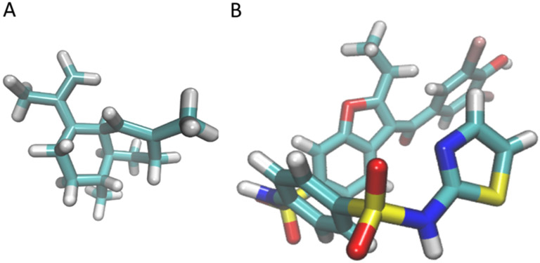
Protein tyrosine phosphatases (PTPs) are promising drug targets for treating a wide range of diseases such as diabetes, cancer, and neurological disorders, but their conserved active sites have complicated the design of selective therapeutics. This study examines the allosteric inhibition of PTP1B by amorphadiene (AD), a terpenoid hydrocarbon that is an unusually selective inhibitor. Molecular dynamics (MD) simulations carried out in this study suggest that AD can stably sample multiple neighboring sites on the allosterically influential C-terminus of the catalytic domain. Binding to these sites requires a disordered α7 helix, which stabilizes the PTP1B-AD complex and may contribute to the selectivity of AD for PTP1B over TCPTP. Intriguingly, the binding mode of AD differs from that of the most well-studied allosteric inhibitor of PTP1B. Indeed, biophysical measurements and MD simulations indicate that the two molecules can bind simultaneously. Upon binding, both inhibitors destabilize the α7 helix by disrupting interactions at the α3-α7 interface and prevent the formation of hydrogen bonds that facilitate closure of the catalytically essential WPD loop. These findings indicate that AD is a promising scaffold for building allosteric inhibitors of PTP1B and illustrate, more broadly, how unfunctionalized terpenoids can engage in specific interactions with protein surfaces.


Figures

Figure 1.
Figure 1.
Chemical structures of (A) AD and (B) BBR are distinct. AD is significantly smaller in size and lacks the h-bond donors and acceptors that allow BBR to form stabilizing h-bonds. Given the structure of AD, it is surprising that AD exhibits a similar level of selectivity for PTP1B over TCPTP and inhibits PTP1B with an IC50 that is only ~6× greater than that of BBR.,
Figure 2.
Figure 2.
(A) Structures of AD and well-studied allosteric (BBR; benzofuran derivative PDB FRJ) and competitive (TCS401) inhibitors. (B) X-ray crystal structure of PTP1B bound to AD (PDB entry 6W30) with the binding sites for BBR and TCS401 overlaid for reference. We aligned structures of the PTP1B–AD, PTP1B–BBR, and PTP1B–TCS401 complexes (PDB entries 6W30, 1T4J, and 5K9W) using the “align” function from PyMol. AD and BBR bind to the allosteric site, which includes residues from the α3, α6, and α7 helices; TCS401 binds to the active site, which is flanked by the WPD and P-loops. (C) Fluorescence-based binding isotherms for BBR measured in the presence and absence of either AD or TCS401. We ensured similar levels of binding by AD and TCS401 using concentrations that produced similar levels of inhibition (~50%; Figure S1). Binding parameters (±SE) for ΔF = (ΔFmax*L)/(Kd + L): Kd = 10.1 ± 2.7 μM and ΔFmax = 227,000 ±13,000 (BBR alone); Kd = 13.1 ± 3.8 μM and ΔFmax = 195,000 ± 12,000 (BBR with AD); and Kd = 31.0 ± 2.8 μM and ΔFmax = 94,000 ± 2000 (BBR with TCS401). The insensitivity of the BBR binding isotherm to the presence of AD suggests that the two inhibitors can bind simultaneously. Error bars denote the standard error for n = 3 technical replicates. (D) Melting temperatures determined with DSF indicate that BBR and Ertiprotafib destabilize PTP1B, while AD and TCS401 do not. Error bars denote standard deviation for n = 3 technical replicates.
Figure 3.
Figure 3.
AD is capable of occupying a diverse set of binding conformations. (A) In MD simulations initialized with a disordered α7 helix, AD samples two adjacent sites with near-equal frequency: the crystallographic site (loc1; blue) and a neighboring site (loc2; green). When the α7 helix is initialized with an ordered conformation or absent, AD moves to two new sites: loc3 (pink) and loc4 (brown). Table S5 provides the percent occupancy of all sites, and Section S1.6 details the procedure used to determine occupancy percentages. (B) During MD trajectories, AD (bound to loc1 and loc2) and BBR interact with the same core set of residues (green) and several residues specific to AD (blue) or BBR (purple). (C) Comparison of rmsds for the COM of AD and BBR in different complexes. AD exhibits significantly larger fluctuations than BBR in complex with PTP1B. In the ternary complex, both AD and BBR experience a stabilizing effect, although it is more significant for AD. This stabilization of both ligands in the ternary complex is likely due to stabilization though additional contacts between AD and BBR. (D) In MD simulations initialized with AD and BBR at their crystallographic binding sites, AD moves to the outside of the α7 helix and remains at this location (blue) for the entire duration of the 1 μs trajectory. In (A–C), the protein and ligand represent centroid structures from the corresponding MD trajectories.
Figure 4.
Figure 4.
Mutations in the helical triad tend to disrupt inhibition by AD and BBR. (A) X-ray crystal structure of PTP1B bound to AD (PTPB entry 6W30) with several other binding sites overlaid: (i) crystallographic binding site for BBR and (ii) two sites sampled by AD in MD simulations (loc1 and loc2) carried out with a disordered α7 helix. To position the alternative sites, we aligned the PTP1B–AD complex (PDB entry 6W30) with the PTP1B–BBR complex (pdb entry 1T4J) and centroid structures from MD simulations (PyMol function “align”). Labels denote residues selected for site-directed mutagenesis with colors by helix. (B) Fractional change in inhibition (F) caused by mutations at the sites highlighted in A. Most mutations decreased the inhibitory effects of AD and BBR. Error bars denote the propagated standard error for n = 4 independent measurements.
Figure 5.
Figure 5.
Mutational effects arise from delocalized structural changes in PTP1B. The influence of mutations on AD-mediated inhibition is not correlated with their effect on enzyme activity (measured kcat/Km for pNPP hydrolysis in the absence of inhibitor), binding affinity (ΔΔG, the difference in free energy of binding between mutants, as calculated from relative free energy simulations using MBAR for analysis), or the mean percent α helicity of the α7 helix. Shaded regions correspond to the wild-type activity of PTP1B (top), ±0.1 kcal/mol (middle), and the percent α helicity of the wild-type enzyme with WPDopen (bottom). Error bars denote the standard error for (top) n > 4 independent measurements and (middle and bottom) 50 ns simulations at 18 alchemical states. See Materials and Methods for a detailed description of our calculation of the standard error for alchemical free energy calculations.
Figure 6.
Figure 6.
(A) h-bond network exists within PTP1B to stabilize the closed conformation. The blue lines denote bonds that form with WPDclosed but do not form with apo WPDopen and are broken with ligand binding. The black line denotes the conserved bond T177–Y152 which connects the active and allosteric sites. (B) Upon binding, AD and BBR disrupt nonbonded interactions between the α3, α6, and α7 helices. Highlights: (black) interactions disrupted in the WPDopen conformation that are also disrupted when AD and BBR bind to the protein, (green) interactions disrupted by both ligands but present in both WPDclosed and WPDopen conformations, and (purple) interactions disrupted by BBR alone (Figure S6). Most of the interactions disrupted by AD, BBR, and WPDopen are located between the α3 and α7 helices. This overlap suggests that the disruption of these interactions is crucial for allosteric inhibition. Disruption of these interactions destabilizes the α7 helix and prevents the formation of h-bonds required for closure of the WPD loop. (C,D) Upon binding, AD (dashed line) and BBR (dotted line) (C) increase the flexibility of the α7 helix and (D) decrease its α helicity to levels that resemble the WPDopen conformation. Destabilization of the α7 helix is faster with AD bound in loc2 compared to the alternative loc4 (Figure S9). In (C,D), all MD trajectories start with the same ordered α7 helix conformation.

References

    1. Östman A; Hellberg C; Böhmer FD Protein-tyrosine phosphatases and cancer. Nat. Rev. Cancer 2006, 6, 307–320. - PubMed
    1. Fuentes F; Zimmer D; Atienza M; Schottenfeld J; Penkala I; Bale T; Bence KK; Arregui CO Protein Tyrosine Phosphatase PTP1B Is Involved in Hippocampal Synapse Formation and Learning. PLoS One 2012, 7, No. e41536. - PMC - PubMed
    1. Zhu Z; Liu Y; Li K; Liu J; Wang H; Sun B; Xiong Z; Jiang H; Zheng J; Hu Z Protein Tyrosine Phosphatase Receptor U (PTPRU) Is Required for Glioma Growth and Motility. Carcinogenesis 2014, 35, 1901–1910. - PubMed
    1. Mustelin T; Vang T; Bottini N Protein Tyrosine Phosphatases and the Immune Response. Nat. Rev. Immunol 2005, 5, 43–57. - PubMed
    1. Zhang C; Wu L; Liu X; Gao J; Liu S; Wu J; Huang D; Wang Z; Su X Discovery of Novel PTP1B Inhibitors Derived from the BH3 Domain of Proapoptotic Bcl-2 Proteins with Antidiabetic Potency. ACS Med. Chem. Lett 2021, 12, 1017–1023. - PMC - PubMed

Publication types