. 2022 Dec 1;323(6):L685-L697.
doi: 10.1152/ajplung.00023.2022. Epub 2022 Oct 12.

Pim-1 kinase is a positive feedback regulator of the senescent lung fibroblast inflammatory secretome

Affiliations

Pim-1 kinase is a positive feedback regulator of the senescent lung fibroblast inflammatory secretome

Ashley Y Gao et al. Am J Physiol Lung Cell Mol Physiol. .

Abstract

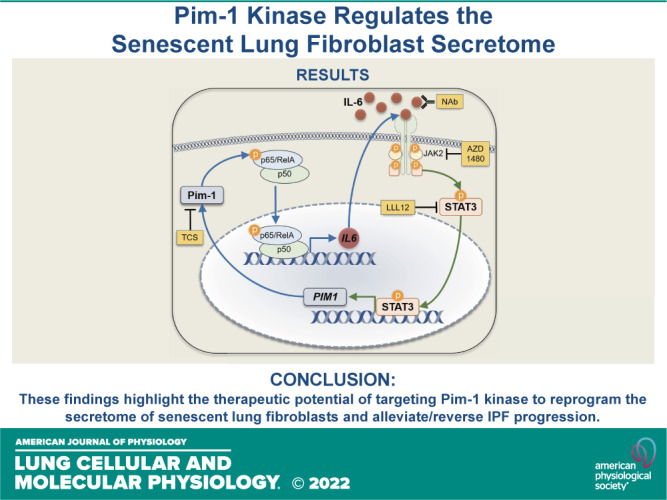
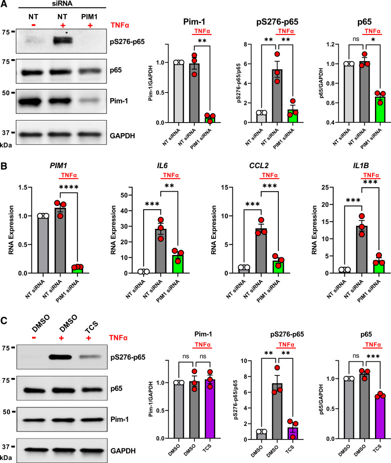
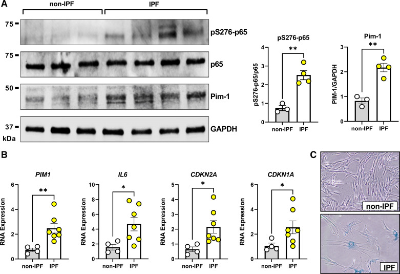
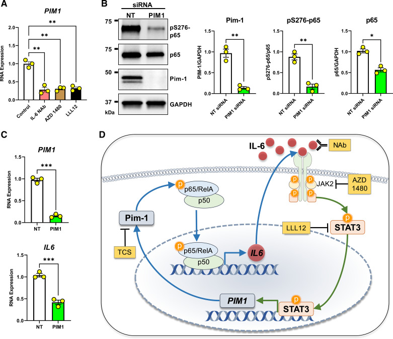
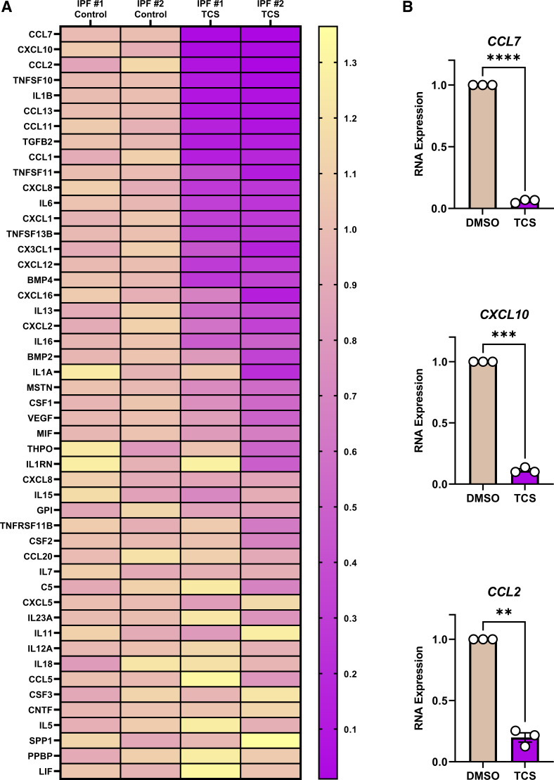
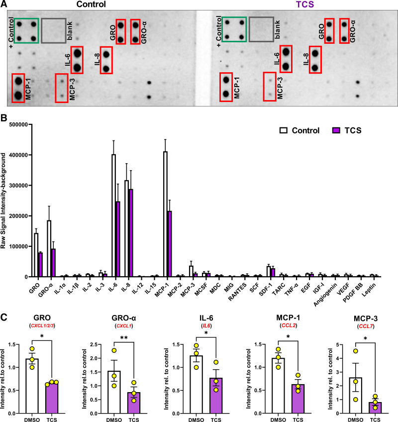
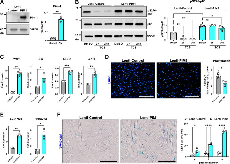
Cellular senescence is emerging as a driver of idiopathic pulmonary fibrosis (IPF), a progressive and fatal disease with limited effective therapies. The senescence-associated secretory phenotype (SASP), involving the release of inflammatory cytokines and profibrotic growth factors by senescent cells, is thought to be a product of multiple cell types in IPF, including lung fibroblasts. NF-κB is a master regulator of the SASP, and its activity depends on the phosphorylation of p65/RelA. The purpose of this study was to assess the role of Pim-1 kinase as a driver of NF-κB-induced production of inflammatory cytokines from low-passage IPF fibroblast cultures displaying markers of senescence. Our results demonstrate that Pim-1 kinase phosphorylates p65/RelA, activating NF-κB activity and enhancing IL-6 production, which in turn amplifies the expression of PIM1, generating a positive feedback loop. In addition, targeting Pim-1 kinase with a small molecule inhibitor dramatically inhibited the expression of a broad array of cytokines and chemokines in IPF-derived fibroblasts. Furthermore, we provide evidence that Pim-1 overexpression in low-passage human lung fibroblasts is sufficient to drive premature senescence, in vitro. These findings highlight the therapeutic potential of targeting Pim-1 kinase to reprogram the secretome of senescent fibroblasts and halt IPF progression.

Keywords: Pim kinase; idiopathic pulmonary fibrosis; lung fibrosis; secretome; senescence.


Conflict of interest statement

No conflicts of interest, financial or otherwise, are declared by the authors.

Figures

None
Graphical abstract
Figure 1.
Figure 1.
Proviral integration site for Moloney murine leukemia virus 1 (Pim-1) regulates phosphorylation and activity of p65/RelA. A: non-idiopathic pulmonary fibrosis (IPF) adult lung fibroblasts were transfected with nontargeting (NT) siRNA or siRNA targeting PIM1 for 72 h. Fifteen minutes before collecting total cell protein for Western blot analysis, the indicated wells were stimulated with 50 ng/mL TNF-α. Representative image is shown and band density for three independent experiments was quantified (*P < 0.05, **P < 0.01 vs. the indicated group). B: non-IPF adult lung fibroblasts were transfected with nontargeting (NT) siRNA or siRNA targeting PIM1 for 72 h. Six hours before collecting RNA for qPCR analysis, the indicated wells were stimulated with 50 ng/mL TNF-α. n = 3 independent experiments (**P < 0.01, ***P < 0.001, ****P < 0.0001 vs. the indicated group). C: non-IPF adult lung fibroblasts were treated for 3 h with TCS Pim-1 1 (TCS) (3 µM) and 15 min before collecting total cell protein for Western blot analysis, the indicated wells were stimulated with 50 ng/mL TNF-α. Representative image is shown and band density for three independent experiments was quantified (**P < 0.01, ***P < 0.001 vs. the indicated group).
Figure 2.
Figure 2.
Senescent idiopathic pulmonary fibrosis (IPF) patient-derived lung fibroblasts express spontaneously elevated Pim-1 and p65/RelA phosphorylation. A: lung fibroblasts from non-IPF or IPF donors were cultured for 24 h in the absence of serum before total cell protein isolation and Western Blot analysis. n = 3 non-IPF and 4 IPF biologically independent samples (**P < 0.01 vs. non-IPF). B: lung fibroblasts from non-IPF or IPF donors were cultured for 24 h in the absence of serum before RNA isolation and qPCR analysis. RNA transcript levels are quantified relative to the same representative non-IPF sample, using GAPDH as a housekeeping gene. n = 4 non-IPF and 7 IPF biologically independent samples (*P < 0.05, **P < 0.01 vs. non-IPF). C: lung fibroblasts from non-IPF or IPF donors were stained for senescence-associated β-galactosidase (SA-β-gal) staining as a marker of cellular senescence. Representative examples for non-IPF and IPF are shown.
Figure 3.
Figure 3.
Proviral Integration site for Moloney murine leukemia virus 1 (Pim-1)/nuclear factor kappa-light-chain-enhancer of activated B cells (NF-κB) and IL-6/JAK2/STAT3 form a positive feedback loop. A: lung fibroblasts derived from patients with idiopathic pulmonary fibrosis (IPF) were cultured for 24 h in the presence of an IL-6 neutralizing antibody (IL-6 NAb, 10 μg/mL), a JAK2 inhibitor (AZD 1480, 10 µM), or a STAT3 inhibitor (LLL12, 1 µM) before RNA isolation and qPCR analysis. RNA transcript levels are quantified relative to the same representative nontargeting (NT) sample, using GAPDH as a housekeeping gene. n = 3 biologically independent and experimentally independent experiments (**P < 0.01 vs. the control treated groups). B: lung fibroblasts derived from patients with IPF were transfected with nontargeting (NT) siRNA or siRNA targeting PIM1 for 72 h before collecting whole cell protein for Western blot analysis. Representative images are shown and band density for three independent experiments was quantified (*P < 0.05, **P < 0.01 vs. the NT control group). C: lung fibroblasts derived from patients with IPF were transfected with nontargeting (NT) siRNA or siRNA targeting PIM1 and cultured for 72 h before collecting RNA for qPCR analysis. RNA transcript levels are quantified relative to the same representative NT sample, using GAPDH as a housekeeping gene. n = 3 independent experiments (***P < 0.001 vs. the indicated group). D: positive feedback model. Pim-1 phosphorylates p65/RelA, supporting NF-κB transcriptional activity that stimulates IL-6 synthesis. IL-6 signals through its receptor system and JAK2/STAT3 to transcribe PIM1. Tools used throughout our studies are highlighted in yellow. Schematic was prepared in part using Motifolio Scientific Illustration Toolkits.
Figure 4.
Figure 4.
Proviral Integration site for Moloney murine leukemia virus 1 (Pim-1) is essential for the expression of a broad range of cytokines and chemokines. A: lung fibroblasts derived from patients with idiopathic pulmonary fibrosis (IPF) were treated for 24 h with the Pim-1 inhibitor (TCS, 3 µM). RNA was isolated and a PCR profiler measuring expression of 84 cytokines and chemokines was performed, amplifying the cDNA 40 cycles, detecting 49 genes. RNA transcript levels were calculated and plotted to a heatmap by normalizing all samples relative to the average delta Ct for the two control-treated samples, using GAPDH as a housekeeping gene. Genes were ranked by the average change in the two TCS-treated samples. B: lung fibroblasts derived from patients with IPF were cultured for 24 h with the Pim-1 inhibitor (TCS, 3 µM) before RNA isolation and qPCR analysis. RNA transcript levels in the TCS treated group were quantified relative to their respective control, using GAPDH as a housekeeping gene. n = 3 biologically independent and experimentally independent experiments (**P < 0.01, ***P < 0.001, ****P < 0.0001 vs. the control-treated groups).
Figure 5.
Figure 5.
Proviral Integration site for Moloney murine leukemia virus 1 (Pim-1) regulates secretion of GRO, IL-6, MCP-1, and MCP-3. Lung fibroblasts derived from patients with idiopathic pulmonary fibrosis (IPF) were treated for 24 h with the Pim-1 inhibitor (TCS, 3 µM) in media without FBS. After incubation, conditioned media was collected and assessed by a cytokine immuno array. A: representative images of the cytokine array from control and TCS-treated samples. +/− controls labeled along with the six cytokines exhibiting the strongest signal intensity. B: raw signal intensity—background quantified for all cytokines included in the array. n = 3 biological and experimentally independent experiments. C: signal intensity for each cytokine was normalized relative to the same DMSO-treated, representative sample. n = 3 biologically and experimentally independent experiments (*P < 0.05, **P < 0.01 vs. DMSO treated groups).
Figure 6.
Figure 6.
Proviral Integration site for Moloney murine leukemia virus 1 (Pim-1) promotes lung fibroblast premature senescence. A: non-idiopathic pulmonary fibrosis (IPF) adult lung fibroblasts were infected with control lentiviral vector or lentiviral vector carrying the human PIM1 gene. Total protein was isolated in three independent Western blot experiments. Representative images are shown and band density was quantified (**P < 0.01 vs. the Lenti-Control group). B: Lenti-Control and Lenti-PIM1 expressing cells were treated for 3 h or 24 h with 3 µM TCS before total protein isolation and Western blot analysis. Representative images are shown and band density for three independent experiments was quantified (**P < 0.01, ***P < 0.001 vs. the indicated group). C and E: qPCR analysis from three independent experiments comparing gene expression between Lenti-Control and Lenti-PIM1 fibroblasts (*P < 0.05, **P < 0.01, ***P < 0.001 vs. the Lenti-Control group). D: Lenti-Control and Lenti-PIM1 expressing fibroblasts were cultured for 0 and 4 days and then stained for DAPI and quantified using automated imaging software. Representative images are shown for the number of DAPI objects (nuclei) on day 4. Quantification of three independent experiments plots the number of cells in each field of view, for each cell line, relative to the number of cells in each field of view immediately after the cells attached (day 0) (*P < 0.05 vs. the Lenti-Control group). The scale bar represents 200 µm. F: senescence-associated β-galactosidase (SA-β-gal) staining comparing Lenti-Control vs. Lenti-PIM1 lung fibroblasts as they progress through cell culture passages 48. Representative images comparing fibroblasts at passage 8. Data are automated quantification of SA-β-gal positive cells relative to the total number of cells in each field of view from three independent experiments (*P < 0.05, ****P < 0.0001 vs. the Lenti-Control group). Scale bar represents 100 µm.

Similar articles

Cited by

References

    1. King TE Jr, Pardo A, Selman M. Idiopathic pulmonary fibrosis. Lancet 378: 1949–1961, 2011. doi: 10.1016/S0140-6736(11)60052-4. - DOI - PubMed
    1. Hutchinson J, Fogarty A, Hubbard R, McKeever T. Global incidence and mortality of idiopathic pulmonary fibrosis: a systematic review. Eur Respir J 46: 795–806, 2015. doi: 10.1183/09031936.00185114. - DOI - PubMed
    1. Khor YH, Ng Y, Barnes H, Goh NSL, McDonald CF, Holland AE. Prognosis of idiopathic pulmonary fibrosis without anti-fibrotic therapy: a systematic review. Eur Respir Rev 29: 190158, 2020. doi: 10.1183/16000617.0158-2019. - DOI - PMC - PubMed
    1. Ley B, Collard HR, King TE Jr.. Clinical course and prediction of survival in idiopathic pulmonary fibrosis. Am J Respir Crit Care Med 183: 431–440, 2011. doi: 10.1164/rccm.201006-0894CI. - DOI - PubMed
    1. Glassberg MK. Overview of idiopathic pulmonary fibrosis, evidence-based guidelines, and recent developments in the treatment landscape. Am J Manag Care 25: S195–S203, 2019. - PubMed

MeSH terms