Review
. 2022 Dec:426:108612.
doi: 10.1016/j.heares.2022.108612. Epub 2022 Sep 20.

Structural and functional diversity of mitochondria in vestibular/cochlear hair cells and vestibular calyx afferents

Affiliations
Review

Structural and functional diversity of mitochondria in vestibular/cochlear hair cells and vestibular calyx afferents

Anna Lysakowski et al. Hear Res. 2022 Dec.

Abstract

Mitochondria supply energy in the form of ATP to drive a plethora of cellular processes. In heart and liver cells, mitochondria occupy over 20% of the cellular volume and the major need for ATP is easily identifiable - i.e., to drive cross-bridge recycling in cardiac cells or biosynthetic machinery in liver cells. In vestibular and cochlear hair cells the overall cellular mitochondrial volume is much less, and mitochondria structure varies dramatically in different regions of the cell. The regional demands for ATP and cellular forces that govern mitochondrial structure and localization are not well understood. Below we review our current understanding of the heterogeneity of form and function in hair cell mitochondria. A particular focus of this review will be on regional specialization in vestibular hair cells, where large mitochondria are found beneath the cuticular plate in close association with the striated organelle. Recent findings on the role of mitochondria in hair cell death and aging are covered along with potential therapeutic approaches. Potential avenues for future research are discussed, including the need for integrated computational modeling of mitochondrial function in hair cells and the vestibular afferent calyx.

Keywords: Computational modeling; Crista ampullaris; Electron tomography; Mitochondrial DNA; Mitochondrial structure; Utricular and saccular macula.


Figures

Figure 1.
Figure 1.
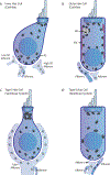
Subcellular anatomy and locations of mitochondria in hair cells and associated afferents and efferents. In all hair cells, mitochondria are found beneath the cuticular plate (CP) and near ribbon synapses (RS) to meet respective energy demands of mechanotransduction and synaptic vesicle release. Mitochondria are also found around the nucleus and in afferent and efferent nerve fibers. A. Cochlear Inner Hair Cells (IHCs). IHC mitochondria follow the general distribution described above and are medium-sized (~0.5 μm in diameter). A distinguishing feature of IHC afferents is that high spontaneous rate (SR) afferent fibers are thicker and possess more mitochondria than low SR fibers, likely to meet the increased need for ATP in neurons firing more action potentials. B. Cochlear Outer Hair Cells (OHCs). In addition to being localized near the CP and ribbon synapses, mitochondria in OHCS are also found adjacent to two specialized structures: Hensen bodies (HB) and sub-surface cisternae (SSC). The exact processes that require a localized supply of ATP in these structures are unknown. Mitochondria in OHCs are generally medium-sized. C. Type I Vestibular Hair Cell. The distinguishing feature of these cells, particularly in the central/striolar region, is that large (~1 μm in diameter) mitochondria are found beneath the CP. These subcuticular mitochondria are globular and restricted to the apical end of the cell. Mitochondria are also found within the entire length of the afferent calyx terminal that envelopes the hair cell. Exact energy demands in the calyx are unknown. D. Type II Vestibular Hair Cell. Here the regional distribution of mitochondria is similar to IHCs. In comparison to type I cells, subcuticular mitochondria are heterogeneous, smaller in volume, cylindrical and not restricted to the apical portion of the cell. Smaller (0.25 μm in diameter), elongated, mitochondria are found in efferent boutons that synapse on hair cells and afferent endings.
Figure 2.
Figure 2.
Mitochondrial substructures discussed in the text. A. Electron microscopic (EM) tomogram of a mitochondrion from a type I vestibular hair cell in which various mitochondrial substructures can be seen. Mitochondria have a double membrane (inner and outer). The inner mitochondrial membranes (IMM) form cristae that contain elements of the electron transport chain for oxidative phosphorylation. These cristae can be either lamellar (arrowheads) or tubular in shape (double arrowheads) and can form crista junctions (arrows) with the IMM. B-E. Same symbol conventions as in A. B. 3-D reconstruction of the same mitochondrion as in A, but with the inner (IMM) and outer (OMM) membranes now transparent. Tubular cristae can be seen at either end of the mitochondrion, while lamellar cristae are found in the center portion, as is typical of neuronal mitochondria. Several crista junctions can be observed as circular openings on the sides of some of the cristae. C. End-on view of the tubular cristae. Two crista junctions are seen as small tubular extensions from the cristae. D. Yellow crista indicated in B, but fenestrations (asterisks) within the crista can now be seen. E. The 3D reconstruction superimposed on the EM tomogram with transparent IMM and OMM.
Figure 3.
Figure 3.
Subcellular organization at the hair cell apex. Reconstruction from EM tomograms of the apical portion of a striolar type I vestibular hair cell shows the organization of structures such as the stereociliary rootlets (green, yellow), mitochondria (aqua), and the striated organelle (blue/pink) at the apex of the hair cell relative to the cuticular plate. The striated organelle constitutes a structure that could directly tether large mitochondria and/or restrict mitochondrial diffusion to and from the subcuticular region.

References

    1. Afzal N, Lederer WJ, Jafri MS, & Mannella CA (2021) Effect of crista morphology on mitochondrial ATP output: A computational study. Curr Res Physiol 4, 163–176. - PMC - PubMed
    1. Anand R, Reichert AS, & Kondadi AK (2021) Emerging roles of the MICOS complex in cristae dynamics and biogenesis. Biology (Basel) 10, 600. 10.3390/biology10070600 - DOI - PMC - PubMed
    1. Anderson S, Bankier AT, Barrell BG, de Bruijn MH, Coulson AR, Drouin J, Eperon IC, Nierlich DP, Roe BA, Sanger F, Schreier PH, Smith AJ, Staden R & Young IG (1981) Sequence and organization of the human mitochondrial genome. Nature 290, 457–65. - PubMed
    1. Beurg M, Nam JH, Chen Q & Fettiplace R (2010) Calcium balance and mechanotransduction in rat cochlear hair cells. J Neurophysiol 104, 18–34. - PMC - PubMed
    1. Bigland MJ, Brichta AM, Smith DW (2018) Effects of ageing on the mitochondrial genome in rat vestibular organs. Curr Aging Sci 11,108–117. - PMC - PubMed

Publication types

Substances

LinkOut - more resources