Review
. 2022 Nov 16;110(22):3667-3687.
doi: 10.1016/j.neuron.2022.09.018. Epub 2022 Oct 11.

Sensing sound: Cellular specializations and molecular force sensors

Affiliations
Review

Sensing sound: Cellular specializations and molecular force sensors

Xufeng Qiu et al. Neuron. .

Abstract

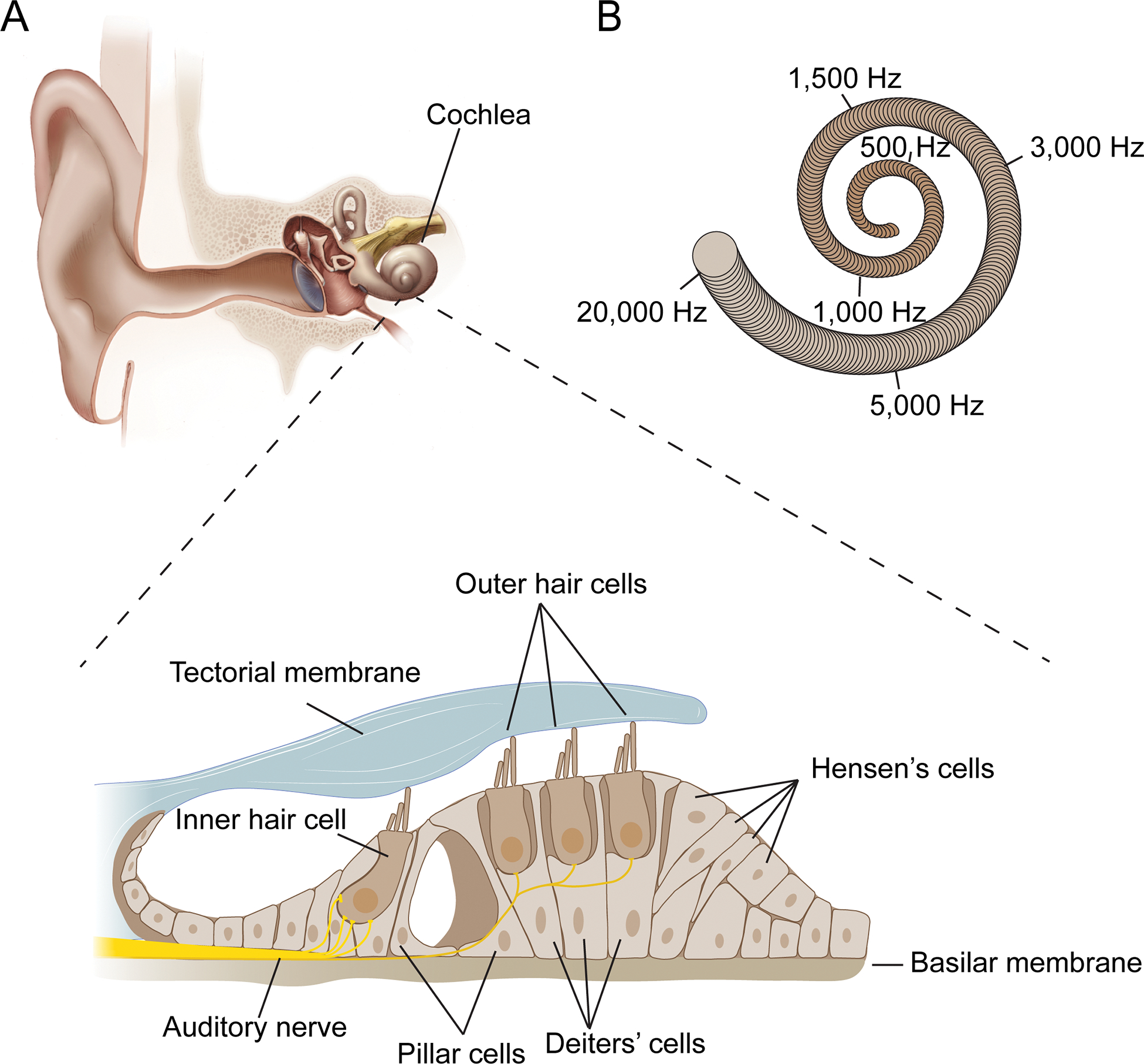
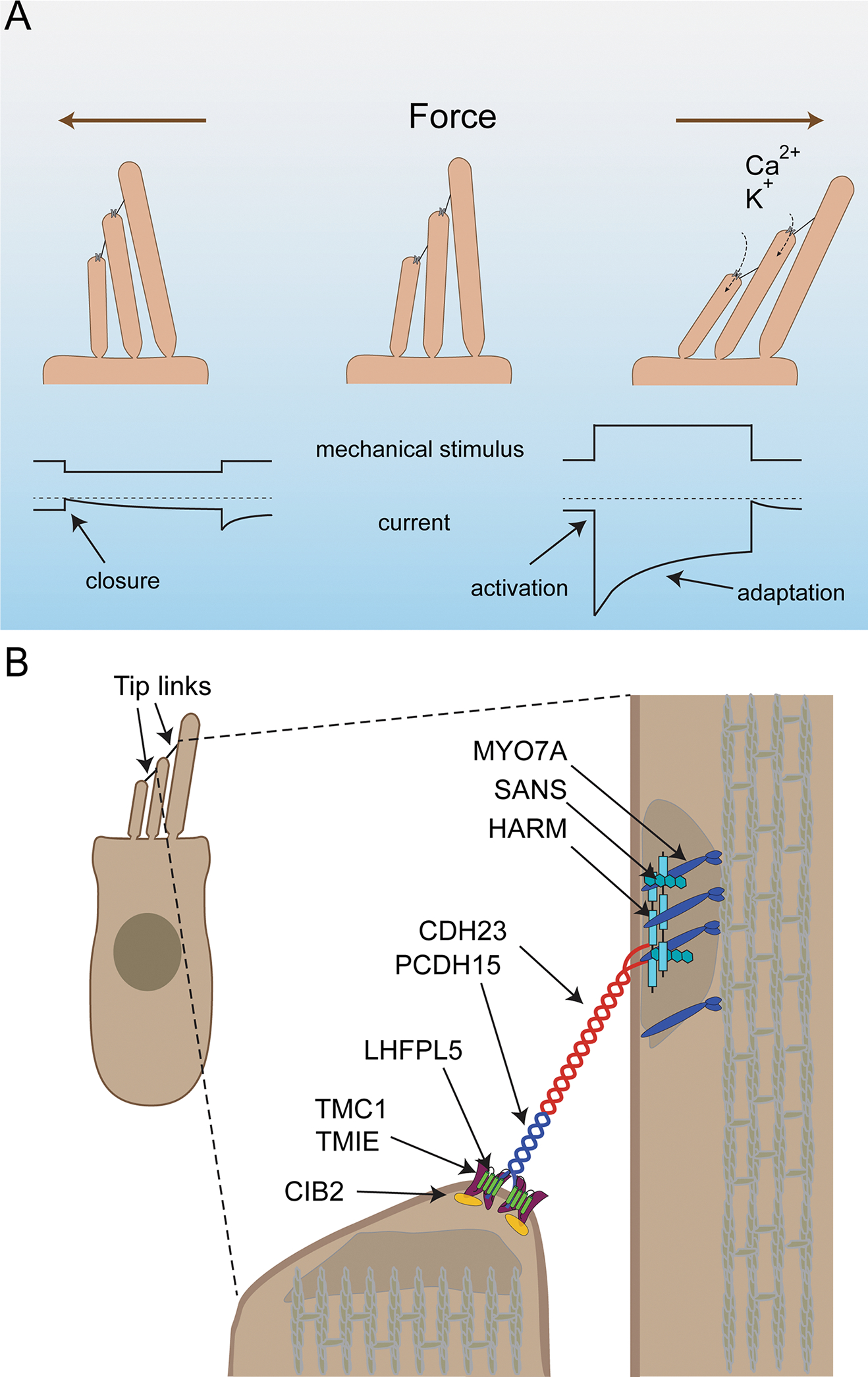
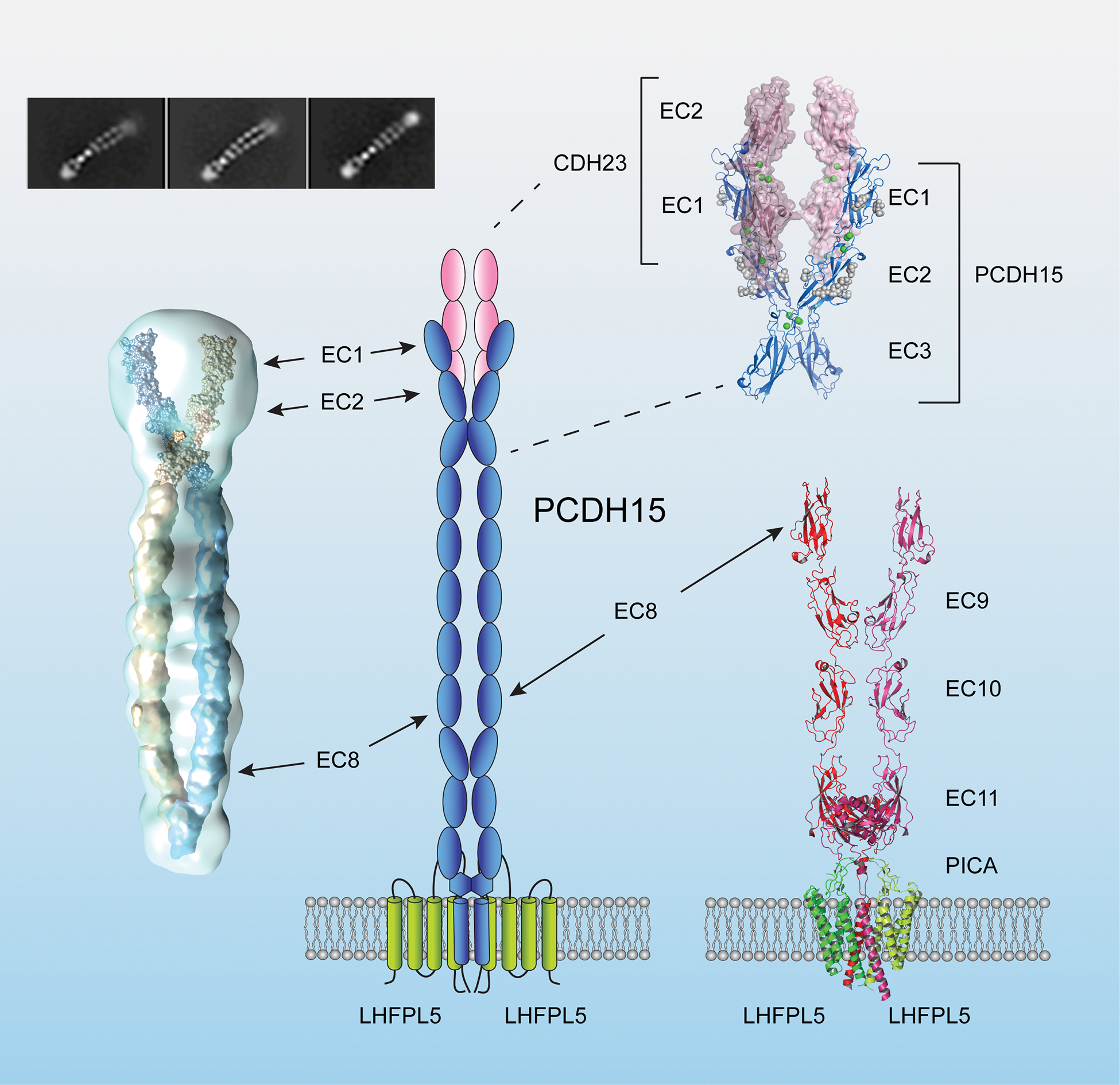
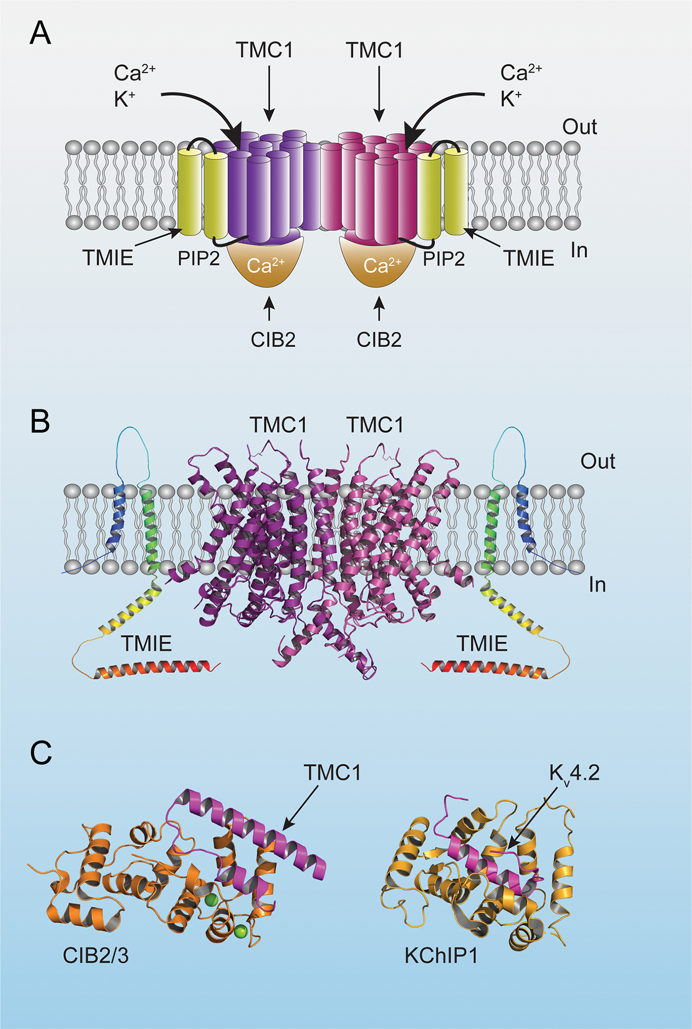
Organisms of all phyla express mechanosensitive ion channels with a wide range of physiological functions. In recent years, several classes of mechanically gated ion channels have been identified. Some of these ion channels are intrinsically mechanosensitive. Others depend on accessory proteins to regulate their response to mechanical force. The mechanotransduction machinery of cochlear hair cells provides a particularly striking example of a complex force-sensing machine. This molecular ensemble is embedded into a specialized cellular compartment that is crucial for its function. Notably, mechanotransduction channels of cochlear hair cells are not only critical for auditory perception. They also shape their cellular environment and regulate the development of auditory circuitry. Here, we summarize recent discoveries that have shed light on the composition of the mechanotransduction machinery of cochlear hair cells and how this machinery contributes to the development and function of the auditory system.


Conflict of interest statement

Declaration of interests U.M. is a co-founder of Decibel Therapeutics.

Figures

Figure 1.
Figure 1.. The auditory sense organ.
(A) Diagram of the outer, middle and inner ear. The bottom shows a cross section of the Organ of Corti that is situated within the snail-shaped cochlea of the inner ear. Sensory hair cells, including one row of inner hair cells (IHCs) and three rows of outer hair cells (OHCs), are indicated. Supporting cells such as Pillar cells, Deiters’ cells and Hensen’s cells separate and support hair cells. The cells of the Organ of Corti are sandwiched between two extracellular matrix assemblies, the tectorial membrane and the basilar membrane. (B) Schematic of the snail-shaped cochlea indicating that different sound frequencies are encoded by hair cells at different tonotopic positions along the cochlear duct. Frequency positions are for the human cochlea.
Figure 2.
Figure 2.. The mechanotransduction machinery of cochlear hair cells.
(A) Diagram of the mechanotransduction process. On top the direction of stereocilia deflection is shown, on the bottom idealized traces from recordings. Upper traces show mechanical deflections of the stereocilia: downward shift (left) indicates a deflection towards the shortest stereocilia, an upward shift (right) deflection towards the longest stereocilia. Deflection towards the shortest stereocilia (left) leads to a closing of mechanotransduction channels that are open at rest followed by slow channel re-opening. Deflection towards the longest stereocilia (right) leads to an increased influx of Ca2+ and K+ into stereocilia. This is reflected in the lower traces by a downward current, which then adapts due to channel closure even during a maintained stimulus. (B) Diagram of a hair cells indicating proteins important for mechanotransduction and their asymmetric localization at the upper and lower ends of the tip link.
Figure 3.
Figure 3.. Structure of the PCDH15-LHFPL5 complex and the PCDH15-CDH23 binding site.
A diagram of the lower end of tip links. The interaction between PCDH15 and CDH23 is mediated by N-terminal EC1 and EC2 domains (upper right, PDB 4APX and 6CV7) (Sotomayor et al., 2012). Dimerization of PCDH15 is mediated by two sites. The first dimerization site is at EC3 and was observed by single particle reconstruction of the entire 11 EC domains (left) and a high-resolution X-ray crystallographic structure of an EC1–3 fragment (upper right: PDB:6CV7) (Dionne et al., 2018). The second dimerization sited is close to the membrane proximal domain observed in crystals of a C-terminal fragment of PCDH15 (Ge et al., 2018). A PCDH15 dimer interacts with an LHFPL5 dimer (bottom right). The PICA domain of PCDH15 forms a collar above the membrane that might have structural flexibility (PDB: 6C13 and 6C14) (Ge et al., 2018).
Figure 4.
Figure 4.. Diagram of the ion channel complex.
(A) Diagram of the mechanotransduction-channel complex based on structural data from C. elegans (Jeong et al., 2022). Transmembrane domains of TMC1 are in purple and burgundy to highlight the dimeric nature of the complex. (B) Structural model of TMC1 based on the structure of TMEM16A (Ballesteros et al., 2018). The structure of TMIE was predicted by alpha-fold (https://alphafold.ebi.ac.uk/). (C) Crystal structure of CIB2/3 and KChiP1 in complex with the first cytoplasmic loop of TMC1 and the N-terminal cytoplasmic domain of Kv4.2, respectively (PDB: 6WUD and 7E84). Note the similar fold for CIB2/3 and KChiP1 with a hydrophobic groove occupied by α-helices of the TMC1/Kv4.2 binding partners (Liang et al. 2021).
Figure 5.
Figure 5.. Diagram of the protein complex at the upper end of the tip link.
Diagram of proteins that are localized to the upper end of the tip link consisting of the adapter protein HARM-B, the ankyrin-domain containing protein SANS, and the motor protein MYO7A. Known protein-protein interaction domains are indicated. Protein domains in the diagram for which structures have been determined are color coded and the structures are shown (PDB:3PVL, 3K1R, 2LSR, 2KBR, 2KBS) (Pan et al., 2009; Wu et al., 2011; Wu et al., 2012; Yan et al., 2010). The structure of the interaction site between the N-terminus of CDH23 and HARM-B was omitted.
Figure 6.
Figure 6.. Mechanotransduction and hair cell development.
(A) Diagram of a hair cell indicating the protein complexes that are stabilized in row 1 and row 2 stereocilia and that are required for normal hair bundle development and/or maintenance. (B) Diagram of the shape of an immature hair bundle and how it matures in wild-type mice, and in mice with defects in mechanotransduction. In the absence of mechanotransduction, a hair bundles containing stereocilia of graded heights still develop, but the dimension of stereocilia is changes and the heights-gradient between stereocilia is reduced.
Figure 7.
Figure 7.. Development of spiral ganglion neurons.
(A) Diagram of the auditory sensory epithelium highlighting type I SGNs and type II SGNs and their innervation specificity onto IHC and OHCs, respectively. (B) Diagram of an IHC showing the innervation specificity of type I SGNs with different spontaneous rates (SRs) along the modiolar-pillar axis of the hair cell. (C) Timeline of the development of SGNs highlighting key events. Mechanotransduction defects affect synaptic maturation and molecular refinement.

Similar articles

Cited by

References

    1. Ahmed ZM, Goodyear R, Riazuddin S, Lagziel A, Legan PK, Behra M, Burgess SM, Lilley KS, Wilcox ER, Riazuddin S, et al. (2006). The tip-link antigen, a protein associated with the transduction complex of sensory hair cells, is protocadherin-15. J Neurosci 26, 7022–7034. 10.1523/JNEUROSCI.1163-06.2006. - DOI - PMC - PubMed
    1. An WF, Bowlby MR, Betty M, Cao J, Ling HP, Mendoza G, Hinson JW, Mattsson KI, Strassle BW, Trimmer JS, and Rhodes KJ (2000). Modulation of A-type potassium channels by a family of calcium sensors. Nature 403, 553–556. 10.1038/35000592. - DOI - PubMed
    1. Appler JM, and Goodrich LV (2011). Connecting the ear to the brain: Molecular mechanisms of auditory circuit assembly. Prog Neurobiol 93, 488–508. 10.1016/j.pneurobio.2011.01.004. - DOI - PMC - PubMed
    1. Araya-Secchi R, Neel BL, and Sotomayor M (2016). An elastic element in the protocadherin-15 tip link of the inner ear. Nat Commun 7, 13458. 10.1038/ncomms13458. - DOI - PMC - PubMed
    1. Ashmore JF (1987). A fast motile response in guinea-pig outer hair cells: the cellular basis of the cochlear amplifier. J Physiol 388, 323–347. 10.1113/jphysiol.1987.sp016617. - DOI - PMC - PubMed

Publication types

MeSH terms

LinkOut - more resources