

Clinical Neuroimaging of Photophobia in Individuals With Chronic Ocular Surface Pain
- PMID: 36223850
- PMCID: PMC10964268
- DOI: 10.1016/j.ajo.2022.09.020
Clinical Neuroimaging of Photophobia in Individuals With Chronic Ocular Surface Pain
Abstract
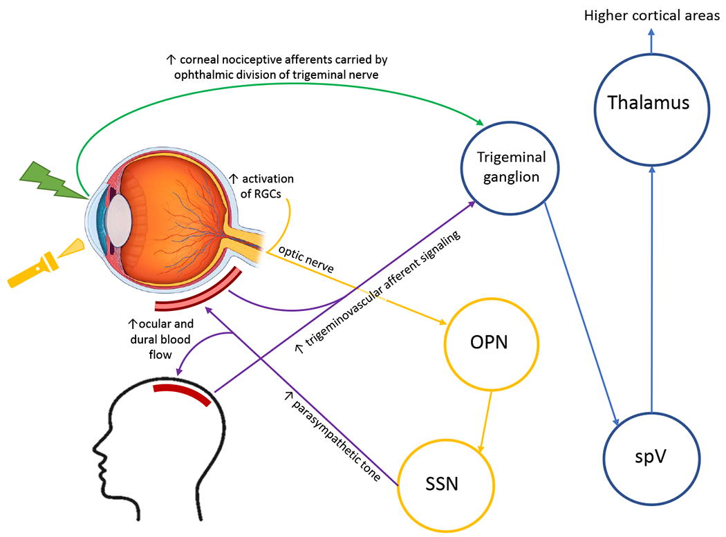
Purpose: To examine neural mechanisms underlying photophobia in individuals with chronic ocular surface pain by using functional magnetic resonance imaging (fMRI).
Design: Cross-sectional case/control analysis.
Methods: A total of 16 individuals from the Miami Veterans Affairs eye clinic underwent comprehensive ocular surface evaluations and were surveyed for ocular surface symptoms. Case patients included patients who reported chronic ocular surface pain symptoms and light sensitivity at least most of the time over 1 week. Controls included persons without chronic ocular surface pain who reported no or minimal light sensitivity. All patients viewed light stimuli during 2 fMRI scans, one before and one after topical anesthetic instillation, and rated their level of pain intensity to the stimulus at the end of each scan. Areas of brain activation in response to light stimuli presentation were correlated with pain responses and examined post- vs pre-anesthesia.
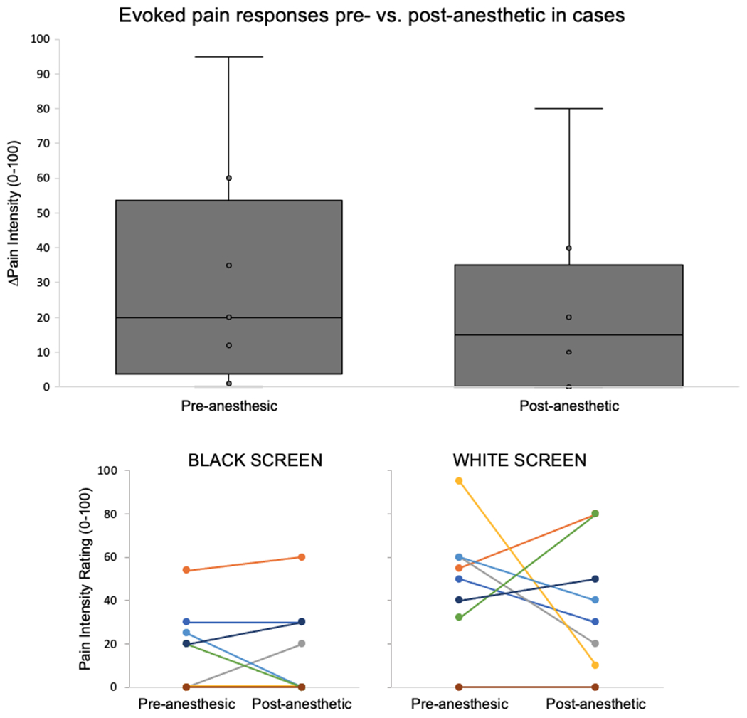
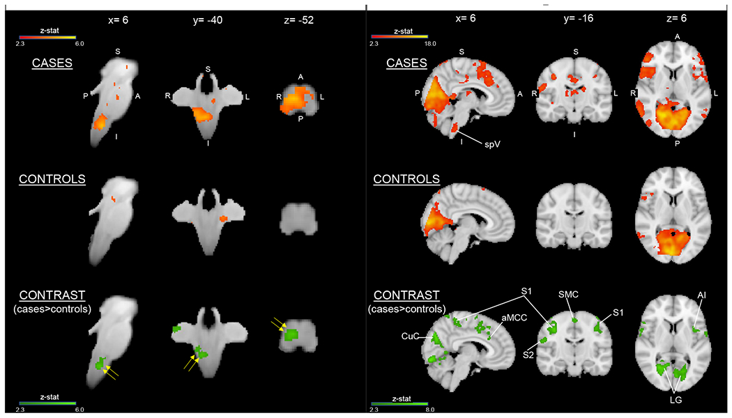
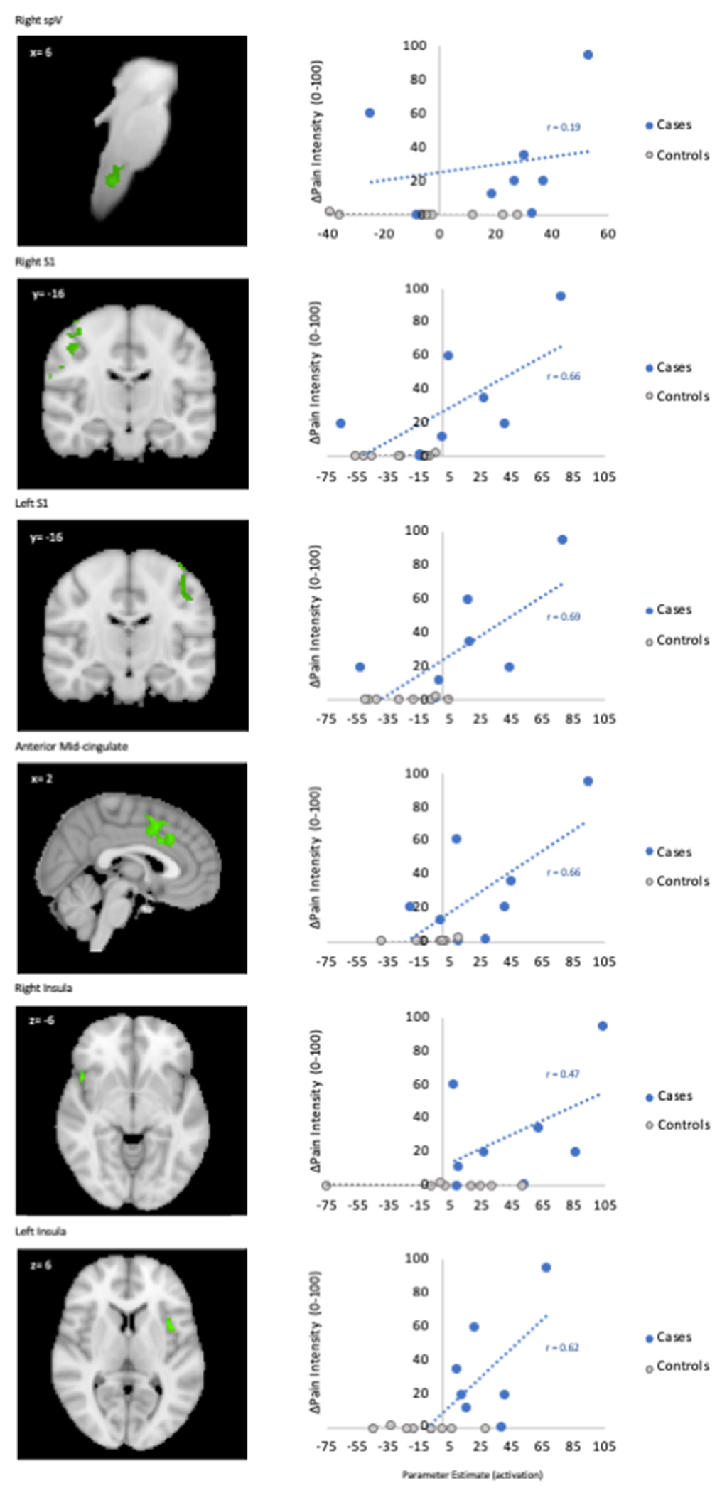
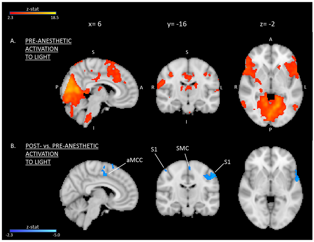
Results: Case patients (n = 8) reported higher pain intensity ratings than controls (n = 8) in response to light stimuli during fMRI. Case patient ratings correlated more with light-evoked activation in pain-related areas within the trigeminal brainstem, primary somatosensory cortex (S1), anterior mid-cingulate cortex (aMCC), and insula than in controls. Topical anesthesia led to varying responses in pain ratings among case patients as well as decreased light-evoked activation in S1 and aMCC.
Conclusions: The trigeminal nociceptive system may contribute to photophobia in individuals with chronic ocular surface pain. We demonstrate modulation of cortical structures in this pathway with topically applied anesthetic to the eyes. Further understanding of modulatory interactions that govern ocular surface pain and photophobia is critical for developing effective, precision-based therapies.
Keywords: Ocular pain; Pain processing; Photophobia; fMRI.
Published by Elsevier Inc.
Figures
Similar articles
-
FL-41 Tint Reduces Activation of Neural Pathways of Photophobia in Patients with Chronic Ocular Pain.Am J Ophthalmol. 2024 Mar;259:172-184. doi: 10.1016/j.ajo.2023.12.004. Epub 2023 Dec 14. Am J Ophthalmol. 2024. PMID: 38101593 Free PMC article.
-
Botulinum toxin A decreases neural activity in pain-related brain regions in individuals with chronic ocular pain and photophobia.Front Neurosci. 2023 Jun 19;17:1202341. doi: 10.3389/fnins.2023.1202341. eCollection 2023. Front Neurosci. 2023. PMID: 37404468 Free PMC article.
-
Noninvasive Electrical Stimulation for the Treatment of Chronic Ocular Pain and Photophobia.Neuromodulation. 2018 Dec;21(8):727-734. doi: 10.1111/ner.12742. Epub 2017 Dec 28. Neuromodulation. 2018. PMID: 29283468 Free PMC article.
-
Neuropathic ocular surface pain: Emerging drug targets and therapeutic implications.Expert Opin Ther Targets. 2022 Aug;26(8):681-695. doi: 10.1080/14728222.2022.2122438. Epub 2022 Sep 20. Expert Opin Ther Targets. 2022. PMID: 36069761 Free PMC article. Review.
-
[Understanding chronic ocular pain].Biol Aujourdhui. 2018;212(1-2):1-11. doi: 10.1051/jbio/2018017. Epub 2018 Oct 26. Biol Aujourdhui. 2018. PMID: 30362450 Review. French.
Cited by
-
Antidepressant Medication Use for Treatment of Chronic Ocular Pain.Cornea. 2024 Nov 1;43(11):1335-1339. doi: 10.1097/ICO.0000000000003646. Epub 2024 Jul 23. Cornea. 2024. PMID: 39348714
-
Neuropathic corneal pain following refractive surgery: risk factors, clinical manifestations, imaging and proteomic characteristics.Br J Ophthalmol. 2025 Jun 23;109(7):747-755. doi: 10.1136/bjo-2024-325996. Br J Ophthalmol. 2025. PMID: 39880672 Free PMC article.
-
Disentangling the neurological basis of chronic ocular pain using clinical, self-report, and brain imaging data: use of K-means clustering to explore patient phenotypes.Front Neurol. 2023 Nov 16;14:1265082. doi: 10.3389/fneur.2023.1265082. eCollection 2023. Front Neurol. 2023. PMID: 38033775 Free PMC article.
-
FL-41 Tint Reduces Activation of Neural Pathways of Photophobia in Patients with Chronic Ocular Pain.Am J Ophthalmol. 2024 Mar;259:172-184. doi: 10.1016/j.ajo.2023.12.004. Epub 2023 Dec 14. Am J Ophthalmol. 2024. PMID: 38101593 Free PMC article.
-
Corneal neuropathic pain: a review to inform clinical practice.Eye (Lond). 2024 Aug;38(12):2350-2358. doi: 10.1038/s41433-024-03060-x. Epub 2024 Apr 16. Eye (Lond). 2024. PMID: 38627548 Free PMC article. Review.
References
-
- Crane AM, Levitt RC, Felix ER, Sarantopoulos KD, McClellan AL, Galor A. Patients with more severe symptoms of neuropathic ocular pain report more frequent and severe chronic overlapping pain conditions and psychiatric disease. Br J Ophthalmol. Feb 2017;101(2):227–231. doi:10.1136/bjophthalmol-2015-308214 - DOI - PMC - PubMed
Publication types
MeSH terms
Grants and funding
LinkOut - more resources
Full Text Sources
