. 2022 Nov 1;128(5):1244-1257.
doi: 10.1152/jn.00259.2022. Epub 2022 Oct 12.

Stretch reflex gain scaling at the shoulder varies with synergistic muscle activity

Affiliations

Stretch reflex gain scaling at the shoulder varies with synergistic muscle activity

Constantine P Nicolozakes et al. J Neurophysiol. .

Abstract

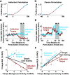
The unique anatomy of the shoulder allows for expansive mobility but also sometimes precarious stability. It has long been suggested that stretch-sensitive reflexes contribute to maintaining joint stability through feedback control, but little is known about how stretch-sensitive reflexes are coordinated between the muscles of the shoulder. The purpose of this study was to investigate the coordination of stretch reflexes in shoulder muscles elicited by rotations of the glenohumeral joint. We hypothesized that stretch reflexes are sensitive to not only a given muscle's background activity but also the aggregate activity of all muscles crossing the shoulder based on the different groupings of muscles required to actuate the shoulder in three rotational degrees of freedom. We examined the relationship between a muscle's background activity and its reflex response in eight shoulder muscles by applying rotational perturbations while participants produced voluntary isometric torques. We found that this relationship, defined as gain scaling, differed at both short and long latencies based on the direction of voluntary torque generated by the participant. Therefore, gain scaling differed based on the aggregate of muscles that were active, not just the background activity in the muscle within which the reflex was measured. Across all muscles, the consideration of torque-dependent gain scaling improved model fits (ΔR2) by 0.17 ± 0.12. Modulation was most evident when volitional torques and perturbation directions were aligned along the same measurement axis, suggesting a functional role in resisting perturbations among synergists while maintaining task performance.NEW & NOTEWORTHY Careful coordination of muscles crossing the shoulder is needed to maintain the delicate balance between the joint's mobility and stability. We provide experimental evidence that stretch reflexes within shoulder muscles are modulated based on the aggregate activity of muscles crossing the joint, not just the activity of the muscle in which the reflex is elicited. Our results reflect coordination through neural coupling that may help maintain shoulder stability during encounters with environmental perturbations.

Keywords: gain scaling; reflex coordination; shoulder; stretch reflex.


Conflict of interest statement

No conflicts of interest, financial or otherwise, are declared by the authors.

Figures

None
Graphical abstract
Figure 1.
Figure 1.
Experimental setup. All participants (n = 10) were seated with their trunk and shoulder secured by straps to minimize motion. Scapular motion was not restrained. The dominant (right) arm of each participant was attached to a rotary motor, with the arm positioned at 90° abduction, 0° extension, and 0° rotation. The glenohumeral joint was perturbed in three degrees of freedom. Participants exerted submaximal isometric torques (5% or 10% of maximum voluntary contraction) in six directions while the perturbations were applied. The glenohumeral joint was also perturbed while participants were instructed to relax and ignore the perturbations. All combinations of torque direction, torque level, and perturbation direction were tested.
Figure 2.
Figure 2.
Methods for estimating reflex responses. Sample data are displayed from a single participant to illustrate methodology. A: perturbations were applied during 60-s trials of submaximal torque production. Positive and negative perturbations were applied in each of the three degrees of freedom tested. Here, the measured motor position is shown for a single flexion and extension ramp applied during extension torque production. B: average rectified EMG of the posterior deltoid during flexion (top) and extension (bottom) perturbations as the participant generated 5% MVC (thin traces) and 10% MVC (thick traces) extension torque trials. EMG traces were averaged across all perturbations in a trial and plotted relative to the average background EMG (0–40 ms before perturbation onset). Short-latency (SLR), middle-latency (MLR), and long-latency reflex (LLR) responses were estimated by averaging the rectified EMG activity in three time-windows postperturbation onset. During flexion perturbations (top), the posterior deltoid is lengthened, and a positive (facilitation) reflex is recorded. In contrast, during extension perturbations (bottom), the posterior deltoid is shortened, and we observed a negative (suppression) reflex. All MLR responses of the posterior deltoid in response to all torque conditions (6 directions at 2 levels) are plotted for the flexion (C) and extension (D) perturbations of a single participant, with the filled dots corresponding to the trials shown in B. The linear relationship between the magnitude of a given muscle’s reflex response and its background activity reflects simple gain scaling, with the slope of this relationship defined as the gain scaling factor. MVC, maximum voluntary contraction.
Figure 3.
Figure 3.
Simple gain scaling of reflex responses organized by muscle, perturbation direction, and reflex window. Each vertical bar represents the gain scaling factor—the linear relationship between reflex response and background activity—in a given condition based on simple gain scaling. Thick vertical black lines represent the 95% confidence interval of the gain scaling factor estimate for each condition. A significant relationship between a given muscle’s background activity and its reflex response was observed in 75/144 conditions (*α = 0.05 with Bonferroni-Holm correction). Results are displayed from n = 10 participants. LLR, long-latency reflex; MLR, middle-latency reflex; MVC, maximum voluntary contraction; SLR, short-latency reflex.
Figure 4.
Figure 4.
Reflexes elicited by a given perturbation varied with torque direction. Data are plotted for the triceps brachii of a single participant during adduction (left column) and flexion (right column) perturbations. A–C: during adduction perturbations, the direction and magnitude of reflex responses differed across torque directions in the MLR window. Facilitative reflexes were observed during abduction torques, whereas suppressive reflexes were observed during adduction torques. D–F: in contrast, during flexion perturbations, similar gain scaling was observed with torque production in all directions. Colored dots reflect reflex responses for the noted torque directions, and gray dots reflect reflex responses for torques in other tested directions. Individual EMGs are plotted relative to the average background activity. MLR, middle-latency reflex; MVC, maximum voluntary contraction.
Figure 5.
Figure 5.
Group results of models describing triceps brachii middle latency reflex responses in two different perturbation directions. In a single muscle among different conditions of perturbation direction, the variation based on torque direction due to torque-dependent gain scaling can be observed. A: in certain cases, such as demonstrated following adduction perturbations, divergence in reflexes between different torque directions was observed. When a divergence in responses was observed, the inclusion of torque direction fixed effects accounted for substantially more variance than modeling the reflex responses without accounting for torque direction. B: in other instances, such as demonstrated following flexion perturbations, little divergence in the reflexes between torque directions was observed. In these cases, the inclusion of torque direction fixed effects did not account for substantially more variance than a simpler model. Shaded areas represent 95% confidence intervals of the gain scaling factor estimates. Results are displayed from n = 10 participants. MLR, middle-latency reflex; MVC, maximum voluntary contraction.
Figure 6.
Figure 6.
Improvements in goodness-of-fit (R2) between simple gain scaling models and torque-dependent gain scaling models in predicting reflex responses. Each layered gray and black bar represents the R2 for the two models in the same condition (muscle, perturbation direction, and reflex window). In certain muscles, such as the triceps brachii and infraspinatus muscles, we observed large improvements in R2 between the simple gain scaling and torque-dependent gain scaling models. In other muscles, such as the anterior and middle heads of the deltoid muscle, R2 increased less between the two models than in all other muscles. All torque-dependent gain scaling models were significantly improved compared with the corresponding simple gain scaling model (log-likelihood ratio test, α = 0.05 with Bonferroni–Holm correction) unless noted as not significant (ns). Results are displayed from n = 10 participants. MLR, middle-latency reflex; MVC, maximum voluntary contraction.
Figure 7.
Figure 7.
Relationships between torque-dependent gain scaling and perturbation directions. A: stretch reflexes in the posterior deltoid, an abductor, when shortened. Enhanced suppression was observed when volitional torque was generated in this muscle’s assistive direction (blue) and excitation—an extreme case of reduced suppression—was observed when volitional torques were generated in the opposite direction (red). Two trials are shown from a typical participant. B: group results (n = 10 participants) showing gain scaling of posterior deltoid stretch reflexes within short, medium, and long latency windows following abduction perturbations. C: stretch reflexes in the infraspinatus, an extensor, when lengthened. Enhanced facilitation was observed when volitional torque was generated in this muscle’s assistive direction (green) and reduced facilitation was observed when volitional torques were generated in the opposite direction (purple). Two additional trials are shown from the same participant. D: group results (n = 10 participants) showing gain scaling of infraspinatus stretch reflexes within short, medium, and long latency windows following flexion perturbations. Vertical black lines represent the 95% confidence interval of the gain scaling estimates. LLR, long-latency reflex; MLR, middle-latency reflex; MVC, maximum voluntary contraction; SLR, short-latency reflex.
Figure 8.
Figure 8.
The modulation of gain scaling when volitional torques and perturbation directions were aligned along one of the three measurement axes. Blue boxes reflect conditions when the gain scaling factor in the torque direction opposing the perturbation direction was higher than in the antagonistic torque direction. Red/hatched boxes reflect conditions when the gain scaling factor in the torque direction opposing the perturbation direction was lower than in the antagonistic torque direction. Asterisks and dark color shades reflect a significant difference between the gain scaling factors in antagonistic torque directions that are in the same measurement axis as a given perturbation direction (α = 0.05 with Bonferroni–Holm correction). White boxes reflect measurement axes in which a muscle did not have assistive/resistive torque directions; these conditions were excluded from the analysis. LLR, long-latency reflex; MLR, middle-latency reflex; SLR, short-latency reflex.

Similar articles

Cited by

References

    1. Peters KM, Kelly VE, Chang T, Weismann MC, Westcott McCoy S, Steele KM. Muscle recruitment and coordination during upper-extremity functional tests. J Electromyogr Kinesiol 38: 143–150, 2018. doi:10.1016/j.jelekin.2017.12.002. - DOI - PMC - PubMed
    1. Labriola JE, Lee TQ, Debski RE, McMahon PJ. Stability and instability of the glenohumeral joint: the role of shoulder muscles. J Shoulder Elbow Surg 14: 32S–38S, 2005. doi:10.1016/j.jse.2004.09.014. - DOI - PubMed
    1. Lipps DB, Baillargeon EM, Ludvig D, Perreault EJ. Quantifying the multidimensional impedance of the shoulder during volitional contractions. Ann Biomed Eng 48: 2354–2369, 2020. doi:10.1007/s10439-020-02509-w. - DOI - PMC - PubMed
    1. Veeger HEJ, van der Helm FCT. Shoulder function: the perfect compromise between mobility and stability. J Biomech 40: 2119–2129, 2007. doi:10.1016/j.jbiomech.2006.10.016. - DOI - PubMed
    1. Reddy AS, Mohr KJ, Pink MM, Jobe FW. Electromyographic analysis of the deltoid and rotator cuff muscles in persons with subacromial impingement. J Shoulder Elbow Surg 9: 519–523, 2000. doi:10.1067/mse.2000.109410. - DOI - PubMed

Publication types

LinkOut - more resources