. 2022 Oct 12;13(1):6017.
doi: 10.1038/s41467-022-33546-9.

Rapid joule heating improves vitrification based cryopreservation

Affiliations

Rapid joule heating improves vitrification based cryopreservation

Li Zhan et al. Nat Commun. .

Abstract

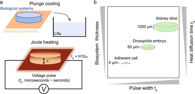
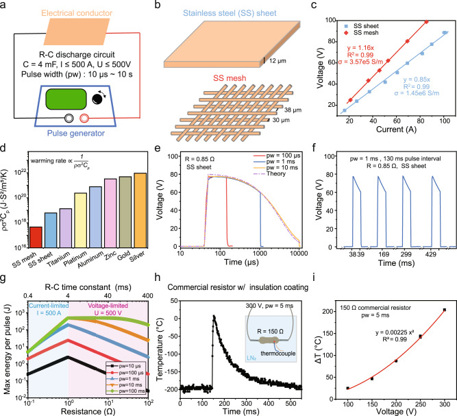
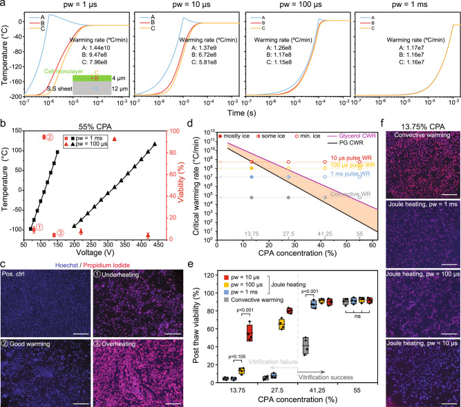
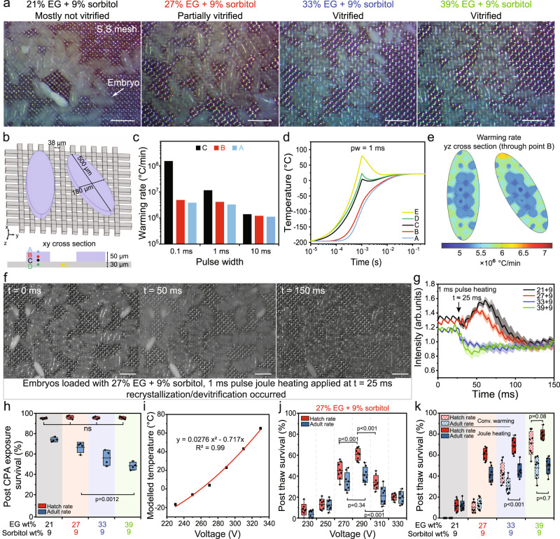
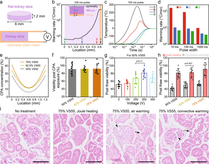
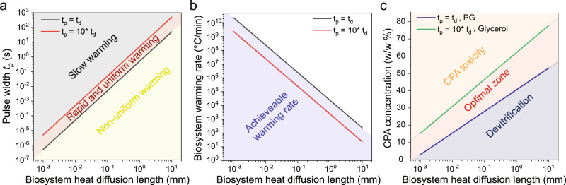
Cryopreservation by vitrification has far-reaching implications. However, rewarming techniques that are rapid and scalable (both in throughput and biosystem size) for low concentrations of cryoprotective agent (CPA) for reduced toxicity are lacking, limiting the potential for translation. Here, we introduce a joule heating-based platform technology, whereby biosystems are rapidly rewarmed by contact with an electrical conductor that is fed a voltage pulse. We demonstrate successful cryopreservation of three model biosystems with thicknesses across three orders of magnitude, including adherent cells (~4 µm), Drosophila melanogaster embryos (~50 µm) and rat kidney slices (~1.2 mm) using low CPA concentrations (2-4 M). Using tunable voltage pulse widths from 10 µs to 100 ms, numerical simulation predicts that warming rates from 5 × 10⁴ to 6 × 10⁸ °C/min can be achieved. Altogether, our results present a general solution to the cryopreservation of a broad spectrum of cellular, organismal and tissue-based biosystems.


Conflict of interest statement

The authors (L.Z., Z.H., Q.S., M.E., and J.B.) have filed a provisional patent application (serial no. 63/358,377) related to this work. The remaining author declares no competing interests.

Figures

Fig. 1
Fig. 1. A rapid and scalable rewarming platform technique using joule heating.
a Schematics of biological systems cryopreservation using plunge cooling and joule heating. The biological systems loaded with cryoprotective agent (CPA) were in contact with the electrical conductor. After removing the excess CPA solution, the biological systems and electrical conductor were plunged together into liquid nitrogen (LN2) for cooling. For rewarming, the electrical conductor was connected to a voltage pulse generator and generated heat via joule heating. The biomaterial was rewarmed via conduction. Drawings not to scale. b Three types of biosystems with different thicknesses were used as the model systems, including adherent cells (4 µm), Drosophila embryos (50 µm), and kidney slices (1.2 mm), as shown in the plot (drawings not to scale). The heat diffusion time (td, right y-axis) across the biosystems is positively correlated with the biosystem thickness.
Fig. 2
Fig. 2. Characterization of joule heating using a pulse generator.
a The R-C discharge circuit is used to generate joule heating by connecting the electrical conductor to a pulse generator. The voltage and pulse width can be adjusted. The capacitance (C) is 4000 µF. Ranges of the current (I), voltage (V), and pulse width (pw) for the low voltage mode (LV, ≤ 500 V) are listed in the figure. b Stainless steel (SS) sheet and mesh were used in this study. The thickness of SS sheet is 12 µm. The wire diameter and aperture size of the SS mesh are 30 and 38 µm, respectively. The SS mesh allows easy removal of excess CPA solution. c The electrical conductivity (σ) of the SS sheet and mesh can be calculated from the resistance by measuring the voltage and current. n = 6 independent experiments. d Comparison of various materials for rapid warming rate. The warming rate is inversely correlated to material density (ρ), electrical conductivity (σ), and heat capacity (Cp). Details in Eq. 7 assuming same electrical current, material dimensions and pulse width. e Voltage profile when different voltage pulse widths were used. The exponential decay of measured voltage matched with the calculated voltage of a R-C discharge circuit. f Voltage profile when multiple voltage pulses were applied. g Maximum energy delivered per pulse depends on the resistance and pulse width. For R ≤ 1 Ω, the max current of 500 A is used for calculation. For R ≥ 1 Ω, the max voltage of 500 V is used for calculation. h Temperature profile of a thin film resistor (150 Ω) when subjected to a voltage pulse of 300 V, 5 ms. A thermocouple was attached to the electrically insulated surface of the resistor. The resistor was submerged in liquid nitrogen (LN2) during the measurement. Sampling resolution was 1 ms. i Temperature change (ΔT) as a function of applied voltage. The same setup shown in h was used. n = 6 independent experiments. For c and i, data presented as mean ± s.d.
Fig. 3
Fig. 3. Adherent cells cryopreservation using joule heating.
a Simulated temperature profile of stainless steel (SS) sheet and cell monolayer under pulse width (pw) ranging from 1 µs to 1 ms (shared y axis). The warming rates at the middle of SS sheet (point A), middle of cells (point B), and top of cells (point C) are displayed. b Simulated temperature (in black) and measured viability (in red) of human dermal fibroblasts (HDFs) for 1 ms and 100 µs pulse width with different voltages. VS55 (i.e., 55 wt%) was used as the cryoprotective agent (CPA). n = 4 independent experiments. Data presented as mean ± s.d. c Merged Hoechst/Propidium Iodide (PI) images of HDF cells. Conditions including underheating, good warming, overheating (for joule heating, as numbered in b), and positive control are shown. The pulse width is 1 ms. Scale bar is 500 µm. d Estimated critical warming rate (CWR) plotted as a function of CPA concentration. Propylene glycol (PG, good glass-forming tendency) and glycerol (poor glass-forming tendency) were used to estimate the range of CWR (orange area) of different CPA concentrations. In this study, 13.75%, 27.5%, 41.25%, and 55% CPA were tested. The measured convective warming rate (WR) were marked in the plot, along with the simulated WR of 1 ms, 100 µs, and 10 µs pulse joule heating. The experimentally tested conditions were labelled as dots at the intersections of CPA concentrations and warming rates. e Post-thaw viability of HDF cells using different CPA concentrations and warming methods. Vitrification failure (i.e., ice formation) was noted for 13.27% and 27% CPA groups. n = 4 independent experiment. Bounds and horizontal line of box represent standard deviation and mean, respectively; whiskers represent max and min. f For 13.75% CPA, merged Hoechst/PI images of HDF cells rewarmed by convective warming, 1 ms, 100 µs, and 10 µs pulse joule heating. Scale bar is 500 µm. One-way ANOVA and Tukey’s post hoc were used for statistical analysis. ns, p > 0.05.
Fig. 4
Fig. 4. Drosophila embryos cryopreservation using joule heating.
a Images of the Drosophila embryos on the stainless steel (SS) mesh after cooling. Embryos were loaded with different ethylene glycol (EG) concentrations. b Geometry and dimensions of the SS mesh and embryos used in the heat transfer modeling. Points A, B, and C represent the top, middle and bottom of the embryo, respectively. Point D and E represent the SS mesh in contact with the embryo and outside the embryo, respectively. c Temperature profile at different locations for 1 ms joule heating. d Warming rate distribution at the middle plane of embryos. e Warming rate of the embryo for different joule heating pulse widths. f Images of embryos on the SS mesh acquired by a high-speed camera at 3000 fps. The cryoprotective agent (CPA) was 27% EG + 9% sorbitol. The voltage was 290 V. g Normalized grayscale intensity of the embryos was plotted from the high-speed camera videos. Ice (i.e., white color) showed a high intensity value. Different CPA concentrations (labelled as EG + sorbitol concentration) were tested. n = 5. Data presented as mean ± s.d. h Survival of embryos after exposure to different CPA concentrations. Hatch and adult rates represent the survival from embryos to larvae and larvae to adults, respectively. n = 4. i Temperature of the embryos after 1 ms joule heating using different voltages. j Survival of embryos after 1 ms joule heating using different voltages. The CPA was 27% EG + 9% sorbitol. n = 8. k Comparison of embryo survival using convective warming and joule heating (290 V, 1 ms) for different CPA concentrations. n = 8. For a and f, scale bar is 500 µm. For gj, n represent independent experiments. For h, j, and k, bounds and horizontal line of box represent standard deviation and mean respectively; whiskers represent max and min. For ce, i modeling results were shown. Multivariate analysis of variance (MANOVA) and Tukey’s post hoc were used for statistical analysis. ns, p > 0.05.
Fig. 5
Fig. 5. Kidney slice cryopreservation using joule heating.
a Rat kidney slices of 1.2 mm thickness and 6 mm in diameter were used. After cryoprotective agent (CPA) loading, the kidney slice was sandwiched using the stainless steel (SS) mesh prior to plunge-cooling in liquid nitrogen, then rewarmed by connecting the SS mesh to a voltage source. b Warming rate distribution within the kidney slice. Point A represents the SS mesh; B, C, and D represent the kidney slice. The detailed locations of points A, B, C, and D were marked in the plot. c Temperature profile at different locations (point A to D) for 100 ms joule heating. d Warming rates at different locations within the kidney slice using 10 ms, 100 ms, and 1000 ms joule heating. e CPA concentration distribution within the kidney slice after loading with different CPA concentrations. f Viability of kidney slices after exposure to different CPA concentrations. n = 9 independent samples. g Post-thaw viability of the kidney slices by joule heating using different voltages. The CPA is 50% VS55. n = 8 independent samples. h Comparison of post-thaw viability of kidney slices using convective warming and joule heating for different CPA concentrations. n = 8 independent samples. i Histology (H & E staining) of kidney slices. Arrowhead showed disrupted glomeruli, and arrow indicated damaged proximal tubules. Scale bar is 500 µm. Modeling results were shown in be. For fh, the viability was measured by alamarBlue assay and normalized by the readings prior to treatment. Date presented as mean ± s.d. One-way ANOVA and Tukey’s post hoc were used for statistical analysis.
Fig. 6
Fig. 6. Design and physical limits of joule heating as the external heat source for rewarming.
a To achieve rapid and sufficiently uniform rewarming (area in red), the pulse width (tp) can be selected based on biosystem heat diffusion length (h). When the pulse width is much smaller than heat diffusion time (td = h2/2α, α is the thermal diffusivity of the biosystem: 10−6 m2/s was used for the calculation), non-uniform warming occurs. For longer pulse widths (i.e., > 10 td), the achieved rewarming rate is slow. We assume sufficiently uniform rewarming can be achieved using tp = 1 ~ 10 td. b Achievable warming rate inside the biosystems as a function of biosystem heat diffusion length. The black and red lines represent the warming rates (calculated as 200 °C / tp) when tp=td and tp= 10 td, respectively. The achieved warming rate decreases with increasing pulse width. c Within the rapid and uniform warming region defined in a, selection of cryoprotective agent (CPA) concentration for optimal cryopreservation of different biosystems using external joule heating source. The upper bound of CPA concentration (green line) is estimated using the longer pulse width (i.e., tp = 10 td) and glycerol as the CPA (i.e., relatively high critical warming rate). The lower bound of CPA concentration (blue line) is estimated using the shorter pulse width (i.e., tp = td) and propylene glycol (PG) as the CPA (i.e., relatively low critical warming rate). The CPA toxicity is the major failure mode of cryopreservation in the upper corner region (i.e., orange color). The devitrification is the major failure mode of cryopreservation in the lower corner region (i.e., blue color).

Similar articles

Cited by

References

    1. Hunt CJ. Cryopreservation of human stem cells for clinical application: a review. Transfus. Med. Hemother. 2011;38:107–123. doi: 10.1159/000326623. - DOI - PMC - PubMed
    1. Karlsson JO, Toner M. Long-term storage of tissues by cryopreservation: critical issues. Biomaterials. 1996;17:243–256. doi: 10.1016/0142-9612(96)85562-1. - DOI - PubMed
    1. Cirino L, et al. First instance of settlement by cryopreserved coral larvae in symbiotic association with dinoflagellates. Sci. Rep. 2019;9:1–8. doi: 10.1038/s41598-019-55374-6. - DOI - PMC - PubMed
    1. Gallichotte EN, et al. Towards a method for cryopreservation of mosquito vectors of human pathogens. Cryobiology. 2021;99:1–10. doi: 10.1016/j.cryobiol.2021.02.001. - DOI - PMC - PubMed
    1. Mazur P, Cole KW, Hall JW, Schreuders PD, Mahowald AP. Cryobiological preservation of Drosophila embryos. Science. 1992;258:1932–1935. doi: 10.1126/science.1470915. - DOI - PubMed

Publication types

Substances

LinkOut - more resources