. 2022 Oct 12;12(1):17068.
doi: 10.1038/s41598-022-21399-7.

The effect of the surgical approach and cochlear implant electrode on the structural integrity of the cochlea in human temporal bones

Affiliations

The effect of the surgical approach and cochlear implant electrode on the structural integrity of the cochlea in human temporal bones

Saad Jwair et al. Sci Rep. .

Abstract

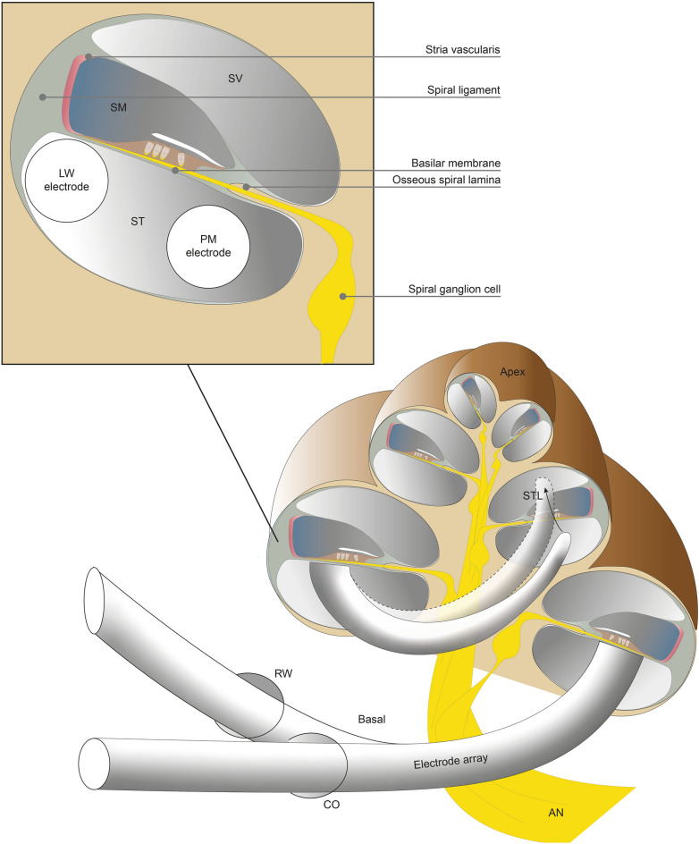
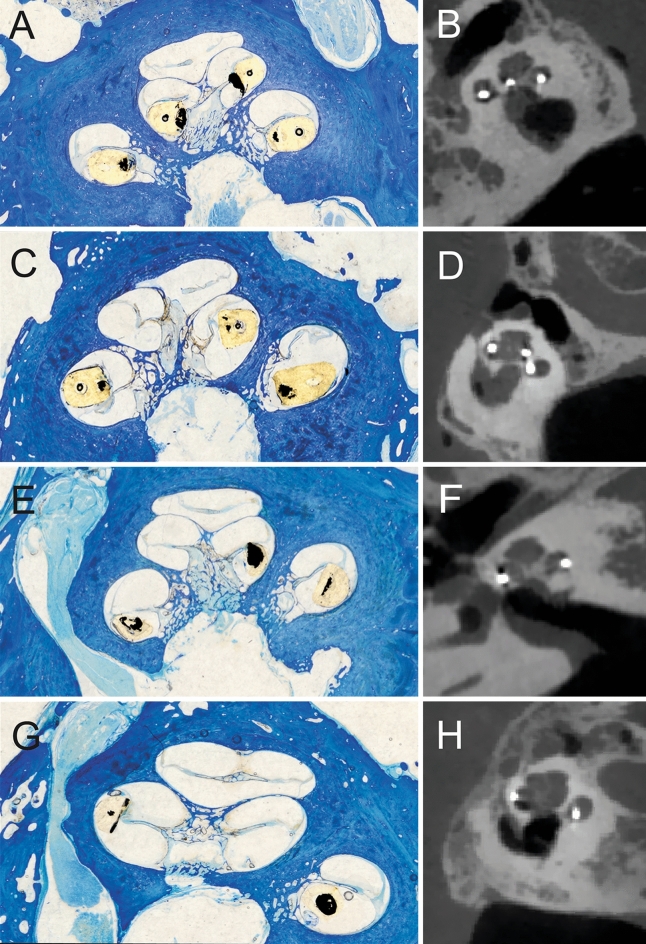
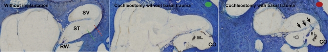
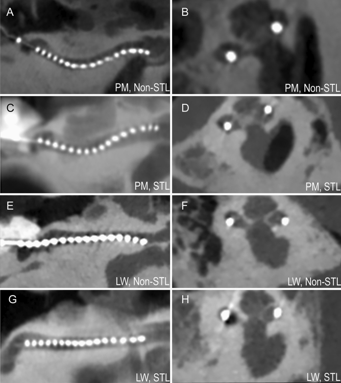
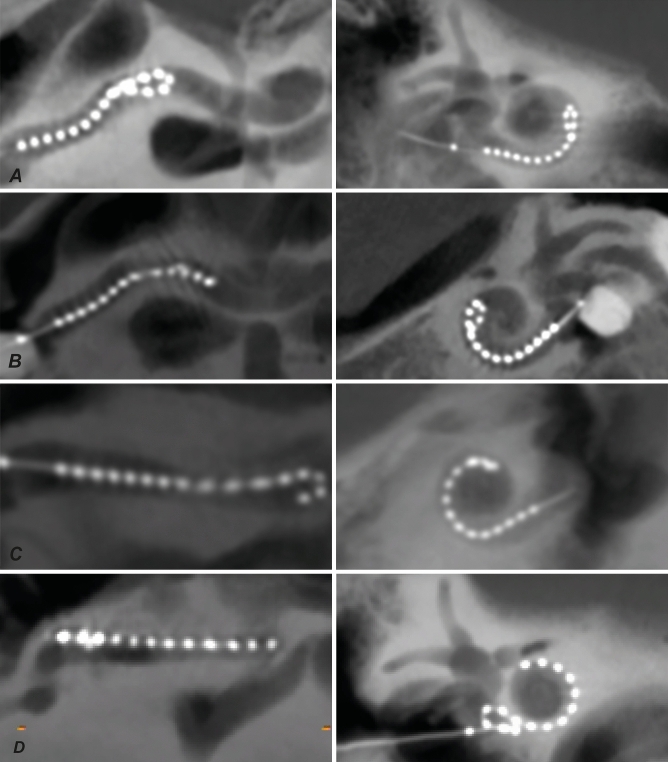
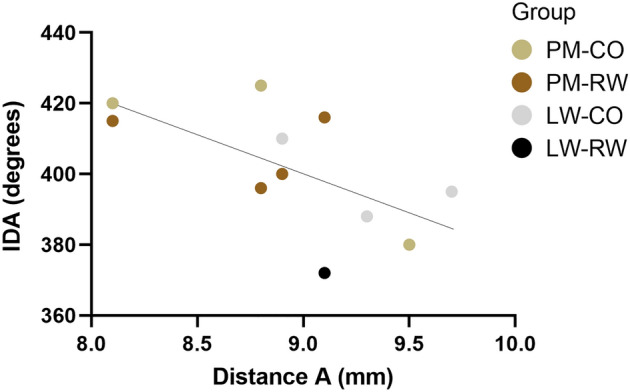
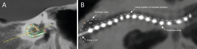
Cochlear implants (CI) restore hearing of severely hearing-impaired patients. Although this auditory prosthesis is widely considered to be very successful, structural cochlear trauma during cochlear implantation is an important problem, reductions of which could help to improve hearing outcomes and to broaden selection criteria. The surgical approach in cochlear implantation, i.e. round window (RW) or cochleostomy (CO), and type of electrode-array, perimodiolar (PM) or lateral wall (LW), are variables that might influence the probability of severe trauma. We investigated the effect of these two variables on scalar translocation (STL), a specific type of severe trauma. Thirty-two fresh frozen human cadaveric ears were evenly distributed over four groups receiving either RW or CO approach, and either LW or PM array. Conventional radiological multiplanar reconstruction (MPR) was compared with a reconstruction method that uncoils the spiral shape of the cochlea (UCR). Histological analysis showed that RW with PM array had STL rate of 87% (7/8), CO approach with LW array 75% (6/8), RW approach with LW array 50% (4/8) and CO approach with PM array 29% (2/7). STL assessment using UCR showed a higher inter-observer and histological agreement (91 and 94% respectively), than that using MPR (69 and 74% respectively). In particular, LW array positions were difficult to assess with MPR. In conclusion, the interaction between surgical approach and type of array should be preoperatively considered in cochlear implant surgery. UCR technique is advised for radiological assessment of CI positions, and in general it might be useful for pathologies involving the inner ear or other complex shaped bony tubular structures.


Conflict of interest statement

The authors declare no competing interests.

Figures

Figure 1
Figure 1
A cross section of the cochlea is depicted with an implanted electrode array. The electrode array is implanted in the scala tympani, using either the round window or a small hole in the cochlea (cochleostomy) for entry. The array follows the spiral curvature of the cochlea from the base of the cochlea towards the apex. Arrays usually reach at least around one turn and half, depending on the exact length of the array. Perimodiolar arrays are positioned more towards the spiral ganglion cells of the auditory nerve and beneath the osseous spiral lamina, and in contrast, lateral wall arrays are positioned laterally towards spiral ligament and beneath basilar membrane. In some cases the array can translocate during insertion (i.e. STL) from ST to SV or SM, which is detrimental for the structures that lie in between. RW round window, CO cochleostomy, AN auditory nerve, STL scalar translocation, ST scala tympani, SV scala vestibule, SM scala media, LW lateral wall, PM perimodiolar.
Figure 2
Figure 2
Histological modiolar plane sections and corresponding CT images were compared. Similar array positions were observed between histology and radiology. In (A,B) non-STL PM array. In (C,D) non-STL LW array. In (E,F) STL PM array. In (G,H) STL LW array. Note: the diameter of the array was increased 30–40% due to swelling of the silicon layer after processing with butyl methacrylate. Contrast of array was increased for visibility reasons.
Figure 3
Figure 3
Group comparison of array scalar positioning. Scala media or scala vestibuli position is assigned if at least one electrode-contact was located in the respective compartment. *p < 0.05, **p < 0.01. ST scala tympani, SM scala media, SV scala vestibuli.
Figure 4
Figure 4
Histological modiolar plane sections. (A) non-STL PM array. (B) STL array with fracture of osseous spiral lamina. (C) non-STL LW array. (D) STL LW array to scala media with displacement of stria vascularis, basilar membrane and spiral ligament.
Figure 5
Figure 5
Histological modiolar plane sections. On the left a non-implanted basal turn of the cochlea. The green and red dotted lines depict angles of cochleostomy site of respectively the middle and right image. Green dotted line represents an antero-inferiorly placed cochleostomy, the red dotted line represents slight displacement of cochleostomy to anterior. In middle image, corresponding to the green line, no basal trauma is observed. In right image, corresponding to the red line, basal trauma is observed: displacement of stria vascularis, basilar membrane and osseous spiral lamina resulting in a crushed scala media compartment. RW round window, ST scala tympani, SV scala vestibuli.
Figure 6
Figure 6
Assessment examples between uncoiled cochlear reconstructions (on the left) and conventional multiplanar reconstructions (on the right). In (A) the PM array is neatly following the scala tympani, which is located in lower half of the uncoiled cochlea. In (C), however, clear kinking of the PM array results in STL from scala tympani to scala vestibuli. This difference of PM non-STL vs. STL is also seen in conventional reconstructions in (B,D), with the array, the white dots, located in STL case of (D) more towards the upper half of the cochlea than in (B). In (E) the LW array follows the scala tympani without interruption, in contrast in (G), the LW array shows an subtle, but still clear kink towards scala vestibuli (i.e. STL). The different position of the LW array is difficult to observe in conventional reconstructions, see (F) vs. (H).
Figure 7
Figure 7
Four fold-overs were observed, depicted on the left with uncoiled cochlear reconstructions and on the right with conventional reconstructions. In (A–C) a PM array was used. Two tip fold-overs occurred at similar position (see A,B), approximately at insertion depth of 180°, while the other tip fold-over occurred deeper at around 270° (C). Finally, in (D) a case with a basal fold-over had occurred using the LW array. In both reconstruction techniques the fold-overs were clearly seen. PM perimodiolar, LW lateral wall.
Figure 8
Figure 8
The insertion depth angles and corresponding cochlear distance A are plotted of the 11 full insertions according to the manufactures guidelines. An inverse correlation is observed between insertion depth angle and distance A (R2 = 0.39, p < 0.05). Distance A is an indirect measure of cochlear duct length. IDA insertion depth angle, PM perimodiolar LW lateral wall, CO cochleostomy, RW round window.
Figure 9
Figure 9
Insertion depth angle is compared between fully scala tympani located arrays and arrays with at least one electrode in scala vestibuli. All insertion depth angles were plotted of arrays with the stop electrode within 2 mm distance from the CO or RW site (n = 22). Median of both groups are shown. IDA insertion depth angle, CO cochleostomy, RW round window.
Figure 10
Figure 10
In the cochlear view reconstruction of CB-CT scan both insertion depth angle and distance A can be measured. The 360° line is drawn perpendicular to a line between round window entry and middle of upper part of the posterior semicircular canal. The insertion depth angle is measured by adding 360° to the angle between the apical electrode and the 360° line. Distance A (dashed line), an indirect measure proportional to cochlear duct length, is measured as the length of the line from the point of the array entering the RW or CO, through the modiolus to the contralateral cochlear wall.
Figure 11
Figure 11
Method for conventional multiplanar reconstructions. The lines in the images represent the position of the shown images relative to the planes. Oblique planes were reconstructed by aligning the red line in sagittal and axial plane to the basal turn of the cochlea, and the blue and green line were aligned to the course of the cochlear modiolus. This resulted in multi reconstructions of three oblique planes: oblique coronal plane, oblique sagittal plane or ‘cochlear view’, and oblique axial plane.
Figure 12
Figure 12
Method for uncoiled cochlear reconstructions (UCR). (A) line is drawn along the implanted array in the oblique ‘cochlear view’ reconstruction with 2 mm thickness. The red circles depict the manually selected points that were used to trace the array. The yellow line shows the cross section perpendicular to this tracing line. (B) Subsequently, these reconstructions generated a planar two-dimensional image with implanted array, UCR. The cochlear structure is therefore viewed from the side, with upper half being the scala vestibuli (SV) and lower part scala tympani (ST). The first basal segment is characterized by the cochlear hook.

Similar articles

Cited by

References

    1. Wilson BS, Tucci DL, Merson MH, O'Donoghue GM. Global hearing health care: New findings and perspectives. Lancet. 2017;390:2503–2515. doi: 10.1016/S0140-6736(17)31073-5. - DOI - PubMed
    1. Carlson ML. Cochlear implantation in adults. N. Engl. J. Med. 2020;382:1531–1542. doi: 10.1056/NEJMra1904407. - DOI - PubMed
    1. Gifford RH, Revit LJ. Speech perception for adult cochlear implant recipients in a realistic background noise: Effectiveness of preprocessing strategies and external options for improving speech recognition in noise. J. Am. Acad. Audiol. 2010;21:441–451. doi: 10.3766/jaaa.21.7.3. - DOI - PMC - PubMed
    1. Snels C, IntHout J, Mylanus E, Huinck W, Dhooge I. Hearing preservation in cochlear implant surgery: A meta-analysis. Otol. Neurotol. 2019;40:145–153. doi: 10.1097/MAO.0000000000002083. - DOI - PubMed
    1. Gifford RH, et al. Cochlear implantation with hearing preservation yields significant benefit for speech recognition in complex listening environments. Ear Hear. 2013;34:413–425. doi: 10.1097/AUD.0b013e31827e8163. - DOI - PMC - PubMed

Publication types