Comparative Study
. 2022 Oct 12;12(1):17058.
doi: 10.1038/s41598-022-21690-7.

A comparative study of spike protein of SARS-CoV-2 and its variant Omicron (B.1.1.529) on some immune characteristics

Affiliations
Comparative Study

A comparative study of spike protein of SARS-CoV-2 and its variant Omicron (B.1.1.529) on some immune characteristics

Ximeng Li et al. Sci Rep. .

Abstract

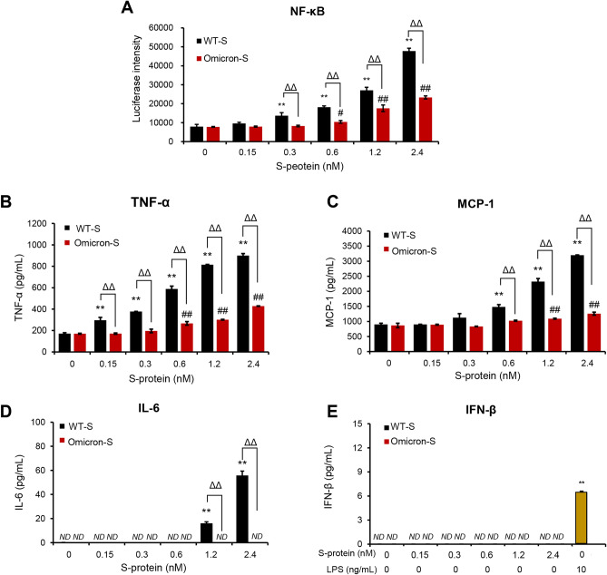
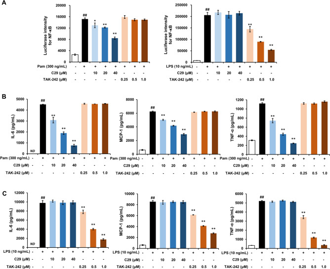
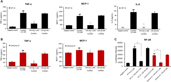
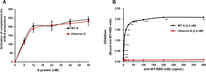
The emergence of Omicron variant raises great concerns because of its rapid transmissibility and its numerous mutations in spike protein (S-protein). S-protein can act as a pathogen-associated molecular pattern and complement activator as well as antigen. We compared some immune characteristics of trimer S-proteins for wild type (WT-S) and B.1.1.529 Omicron (Omicron-S) to investigate whether the mutations have affected its pathogenicity and antigenic shift. The results indicated that WT-S and Omicron-S directly activated nuclear factor-κB (NF-κB) and induced the release of pro-inflammatory cytokines in macrophages, but the actions of Omicron-S were weaker. These inflammatory reactions could be abrogated by a Toll-like receptor 4 antagonist TAK-242. Two S-proteins failed to induce the production of antiviral molecular interferon-β. In contrast to pro-inflammatory effects, the ability of two S-proteins to activate complement was comparable. We also compared the binding ability of two S-proteins to a high-titer anti-WT-receptor-binding domain antibody. The data showed that WT-S strongly bound to this antibody, while Omicron-S was completely off-target. Collectively, the mutations of Omicron have a great impact on the pro-inflammatory ability and epitopes of S-protein, but little effect on its ability to activate complement. Addressing these issues can be helpful for more adequate understanding of the pathogenicity of Omicron and the vaccine breakthrough infection.


Conflict of interest statement

The authors declare no competing interests.

Figures

Figure 1
Figure 1
Comparison of the effects of trimer WT-S and Omicron-S on inflammatory responses in RAW264.7 macrophages. (A) Effects of trimer WT-S and Omicron-S on NF-κB activity. The cells stably transfected with NF-κB-TA-luc plasmid were stimulated with trimer WT-S or Omicron-S at the indicated concentrations. Four hours later, the luciferase activities were detected. (B–E) Effects of trimer WT-S and Omicron-S on supernatant TNF-α, MCP-1, IL-6 and IFN-β in RAW264.7 macrophages. Cells were treated with trimer WT-S or Omicron-S at the indicated concentrations. Twenty-four hours later, supernatant TNF-α, MCP-1, IL-6 and IFN-β were determined. LPS was used as a positive control for INF-β. Mean ± SD (n = 3). **P < 0.01 vs. 0 nM of WT-S (negative control); #P < 0.05 and ##P < 0.01 vs. 0 nM of Omicron-S (negative control); ΔΔP < 0.01. ND, not detected.
Figure 2
Figure 2
Effects of C29 and TAK-242 on Pam- or LPS-induced pro-inflammatory reactions in RAW264.7 macrophages. (A) Effects of C29 and TAK-242 on Pam- or LPS-induced NF-κB activation in RAW264.7 macrophages. (B) Effects of C29 and TAK-242 on Pam-elevated supernatant IL-6, MCP-1 and TNF-α in RAW264.7 macrophages. (C) Effects of C29 and TAK-242 on LPS-elevated supernatant IL-6, MCP-1 and TNF-α in RAW264.7 macrophages. Mean ± SD (n = 3). ##P < 0.01 vs. negative control; *P < 0.05 and **P < 0.01 vs. Pam or LPS alone. ND, not detected.
Figure 3
Figure 3
Effects of TAK-242 and C29 on the pro-inflammatory reactions induced by two trimer S-proteins. (A–B) Effects of TAK-242 and C29 on supernatant TNF-α, MCP-1 and IL-6 induced by WT-S (A) or Omicron-S (B) in RAW264.7 macrophages. The cells were treated with trimer WT-S or Omicron-S (2.4 nM) in the presence of TAK-242 (1 μM) or C29 (40 μM). Twenty-four hours later, supernatant TNF-α, MCP-1 and IL-6 were determined. ND, not detected. (C) Effects of TAK-242 and C29 on trimer WT-S- or Omicron-S-induced NF-κB activation. RAW264.7 cells stably transfected with NF-κB-TA-luc plasmid were treated with C29 (40 μM) or TAK-242 (1 μM) and then stimulated by trimer WT-S or Omicron-S (2.4 nM). Four hours later, the luciferase activities were detected. Mean ± SD (n = 3). ##P < 0.01 vs. negative control; **P < 0.01 vs. WT-S or Omicron-S alone.
Figure 4
Figure 4
Comparison of the ability of trimer WT-S and Omicron-S to activate complement and bind to anti-WT-RBD mAb. (A) Comparison of the ability of trimer WT-S and Omicron-S to activate complement. Normal human serum complement was added into the plate coated with trimer WT-S or Omicron-S at different concentrations (0–48 nM). After 1 h incubation, complement activation was terminated by washing the plate four times. The produced membrane attack complex C5b-9 was combined with a mouse anti-human C5b-9 IgG2b antibody which further combined with an HRP-conjugated goat anti-mouse IgG antibody. The chromogenic reaction was developed by adding TMB substrate and then terminated by 2 M H2SO4. OD values at 450 nm, which were positively correlated with the C5b-9 production, were determined. Activation percentage of complement was calculated relative to the negative control. Mean ± SD (n = 3). (B) Comparison of the binding ability of two trimer S-proteins to anti-WT-RBD mAb. The purified anti-WT-RBD mAb at different concentrations was added into the plate coated with 2.4 nM of trimer WT-S or Omicron-S. After 1 h incubation, the unbound mAb was washed away by PBST and the bound mAb could be detected by HRP-conjugated goat anti-mouse IgG. The chromogenic reaction was developed by adding TMB substrate and then terminated by 2 M H2SO4. OD values at 450 nm, which were positively correlated with the binding ability (affinity) of S-protein to anti-WT-RBD mAb, were determined. Mean ± SD (n = 3). **P < 0.01 vs. Omicron-S.

Similar articles

Cited by

References

    1. Chen J, et al. Omicron Variant (B.1.1.529): Infectivity, vaccine breakthrough, and antibody resistance. J. Chem. Inf. Model. 2022;62:412–422. doi: 10.1021/acs.jcim.1c01451. - DOI - PMC - PubMed
    1. Solanki K, et al. Structural analysis of spike proteins from SARS-CoV-2 variants of concern highlighting their functional alterations. Future Virol. 2022 doi: 10.2217/fvl-2022-0003. - DOI - PMC - PubMed
    1. Hu J, et al. Increased immune escape of the new SARS-CoV-2 variant of concern Omicron. Cell Mol. Immunol. 2022;19:293–295. doi: 10.1038/s41423-021-00836-z. - DOI - PMC - PubMed
    1. Cao Y, et al. Omicron escapes the majority of existing SARS-CoV-2 neutralizing antibodies. Nature. 2021;602:657–663. doi: 10.1038/s41586-021-04385-3. - DOI - PMC - PubMed
    1. Planas D, et al. Considerable escape of SARS-CoV-2 Omicron to antibody neutralization. Nature. 2021;602:671–675. doi: 10.1038/s41586-021-04389-z. - DOI - PubMed

Publication types

Supplementary concepts