. 2022 Oct;610(7932):575-581.
doi: 10.1038/s41586-022-05324-6. Epub 2022 Oct 12.

Structure of the OMEGA nickase IsrB in complex with ωRNA and target DNA

Affiliations

Structure of the OMEGA nickase IsrB in complex with ωRNA and target DNA

Seiichi Hirano et al. Nature. 2022 Oct.

Abstract

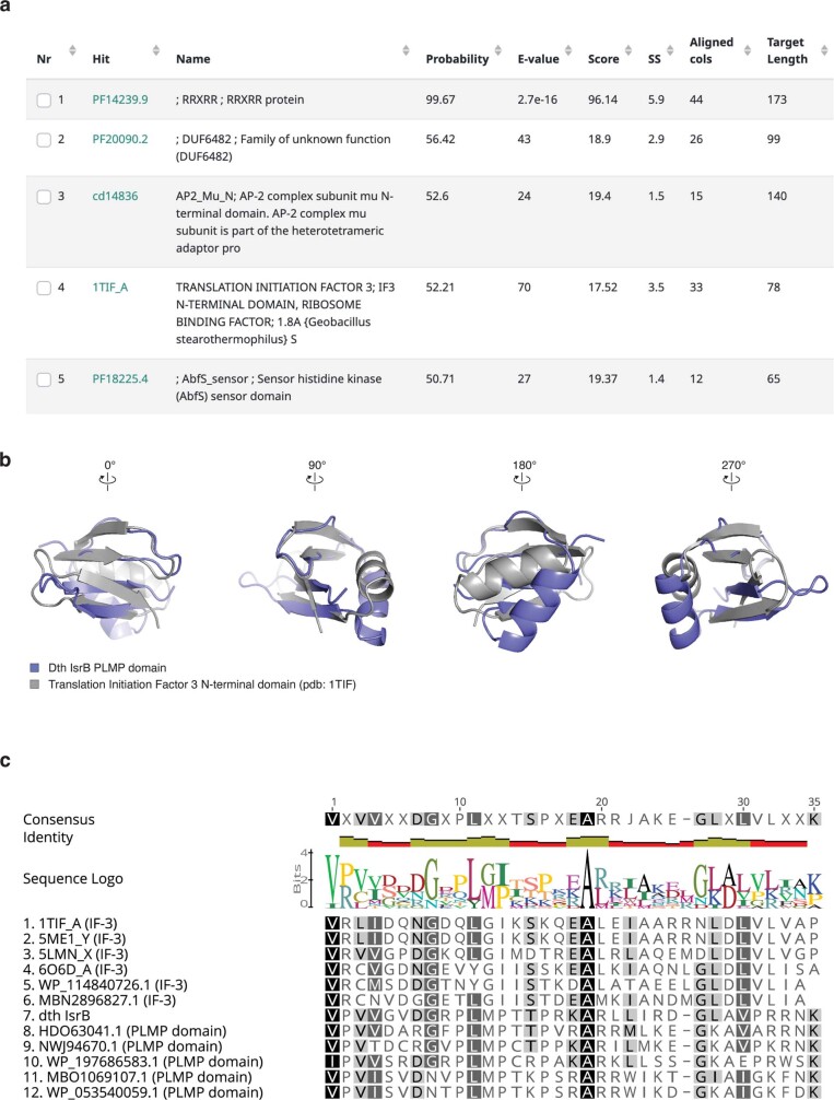
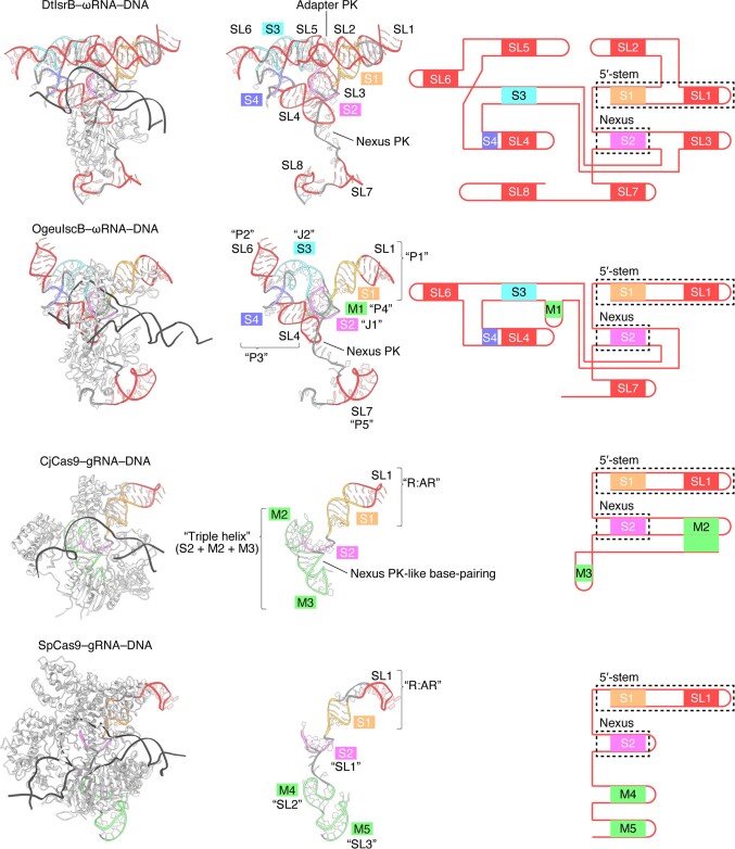
RNA-guided systems, such as CRISPR-Cas, combine programmable substrate recognition with enzymatic function, a combination that has been used advantageously to develop powerful molecular technologies1,2. Structural studies of these systems have illuminated how the RNA and protein jointly recognize and cleave their substrates, guiding rational engineering for further technology development3. Recent work identified a new class of RNA-guided systems, termed OMEGA, which include IscB, the likely ancestor of Cas9, and the nickase IsrB, a homologue of IscB lacking the HNH nuclease domain4. IsrB consists of only around 350 amino acids, but its small size is counterbalanced by a relatively large RNA guide (roughly 300-nt ωRNA). Here, we report the cryogenic-electron microscopy structure of Desulfovirgula thermocuniculi IsrB (DtIsrB) in complex with its cognate ωRNA and a target DNA. We find the overall structure of the IsrB protein shares a common scaffold with Cas9. In contrast to Cas9, however, which uses a recognition (REC) lobe to facilitate target selection, IsrB relies on its ωRNA, part of which forms an intricate ternary structure positioned analogously to REC. Structural analyses of IsrB and its ωRNA as well as comparisons to other RNA-guided systems highlight the functional interplay between protein and RNA, advancing our understanding of the biology and evolution of these diverse systems.


Conflict of interest statement

F.Z. is a scientific adviser and cofounder of Editas Medicine, Beam Therapeutics, Pairwise Plants, Arbor Biotechnologies and Proof Diagnostics.

Figures

Fig. 1
Fig. 1. Cryogenic-electron microscopy (cryo-EM) structure of the IsrB–ωRNA-target DNA complex.
a, Locus architecture and guide RNAs for IsrB (left) and Cas9 (right). b, Domain architecture of Streptococcus pyogenes SpCas9 (top) and D. thermocuniculi IsrB (DtIsrB) (bottom). c, Schematic of IsrB in complex with the ωRNA and the target DNA. The partial DNA duplex containing the TAM and target sequences used for the structural study are shown in sequence letters. d,e, Cryo-EM-density map (d) and structural model (e) of the IsrB–ωRNA-target DNA complex. Dashed lines represent poorly resolved regions of ωRNA. TE, transposon end; DR, direct repeat; NUC, nuclease; PI, PAM-interacting; PLL, phosphate-lock loop; TI, TAM-interacting; TS, target strand; NTS, non-target strand.
Fig. 2
Fig. 2. Model of the DtIrsB ωRNA structure.
a,b, Schematic (a) and structural model (b) of the ωRNA scaffold (residues 21–282). S1–4, stem 1–4; SL1–8, stem loop 1–8; PK, pseudoknot. In a, canonical and non-canonical base pairs are depicted by solid black lines. Poorly resolved regions are enclosed in a dashed box. In b, the guide segment is omitted for clarity. c, A base-triple formation in the adaptor pseudoknot. Hydrogen bonds are shown as dashed lines. d, In vitro reconstituted DtIsrB-ωRNA RNP nicking of dsDNA substrates (with TTGA TAM) with full-length ωRNA or truncated ωRNA. n = 3 independent technical replicates. Δ34–67, ωRNA in which nucleotides 34–67 were replaced with GAAA; 165-AGCG-168, ωRNA in which nucleotides 165–168 were replaced with AGCG; 194-GCGG-197, ωRNA in which nucleotides 194–197 were replaced with GCGG; 194-GCGG-197/81-CCGC-84, ωRNA in which nucleotides 81–84 and 194–197 were replaced with CCGC and GCGG, respectively.
Fig. 3
Fig. 3. DNA targeting and nicking mechanism of IsrB.
Inset shows the location of zoomed in panels. a, Heteroduplex recognition by the adaptor pseudoknot. b, Heteroduplex recognition by SL4, S4 and RECL. The volumes of RNA and DNA are generated from atomic coordinates, using Chimera X. c, In vitro reconstituted DtIsrB-ωRNA RNP nicking of dsDNA substrates (with TTGA TAM) with wild-type (WT) or mutant DtIsrB. = 3 independent technical replicates. ΔHNHL, IsrB mutant in which residues 161–174 were replaced with a GSG-linker. Δβ7, IsrB mutant in which residues 341–353 were deleted. ΔPLMP, IsrB mutant in which residues 1–52 were deleted. To confirm the protein stability of deletion mutants, we checked the protein expression in bacterial lysate overexpressing the deletion mutants (Extended Data Fig. 5b). d, Heteroduplex recognition by HNHL. e, Recognition of the +1 phosphate (phosphodiester bond between nucleotides dG1 and dA(−1) of target strand DNA) by the phosphate-lock loop. f, Recognition of the guide segment by BH. g, TAM recognition by the TI domain. h, TAM specificity of DtIsrB.  In vitro reconstituted DtIsrB-ωRNA RNP nicking of dsDNA substrates (with TTGA/ATGA/TTGG/ATGG TAMs) with WT or mutant DtIsrB. = 3 independent technical replicates.
Fig. 4
Fig. 4. IsrB diversity.
a, Phylogenetic tree of selected IsrB orthologues. Protein sizes are indicated, with domains highlighted in coloured boxes and conserved sequences in black. Cognate RNA sizes and groups (Fig. 4d) are indicated. b, TAM sequences for six IsrB orthologues using in vitro cleavage of a plasmid library containing randomized TAMs and the target sequence. c, In vitro reconstituted IsrB-ωRNA RNP nicking of dsDNA substrates with five IsrB orthologues. For CwIsrB, CsIsrB and K2IsrB, the target DNA contained a TTGA TAM. For DsIsrB and BbIsrB, the target DNA contained an ATGG TAM. = 3 independent technical replicates. d, Structural models of the ωRNA scaffolds for six IsrB orthologues based on secondary structure predictions. The predicted ωRNA scaffolds are classified into groups A (subgroup A1, CsIsrB and K2IsrB; subgroup A2, BbIsrB) and B (subgroup B1, DtIsrB; subgroup B2, CwIsrB and DsIsrB). In group A, SL2 and SL4 form pseudoknots, and SL5 and the intermediate region between S2 and SL7 form pseudoknots. Connecting regions that differ from group B are coloured pink. The intermediate region between SL5 and S3 as well as the terminal region after SL7 (‘no motif’, grey) are predicted to be unpaired nucleotides. In group B, SL2 and SL5 form pseudoknots, and SL4 and the intermediate region between S2 and SL7 form pseudoknots. Connecting regions (red) are as in group A. The intermediate region between SL5 and S3 as well as the terminal region after SL7 are predicted to be stem loops (SL6 and SL8, grey). In subgroups A1 and B1, the intermediate region between S2 and S3 is predicted to be a stem loop (SL3, dark grey), whereas in subgroups A2 and B2, that region is predicted to be unpaired nucleotides (‛no motif’, dark grey).
Fig. 5
Fig. 5. Model of IsrB evolution.
Structural determinants of the evolution from ancestral RuvC nucleases to IsrB and then Cas9. Examples from modern descendants (extants) of each family are shown beginning with T. thermophilus RuvC (TtRuvC, PDB 6S16), DtIsrB, CjCas9 (PDB 5X2G) and SpCas9 (PDB 7S4X). Critical stages in the proposed evolutionary process are shown, including the insertions of the TI, PLMP and BH domains, interaction with ωRNA, insertion of the HNH domain, loss of the PLMP domain and replacement of various parts of the ωRNA with REC regions (domain replacements are shown with a colour key). The portion of REC2 in CjCas9 and SpCas9 that replace SL2 in the DtIsrB ωRNA are coloured in a dark grey. Connected base pairing is shown only for the guide–DNA duplex. Disconnected base pairing is shown for the ωRNA adaptor pseudoknot to highlight its position near the RNA–DNA duplex.
Extended Data Fig. 1
Extended Data Fig. 1. Cryo-EM data processing for the IsrB-ωRNA-DNA complex (complex A).
(a) Cryo-EM data processing schematic for single particle analysis of the complex A. Unsharpened (Left) and sharpened (Right) maps in the final 3D refinement. Particle orientation distribution (Center). (b) Final refined map, colored by local resolution, calculated in RELION-4.0 with FSC threshold 0.5.(c) FSC curves calculated between the half maps of complex A from the final round of the refinement in RELION-4.0. (d) FSC curves calculated between the model and the final refined map, using phenix.validation_cryoem. (e) Q-scores for each residue of IsrB-ωRNA-target strand DNA-non-target strand DNA model in 3.1 Å map of the IsrB-ωRNA-DNA complex. The dashed black and grey lines in the plot represent the expected Q-scores based on the global map resolution (3.1 Å) and the local map resolution (4.5 Å), respectively. Q-scores for the RNA and DNA residues are consistent with the expected values based on the local map resolution.
Extended Data Fig. 2
Extended Data Fig. 2. Cryo-EM density maps.
Cryo-EM density maps for residues represented in main figures.
Extended Data Fig. 3
Extended Data Fig. 3. Details of the IsrB protein structure.
(a) Close-up view of the IsrB protein structure. (b) Structural comparison between DNA-bound IsrB-TI domain and SpCas9-PI domain (PDB: 7S4X). In the Cas9 structure, the subdomain inserted between β6 and β7 is omitted for clarity.
Extended Data Fig. 4
Extended Data Fig. 4. PLMP domain homology.
(a) Top five hits from HHPred search using seed sequence SITRVPVVGVDGRPLMPTTPRKARLLIRDGLAVPRRNKLGLFYIQMLRPVGTRTQ corresponding to the PLMP domain from DtIsrB. (b) Structural comparison of the PLMP domain from DtIsrB and the N-terminal domain of Translation Initiation Factor 3 (IF-3) from Geobacillus stearothermophilus (PDB: 1TIF). (c) Alignment of representative IF-3 N-terminal domains and OMEGA-related PLMP domains.
Extended Data Fig. 5
Extended Data Fig. 5. Uncropped gel images used in this study.
(a) Denatured PAGE gels for resolving nicked DNA products. (b) An SDS-PAGE gel for expression check of the deletion mutants. Related to Fig. 3c.
Extended Data Fig. 6
Extended Data Fig. 6. Cryo-EM structure of the λN-IsrB-ωRNA mutant-DNA complex (complex B).
(a) Cleavage sites in the target DNA as assay by Sanger sequencing. The nicking sites are marked by black triangles. The additional non-templated adenine is indicated by an asterisk in the Sanger sequencing trace. (b) Domain structure of the λN-IsrB fusion protein (left) and schematic of the ωRNA mutant and target DNA (right). In the ωRNA mutant, residues 34–67 were replaced with GAAA and boxB RNA was appended to the 3′ end of the ωRNA scaffold. (c) Cryo-EM data processing schematic for single particle analysis of complex B (Left). Final refined map (Right). (d) FSC curves calculated between the half maps of complex B from the final round of the refinement in cryoSPARC v3.3. (e) Cryo-EM density map of complex B. Based on the superposition of complex B map and complex A model, regions of protein, RNA, and DNA were assigned. Extra density was observed in the vicinity of the ωRNA SL8 region and assigned to the λN-boxB complex, consistent with the SL8-boxB connectivity and the λN-boxB volume (PDB: 1QFQ). TS, target strand; NTS, non-target strand. (f and g) Cryo-EM density maps of complex A (f) and SpCas9 in complex with its cognate RNA and target DNA (EMD: 24838) (g).
Extended Data Fig. 7
Extended Data Fig. 7. Structural prediction of IsrB orthologs and their cognate ωRNAs.
(a) Secondary structure and pseudoknot prediction of the ωRNA scaffolds based on covariance model. In CwIsrB/DsIsrB/BbIsrB ωRNAs, SL3 motifs are replaced with unpaired nucleotides. In CsIsrB/K2IsrB/BbIsrB ωRNAs, SL6 motifs are degenerated and SL8 motifs are replaced with unpaired nucleotides. (b) Superposition of AlphaFold (AF) and cryo-EM (EM) structures of DtIsrB. (c) Superposition of AlphaFold structures of six IsrB orthologs. CwIsrB/DsIsrB have β-hairpin and loop insertions in the RuvC domain and RECL, respectively.
Extended Data Fig. 8
Extended Data Fig. 8. Evolutionary snapshot of Cas9 ancestors.
Structural comparison between DtIsrB, OgeuIscB (8CSZ), YnpsCas9-1 (AF2 model), and SpCas9 (PDB: 4OO8).
Extended Data Fig. 9
Extended Data Fig. 9. Evolutionary snapshot of tracrRNA ancestors.
Structural comparison between cognate RNAs of DtIsrB, OgeuIscB (8CSZ), CjCas9 (5X2G), and SpCas9 (7S4X) in their protein/DNA-bound states. Overall structures (left). RNA structures (center). RNA schematic diagrams (right).
Extended Data Fig. 10
Extended Data Fig. 10. Models of RNA-guided DNA nicking/cleavage by IsrB/Cas9.
Schematic highlighting the mechanistic similarities and differences between IsrB and Cas9.

References

    1. Zhang F. Development of CRISPR–Cas systems for genome editing and beyond. Q. Rev. Biophys. 2019;52:e6–e6. doi: 10.1017/S0033583519000052. - DOI
    1. Doudna JA. The promise and challenge of therapeutic genome editing. Nature. 2020;578:229–236. doi: 10.1038/s41586-020-1978-5. - DOI - PMC - PubMed
    1. Nishimasu H, Nureki O. Structures and mechanisms of CRISPR RNA-guided effector nucleases. Curr. Opin. Struct. Biol. 2017;43:68–78. doi: 10.1016/j.sbi.2016.11.013. - DOI - PubMed
    1. Altae-Tran H, et al. The widespread IS200/IS605 transposon family encodes diverse programmable RNA-guided endonucleases. Science. 2021;374:57–65. doi: 10.1126/science.abj6856. - DOI - PMC - PubMed
    1. Kapitonov VV, Makarova KS, Koonin EV. ISC, a novel group of bacterial and archaeal DNA transposons that encode Cas9 homologs. J. Bacteriol. 2015;198:797–807. doi: 10.1128/JB.00783-15. - DOI - PMC - PubMed

Publication types

MeSH terms

Supplementary concepts