. 2022 Oct 12;21(1):198.
doi: 10.1186/s12943-022-01653-2.

The FUS/circEZH2/KLF5/ feedback loop contributes to CXCR4-induced liver metastasis of breast cancer by enhancing epithelial-mesenchymal transition

Affiliations

The FUS/circEZH2/KLF5/ feedback loop contributes to CXCR4-induced liver metastasis of breast cancer by enhancing epithelial-mesenchymal transition

Peng Liu et al. Mol Cancer. .

Abstract

Background: Metastasis of breast cancer have caused the majority of cancer-related death worldwide. The circRNAs are associated with tumorigenesis and metastasis in breast cancer according to recent research. However, the biological mechanism of circRNAs in liver metastatic breast cancer remains ambiguous yet.

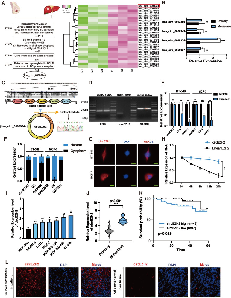
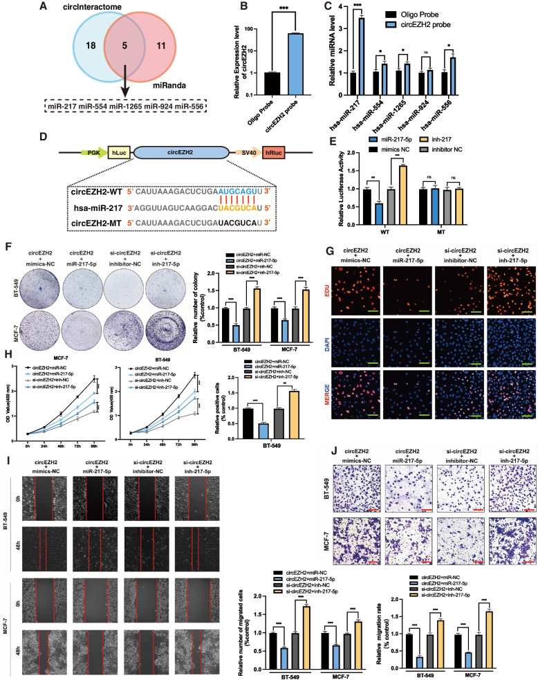
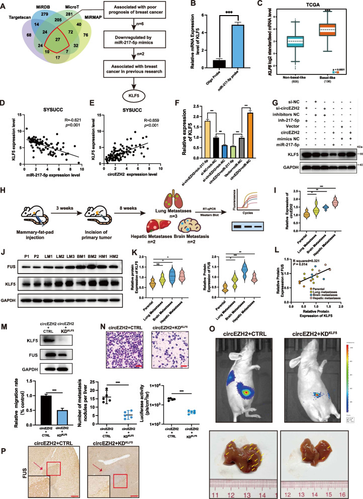
Methods: Microarray analysis of three pairs of primary BC tissues and matched hepatic metastatic specimens identified circEZH2. We used RT-qPCR and FISH assays to confirm circEZH2 existence, characteristics, and expression. Both in vivo and in vitro, circEZH2 played an oncogenic role which promoted metastasis as well. A range of bioinformatic analysis, Western blot, RNA pull-down, RIP, ChIP, and animal experiments were used to define the feedback loop involving FUS, circEZH2, miR-217-5p, KLF5, FUS, CXCR4 as well as epithelial and mesenchymal transition.

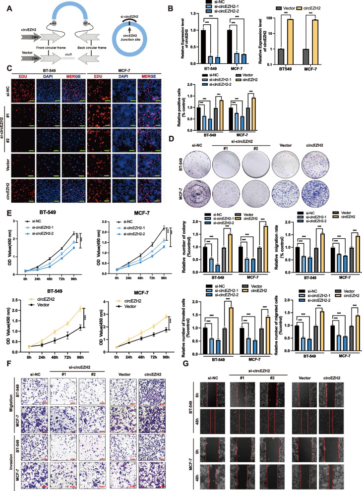
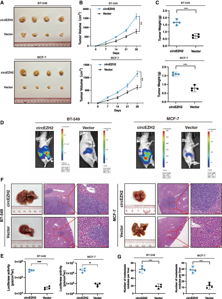
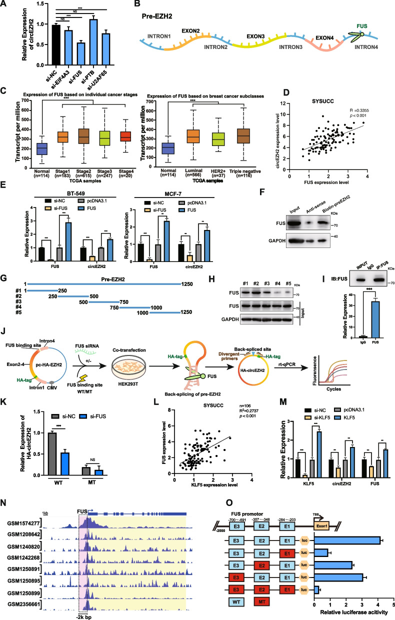
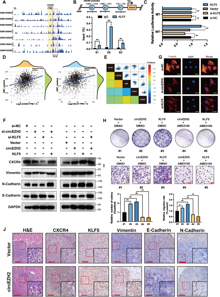
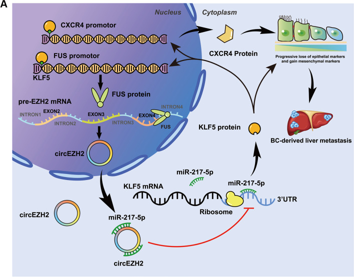
Results: In our research, circEZH2 was proved to be upregulated in liver metastases in BC and predicted the worse prognosis in breast cancer patients. Overexpression of circEZH2 notably accentuated the vitality and invasion of BC cells, whereas knockdown of circEZH2 elicited the literally opposite effects. Besides, overexpressed circEZH2 promoted tumorigenesis and liver metastasis in vivo. Moreover, circEZH2 could adsorb miR-217-5p to upregulate KLF5 thus leading to activate FUS transcription which would facilitate the back-splicing program of circEZH2. Meanwhile, KLF5 could upregulated CXCR4 transcriptionally to accelerate epithelial and mesenchymal transition of breast cancer.

Conclusions: Consequently, a novel feedback loop FUS/circEZH2/KLF5/CXCR4 was established while circEZH2 could be novel biomarker and potential target for BC patients' therapy.

Keywords: Breast cancer; EMT; Feedback loop; Metastasis; circRNAs.


Conflict of interest statement

The authors have disclosed no competing interests.

Figures

Fig. 1
Fig. 1
circEZH2 is identified and characterized in BC. a Left, flowchart showing the screening criteria of upregulated circRNAs enriched in BCLM; right, heatmap revealing that 32 circRNAs was selected according to the step2 criteria and hsa_circ_0008324 was marked by red box. b RT-qPCR was used to testify the expression of four circRNA candidates among BC liver metastases and BC primary tumors. c Top, three exons of circEZH2 were highly conservative in different species according to NCBI; bottom, the consitutions of circEZH2 and Sanger-sequencing analysis identified the back-spliced site of circEZH2. d Divergent and convergent primers were used for amplification of circEZH2 in cDNA and gDNA while linear EZH2 and GAPDH were used as controls through PCR. e Following digestion with Rnase R, RT-qPCR was used to measure the change in circEZH2 and EZH2. f Nuclear-cytoplasmic fraction assays were performed to determine the subcellular expression of circEZH2. g FISH assays were performed to determine that circEZH2 was localized in the cytoplasm (Scale bar 20 μm). h The expression of circEZH2 and linear EZH2 were identified after treated with actinomycin D for 4 h, 8 h, 12 h, 24 h. i The expressions of circEZH2 among different BC cells were identified by RT-qPCR. j In primary BC tissues (n = 20) and liver metastatic tissues (n = 11), RT-qPCR was used to verify the expression of circEZH2. The results were analyzed by unpaired Student’s t test. k To determine the Kaplan-Meier survival of BC patients (n = 115), log-rank tests were used. l In BCLM sample, FISH analysis showed circEZH2 overexpression significantly (magnification, X4 scale bar, 200 μm and X20 scale bar, 50 μm). The data was revealed as the mean ± SD and all experiments were repeated at least three times, ns: no significant; * p < 0.05; ** p < 0.01; *** p < 0.001
Fig. 2
Fig. 2
CircEZH2 accentuate vitality, migration, and invasion of BC cells in vitro. a Here is a diagram of mechanisms about circEZH2 overexpression with plasmid and knockdown with siRNA. b We determined the success of knockdown as well as overexpression of circEZH2 using RT-qPCR. c-e To evaluate the vitality in BC cells, EdU assays (Scale bar, 50 μm), colony formation assays and CCK8 assays were conducted. f and g Migration, invasion of transwell (Scale bar 100 μm) and wound-healing assays (Scale bar 200 μm) were conducted to determine the ability of metastasis in BC cells. The data was revealed as the mean ± SD and all experiments were repeated at least three times, * p < 0.05; ** p < 0.01; *** p < 0.001
Fig. 3
Fig. 3
circEZH2 accentuates tumorigenesis and liver metastasis of BC cells in vivo. a Pictures of xenograft tumors were shown in the circEZH2 overexpression group and relative control group (n = 4). b The growth curves of each group of xenograft tumors were displayed. c The xenograft tumor weight was measured and analyzed. d and e Inferior hemi-spleen implantation mice models in vivo were analyzed by IVIS to identify the potential of circEZH2 in promoting liver metastasis. f and g Metastatic sites in the liver were noted to be increased in the overexpressed circEZH2. On the H&E staining liver slides, metastases were marked with arrows. (Magnification, X4 scale bar 200 μm; X20 scale bar 40 μm). The data was displayed as the mean ± SD and all experiments were repeated at least three times, **P < 0.01, ***P < 0.001
Fig. 4
Fig. 4
FUS promotes back-splicing program of circEZH2 and KLF5 activates FUS transcription. a RT-qPCR was used to determine the expression of circEZH2 after transfected with siRNA targeting EIF4A3, FUS, PTBP1, U2AF65 and control. b Schematic diagram showed that FUS binding downstream intron4 region of pre-EZH2. c TCGA analysis of FUS expression in different stages and subtypes of BRCA.. d Clinical BC samples from SYSUCC were used by RT-qPCR analysis revealing the spearman correlation evaluation between circEZH2 and FUS transcripts. e RT-qPCR was performed to reveal the assoicated expression of circEZH2 when FUS was overexpressed or knocked down in BC cells. f Western blots were used to determine the resultants of RNA pull-down assay using biotin-preEZH2-probe. g Diagram revealing five truncated biotinylated pre-EZH2 probes were constructed. h RNA pull down assays were performed by five truncated probes and the resultants analyzed by western-blot were displayed. i Lysates of BT-549 were subjected to RIP by anti-FUS antibody and anti-IgG antibody and the enrichments of IP and IgG groups were displayed. j Flow illustration showed that firstly pc-HA-EZH2, a novel back-splicing formation validation vector by adding a HA label to the 5′-second EZH2 exon to differentiate the internal circEZH2 was constructed. Secondly, FUS binding region was mutated of pc-HA-EZH2 as mutant type (MT). After that MT and WT vector were transfected into HEK293T with/without knockdown of FUS. Eventually, the back-splicing efficiency of circEZH2 was deternmined by divergent primers across HA tag region and RT-qPCR while linear HA-EZH2 was used as reference. k The back-splicing efficiency of circEZH2 was identified by RT-qPCR in WT and MT groups. l In SYSUCC clinical human BC tissues, expression of KLF5 was positively correlated with FUS as determined by Spearman’s correlation analysis. m RT-qPCR was used to find the associated expression of KLF5, circEZH2 and FUS in change the expression of KLF5. n Eight ChIP-seq datasets showed that there were binding peaks within 2k bp upstream promotor of FUS by KLF5. o Top, JASPAR predicted potential three binding regions between KLF5 and the promotor of FUS. Bottom, each of three binding sites were mutated in FUS promotor dual-luciferase plasmid while wild type without mutation was positive control and all mutated group was negative control. The results of each group were quantitated by dual-luciferase assays. The data was showed as the mean ± SD and all experiments were repeated at least three times, ns no significance, **P < 0.01, ***P < 0.001
Fig. 5
Fig. 5
CircEZH2 adsorbs miR-217-5p. a CircEZH2 exhibits potential binding regions for miRNAs in the intersection between CircInteractome and miRanda databases, as indicated on the Venn diagram. The illustration revealed the potential binding miRNAs with circEZH2. b-c Right, CircEZH2 RNA pull down assay was performed to find that hsa-miR-217-5p was the most enriched one. Left, the biotin-miR-217-5p probe could enrich circEZH2 effectively. d and e Left, the diagram states the dual-luciferase plasmids with mutant (MT) and wild (WT) containing binding regions of miR-217-5p. Right, the relative luciferase activities were determined after co-transfection of circEZH2-MT or circEZH2-WT and miR-217-5p inhibitors or mimics with control respectively. f-h Co-transfections of BC cells with indicated vector, siRNA, miRNA, or inhibitor were performed to validate their viability by colony formation assay, EdU assay (Scale bar, 50 μm), and CCK8 assay, respectively. i and j The migration ability in BC cells co-transfected with described vector, siRNA, miRNA or inhibitor was analyzed by migration transwell (Scale bar 100 μm), and wound healing assay (Scale bar 200 μm), respectively. The data was displayed as the mean ± SD and all experiments were repeated at least three times, ns no significant, **P < 0.01, ***P < 0.001
Fig. 6
Fig. 6
KLF5 is an distinct target of miR-217-5p. a Left, The Venn diagram revealed the potential targeted gene of miR-217-5p among miRDB, microT, Targetscan, and miRMAP four databases; Right, the flow chart to filter for target gene of miR-217-5p step by step.. b RNA pull down assays were performed by biotin-miR-217-5p-probe to determine enrichment of KLF5 through RT-qPCR analysis. c Through TCGA analysis by bc-GenExMiner, the KLF5 expression was upregulated in basal-like subtype of BC d and e Right, miR-217-5p expression was negative correlated with KLF5 in BC samples in transcript analysis of clinical BC samples from SYSUCC. Left, the expression of circEZH2 was positive correlated with KLF5 in clinical BC tissues from SYSUCC in mRNA level. Both were conducted by spearman correlation analysis. f and g RT-qPCR, and western blots were used to analyze KLF5 expression after co-transfection with siRNA, vector, mimic or inhibitor as indicated. h Flow diagram indicated that establishment of xenograft-induced metastatic mice model and multi-organ metastases were analyzed by western blot and rt-qPCR. I RT-qPCR was conducted to reveal the expression pattern in different metastases compared with parental cells. j and k The expression FUS and KLF5 in different metastases were determined by western blot. P = parental; LM = lung metastases; BM = brain metastases; HM = hepatic metastases. l The pearson correlation analysis was performed to identify the protein expression association between FUS and KLF5 in metastases. m The doxycycline-induced knockdown of KLF5 in circEZH2 overexpresed cells was verified by westernblot. n and o Migration Transwell rescue assay and liver metastatic mice rescue models were performed to prove the influence of KLF5 knockdown on circEZH2 overexpression cells. The scale bar of Transwell was 100µm. The liver metastases were quantitated by IVIS and number of liver metastases were marked with yellow arrows. p Immunohistochemistry (IHC) of FUS was performed in metastases of KLF5 knockdown group and its control group (Low power field: Scale bar 100 μm; High power field: Scale bar 20µm). The data was showed as the mean ± SD and all experiments were repeated at least three times, ns no significant, *P < 0.05 **P < 0.01, ***P < 0.001
Fig. 7
Fig. 7
FUS/circEZH2/KLF5 loop promotes CXCR4-induced EMT program. a Seven ChIP datasets showed that there were KLF5 peaks within 2k bp promotor of CXCR4. b Top, the potential binding sites on the promotor of CXCR4 by KLF5 according to JASPAR. c Dual luciferase assays were conducted in FUS promotor wild type group and E2 mutated group. d According to TCGA GSVA analysis, KLF5 and CXCR4 were positive correlated with EMT markers respectively e Based on STARBASE database, regression correlation rates among KLF5, CXCR4, VIM, CDH1 and CDH2 transcripts were displayed in heatmap. f Western blot was conducted to reveal the change of CXCR4, Vimentin, N-cadherin and E-cadherin after indicated siRNA or Vector. g IF was conducted to find that F-actin were vividly downregulated after knockdown circEZH2 and KLF5 respectively. h and i The CXCR4-rescue colony formation assay and CXCR4-rescue migration Transwell assay were conducted within indicated Vector or drug (Scale bar 100 μm) j IHC were performed in circEZH2 overexpressed group of BCLM nude mice and control group with indicated antibody (Low power field: Scale bar 100 μm; High power field: Scale bar 20µm. ). The data are showed as the mean ± SD and all experiments were repeated at least three times, ns no significant, *P < 0.05 **P < 0.01, ***P < 0.001
Fig. 8
Fig. 8
a The diagram explains the biological mechanism of a novel positive feedback loop FUS/circEZH2/KLF5 through which liver metastasis of breast cancer was induced by enhancing epithelial-mesenchymal transition (EMT) via up-regulation of CXCR4

Similar articles

Cited by

References

    1. Siegel RL, Miller KD, Jemal A. Cancer statistics, 2020. CA Cancer J Clin. 2020;70:7–30. doi: 10.3322/caac.21590. - DOI - PubMed
    1. He Z-Y, Wu S-G, Peng F, Zhang Q, Luo Y, Chen M, et al. Up-regulation of RFC3 promotes triple negative breast Cancer metastasis and is associated with poor prognosis via EMT. Transl Oncol. 2017;10:1–9. doi: 10.1016/j.tranon.2016.10.004. - DOI - PMC - PubMed
    1. Adam R, Aloia T, Krissat J, Bralet M-P, Paule B, Giacchetti S, et al. Is liver resection justified for patients with hepatic metastases from breast cancer? Ann Surg. 2006;244:897–907. doi: 10.1097/01.sla.0000246847.02058.1b. - DOI - PMC - PubMed
    1. Pivot X, Asmar L, Hortobagyi GN, Theriault R, Pastorini F, Buzdar A. A retrospective study of first indicators of breast cancer recurrence. Oncology. 2000;58:185–190. doi: 10.1159/000012098. - DOI - PubMed
    1. Leung AM, Vu HN, Nguyen K-A, Thacker LR, Bear HD. Effects of surgical excision on survival of patients with stage IV breast cancer. J Surg Res. 2010;161:83–88. doi: 10.1016/j.jss.2008.12.030. - DOI - PubMed

Publication types