. 2022 Oct 12;21(1):197.
doi: 10.1186/s12943-022-01667-w.

Two circPPFIA1s negatively regulate liver metastasis of colon cancer via miR-155-5p/CDX1 and HuR/RAB36

Affiliations

Two circPPFIA1s negatively regulate liver metastasis of colon cancer via miR-155-5p/CDX1 and HuR/RAB36

Haein Ji et al. Mol Cancer. .

Abstract

Background: Circular RNAs (circRNAs) play a critical role in colorectal cancer (CRC) progression, including metastasis. However, the detailed molecular mechanism is not fully understood.

Methods: Differentially expressed circRNAs between primary KM12C and liver metastatic KM12L4 colon cancer cells were identified by microarray. The expression of circRNAs was measured by semi-quantitative (semi-qPCR) and real time-quantitative PCR (RT-qPCR). Metastatic potential including invasive and migratory abilities, and liver metastasis were examined by transwell assays and intrasplenic injection, respectively. CircPPFIA1-associated microRNA (miRNA) and RNA-binding protein (RBP) were screened by an antisense oligonucleotide (ASO) pulldown experiment. The effects of circPPFIA1 on target gene expression were evaluated by RT-qPCR and western blot analyses.

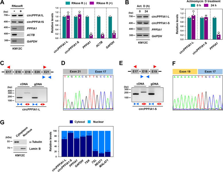
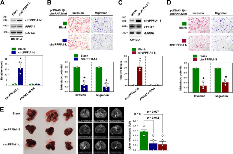
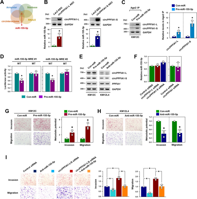
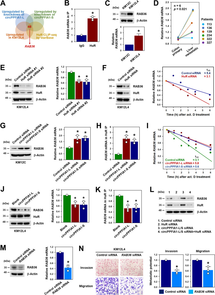
Results: By analyzing circRNA microarray data, we identified two anti-metastatic circRNAs generated from PPFIA1 with different length, which named circPPFIA1-L (long) and -S (short). They were significantly downregulated in liver metastatic KM12L4 cells compared to primary KM12C cells. The knockdown of circPPFIA1s in KM12C enhanced metastatic potential and increased liver metastasis. Conversely, overexpression of circPPFIA1s weakened metastatic potential and inhibited liver metastasis. circPPFIA1s were found to function as sponges of oncogenic miR-155-5p and Hu antigen R (HuR) by an ASO pulldown experiment. circPPFIA1s upregulated tumor-suppressing CDX1 expression and conversely downregulated oncogenic RAB36 by decoying miR-155-5p and by sequestering HuR, respectively.

Conclusion: Our findings demonstrate that circPPFIA1s inhibit the liver metastasis of CRC via the miR-155-5p/CDX1 and HuR/RAB36 pathways.

Keywords: CDX1; Colorectal cancer; HuR; Liver metastasis; RAB36; circPPFIA1; miR-155-5p.


Conflict of interest statement

The authors have no conflicts of interest to declare.

Figures

Fig. 1
Fig. 1
circPPFIA1 is downregulated in liver metastatic colorectal cancer. A Transwell invasion and migration assays were carried out to examine the invasive and migratory abilities of KM12C and KM12L4 cells. Metastatic potential was determined by counting the number of invaded and migrated cells, and the representative images are shown. B Liver metastasis was examined through in vivo intrasplenic injection. At four weeks post-injection, the optical and MRI images were obtained. The degree of liver metastasis (n = 5) was calculated by giving scores in arbitrary units (0–3). C Volcano plot illustrating differentially expressed circRNAs in KM12C versus KM12L4. Blue and red dots represent circPPFIA1-L and -S, respectively. D Expression level of circPPFIA1-L and -S in KM12C and KM12L4 cells. E Schematic illustration and detailed information of circPPFIA1-L and -S. F, G Expression levels of circPPFIA1-L, -S, and linear PPFIA1 mRNA were determined by RT-qPCR (F) and semi-qPCR (G). H, The levels of circPPFIA1-L and -S in primary and liver metastatic colon cancer tissues were determined by RT-qPCR (H) and semi-qPCR (I). Statistical significance was calculated from three independent experiments using the Student’s t-test (*p < 0.05). All data represent mean ± standard deviation (SD)
Fig. 2
Fig. 2
Characterization of circPPFIA1-L and -S. A, B The stability of circPPFIA1-L and -S was examined by RNase R resistance (A) and actinomycin D experiments (B). The level of remaining circPPFIA1-L and -S was determined by semi-qPCR and RT-qPCR. Linear PPFIA1 mRNA, GAPDH, and ACTB were included as controls. C, E Complementary (cDNA) and genomic DNA (gDNA) were prepared using RNA isolated from KM12C cells. The level of circPPFIA1-L (C) and -S (E) was determined by semi-qPCR. D, F To verify the divergent region of circPPFIA1-L (D) and -S (F), Sanger sequencing was performed. (G) To determine the cellular localization of circPPFIA1s, the fractionation experiment was performed using digitonin. The level of a-Tubulin and lamin B were checked for verification of cytoplasmic and nuclear extracts, respectively. The levels of circPPFIA1-L, -S, and linear PPFIA1 mRNA were determined by RT-qPCR. GAPDH mRNA and 7SK were used as a marker of cytoplasmic RNA and NEAT1, MALAT1, and 7SL were used for nuclear RNA. Statistical analyses were performed using the Student’s t-test using three independent experiments (*p < 0.05). All data represent mean ± standard variation (SD)
Fig. 3
Fig. 3
Knockdown of circPPFIA1s enhances the metastatic potential of KM12C cells. A–D The effect of circPPFIA1-L and -S silencing on the metastatic potential of KM12C cells was examined. The level of circPPFIA1-L (A) and -S (C) in KM12C cells transfected with the indicated siRNA was determined by semi-qPCR (upper) and RT-qPCR (lower). Invasive and migratory abilities of transfected cells (knockdown of circPPFIA1-L (B) and -S (D)) were examined by transwell invasion and migration assays. E, The increase in invasive and migratory abilities with knockdown of both circPPFIA1-L and circPPFIA1-S was determined by transwell invasion (E) and migration (F) assays. G, H Intrasplenic injection model (n = 7) confirmed that knockdown of circPPFIA1-L (G) and -S (H) significantly promoted liver metastasis of KM12C cells. Statistical analyses were performed using the Student’s t-test using three independent experiments (*p < 0.05). All data represent mean ± standard variation (SD)
Fig. 4
Fig. 4
Overexpression of circPPFIA1s attenuates metastatic potential of KM12L4 cells. AD To investigate the roles of circPPFIA1s overexpression on the metastatic potential of KM12L4 cells, overexpression vectors for circPPFIA1-L and -S were introduced into KM12L4 cells. The relative levels of circPPFIA1-L (A) and -S (C) were determined by semi-qPCR (upper) and RT-qPCR (lower). Transwell invasion and migration assays showed that overexpression of circPPFIA1-L (B) and -S (D) significantly reduced the invasive and migratory abilities of KM12L4 cells. E Intrasplenic injection model (n = 5) confirmed that overexpression of circPPFIA1-L and -S significantly reduced liver metastasis of KM12L4 cells. Statistical analyses were performed using Student’s t-test using three independent experiments (*p < 0.05). All data represent mean ± standard variation (SD)
Fig. 5
Fig. 5
Oncogenic miR-155-5p is a sponging target of circPPFIA1s. A Venn diagram showing miR-155-5p predicted as a putative sponging target of circPPFIA1s in four databases (Arraystar, Starbase, circInteractome, and RNA22). B To verify that miR-155-5p binds to circPPFIA1s, an ASO pulldown experiment was performed. The pulldown efficacies of circPPFIA1s ASO were examined by semi-qPCR (upper) and the level of miR-155-5p in pulldown materials was checked by RT-qPCR (lower). C Enrichment of circPPFIA1-L and -S in miRISC was analyzed by Ago2 RIP with KM12C cells transfected with pre-miR-155-5p. D The levels of circPPFIA1-L and -S were determined by semi-qPCR in KM12C and KM12L4 cells transfected with pre-miR-155-5p or anti-miR-155-5p, respectively. E To examine the direct interaction between circPPFIA1s and miR-155-5p, the luciferase reporter vectors containing the wild-type or mutant sequence of miR-155-5p MRE were constructed. Following overexpression of miR-155-5p, a luciferase activity assay was carried out in KM12C cells. F After transfection of circPPFIA1 siRNAs, the expression level of miR-155-5p was determined by RT-qPCR. G, H Following transfection of pre-miR-155-5p or anti-miR-155-5p into KM12C (G) and KM12L4 (H) cells, the Invasive and migratory abilities were measured by transwell invasion and migration assays, respectively. I To check whether miR-155-5p is required for increased metastatic potential by knockdown of circPPFIA1s, rescue experiments were conducted. Invasive and migratory abilities were examined by transwell invasion and migrations assays, respectively. Statistical analyses were performed using the Student’s t-test using three independent experiments (*p < 0.05). All data represent mean ± standard variation (SD)
Fig. 6
Fig. 6
CDX1 is a target of miR-155-5p. A CDX1 is predicted as a target of miR-155-5p by comparing the lists of downregulated mRNAs obtained from RNA sequencing results with databases (miRDB and TargetScan). B The expression levels of CDX1 protein and mRNA in KM12C and KM12L4 were compared by western blot and RT-qPCR analyses. C The expression level of CDX1 in primary and liver metastatic colon cancer tissues was determined by RT-qPCR. D Following transfection of KM12C and KM12L4 cells with pre-miR-155-5p and anti-miR-155-5p respectively, the levels of CDX1 protein and mRNA were assessed by western blot and RT-qPCR analyses, respectively. E, F Direct interaction of miR-155-5p with the 3’UTR of CDX1 mRNA, Ago2 RIP (E), and luciferase activity assays (F) were performed. G–I Following transfection of KM12C cells with siRNAs targeting circPPFIA1-L or circPPFIA1-S, the expression levels of CDX1 protein and mRNA (G), the enrichment of CDX1 mRNA in Ago2 RIP (H), and luciferase assay (I) were performed as described above. J, K To examine whether overexpression of circPPFIA1s increases CDX1 expression, KM12C cells were transfected with blank or overexpression vectors. The expression levels of CDX1 protein and mRNA were determined by western blot and RT-qPCR, respectively (J). The effect of circPPFIA1s overexpression on the interaction between miR-155-5p and CDX1 mRNA was examined by Ago2 RIP (K). L To examine whether the inhibition of miR-155-5p restores downregulation of CDX1 by knockdown of circPPFIA1s, western blot analysis was performed. M, N Following the transfection of KM12C cells with CDX1 siRNA, the efficacy of CDX1 siRNA was examined by western blot and RT-PCR (M). The effect of CDX1 silencing on the metastatic potential of KM12C cells was checked by transwell invasion and migration assays (N). Statistical analyses were performed using the Student’s t-test using three independent experiments (*p < 0.05). All data represent mean ± standard variation (SD)
Fig. 7
Fig. 7
HuR is a sponging target of circPPFIA1s and is required for regulation of metastatic potential. A Venn diagram demonstrated the overlapping gene of the interacting RBPs with circPPFIA1 predicted by databases (circInteractome, RBPDB, and Starbase). B Validation of interaction of HuR with circPPFIA1s. The level of HuR in ASO pulldown materials was determined by western blot analysis. C To demonstrate the interaction between circPPFIA1s and HuR, the level of circPPFIA1-L and -S in HuR RIP was determined by semi-qPCR. D, E The expression level of HuR in KM12C and KM12L4 cells was determined by western blot analysis (D). Localization of HuR was examined by Western blot analysis followed by cellular fractionation (E). F, G The effect of circPPFIA1s silencing on the expression (F) and localization (G) of HuR was examined by western blot analysis. H The expression levels of circPPFIA1-L and -S were determined by semi-qPCR in HuR-silenced KM12L4 cells. I Transwell invasion and migration assays were carried out to check whether HuR regulates metastatic potential of KM12L4 cells. J Requirement of HuR in circPPFIA1s-regulated metastatic potential. KM12C cells were transfected with indicated siRNAs and the invasive and migratory abilities were examined by transwell invasion and migration assays, respectively. Statistical analyses were performed using the Student’s t-test using three independent experiments (*p < 0.05). All data represent mean ± standard variation (SD)
Fig. 8
Fig. 8
CircPPFIA1 negatively regulates RAB36 expression by sequestering HuR. A Venn diagram representing overlapping genes by comparing RNA sequencing data with HuR CLIP-seq data. B Association of HuR with RAB36 mRNA was checked by HuR RIP. The enrichment of RAB36 mRNA in HuR RIP was determined by RT-qPCR. C The expression levels of RAB36 protein and mRNA were compared in KM12C and KM12L4 cells by western blot and RT-qPCR analyses, respectively. D The expression level of RAB36 in primary and liver metastatic colon cancer tissues was determined by RT-qPCR. E Western blot and RT-qPCR analyses were used to detect RAB36 protein and mRNA upon HuR silencing by two independent siRNAs. F The effect of HuR silencing on the stability of RAB36 mRNA was examined in KM12L4 treated with actinomycin D at the indicated time point. Half-lives of RAB36 mRNA were determined by calculating the time (t1/2) with 50% mRNA remaining. GI The effect of circPPFIA1s silencing on RAB36 expression was examined by western blot analysis (G), HuR RIP (H), and the stability assay (I) using actinomycin as described above. J, K To examine whether overexpression of circPPFIA1s decreases RAB36 expression, KM12L4 cells were transfected with blank or overexpression vectors. The expression levels of RAB36 protein and mRNA were determined by western blot and RT-qPCR, respectively (J). The effect of circPPFIA1s overexpression on the interaction between HuR and RAB36 mRNA was examined by HuR RIP (K). To examine whether knockdown of HuR reverses upregulation of RAB36 by knockdown of circPPFIA1s, western blot analysis was performed. M, N Following the transfection of KM12L4 cells with RAB36 siRNA, the efficacy of CDX1 siRNA was examined by western blot and RT-PCR (M). The effect of RAB36 silencing on the metastatic potential of KM12L4 cells was checked by transwell invasion and migration assays (N). Statistical analyses were performed using the Student’s t-test using three independent experiments (*p < 0.05). All data represent mean ± standard variation (SD)
Fig. 9
Fig. 9
Summarized schematic illustration showing the inhibitory effect of circPPFIA1s on the metastatic potential of CRC

Similar articles

Cited by

References

    1. Bray F, Ferlay J, Soerjomataram I, Siegel RL, Torre LA, Jemal A. Global cancer statistics 2018: GLOBOCAN estimates of incidence and mortality worldwide for 36 cancers in 185 countries. CA Cancer J Clin. 2018;68(6):394–424. - PubMed
    1. Engstrand J, Nilsson H, Stromberg C, Jonas E, Freedman J. Colorectal cancer liver metastases - a population-based study on incidence, management and survival. BMC Cancer. 2018;18(1):78. doi: 10.1186/s12885-017-3925-x. - DOI - PMC - PubMed
    1. Manfredi S, Lepage C, Hatem C, Coatmeur O, Faivre J, Bouvier AM. Epidemiology and management of liver metastases from colorectal cancer. Ann Surg. 2006;244(2):254–9. doi: 10.1097/01.sla.0000217629.94941.cf. - DOI - PMC - PubMed
    1. Siegel R, Naishadham D, Jemal A. Cancer statistics, 2012. CA Cancer J Clin. 2012;62(1):10–29. doi: 10.3322/caac.20138. - DOI - PubMed
    1. Siegel RL, Miller KD, Fedewa SA, Ahnen DJ, Meester RGS, Barzi A, et al. Colorectal cancer statistics, 2017. CA Cancer J Clin. 2017;67(3):177–93. doi: 10.3322/caac.21395. - DOI - PubMed

Publication types

MeSH terms