

Hernia Following Rectus Sheath Hematoma
- PMID: 36225418
- PMCID: PMC9534222
- DOI: 10.7759/cureus.28795
Hernia Following Rectus Sheath Hematoma
Abstract
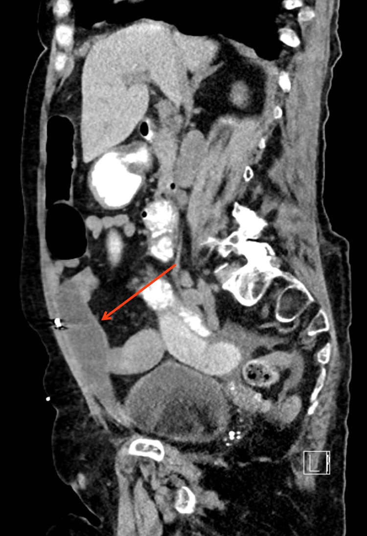
Rectus sheath hematomas (RSH) are increasing in prevalence, presumably correlating with increased use of anticoagulation medications and an aging population. Comorbidities such as blood dyscrasias, atherosclerosis, and hypertension are associated with an increased risk of developing an RSH. Iatrogenic origin of RSH, secondary to treatment of various abdominal pathologies, is not uncommon. Due to its exceptionally non-specific array of clinical signs and symptoms, RSH can be challenging to diagnose in the clinical setting without the aid of radiological images. Abdominal computed tomography (CT) is generally the modality of choice through which the RSH can be successfully identified and characterized. CT imaging can play an important role in the planning of RSH management, as effective management varies depending on the size and position of the RSH. Recurrent bleeding, hypovolemic shock, abdominal compartment syndrome, myonecrosis, and infection have been traditionally considered as the more prominent complications of RSH. However, with more cases occurring, more complications are being described in the literature. The following case presents a previously unreported complication of RSH, that of bowel herniation into a potential space created by a previously treated RSH.
Keywords: abdominal wall hernia; posterior rectus sheath hematoma; rectus hematoma; rectus sheath hematoma; rectus sheath hernia.
Copyright © 2022, Dulberger et al.
Conflict of interest statement
The authors have declared that no competing interests exist.
Figures


References
-
- The changing nature of rectus sheath haematoma: case series and literature review. Fitzgerald JE, Fitzgerald LA, Anderson FE, Acheson AG. Int J Surg. 2009;7:150–154. - PubMed
-
- Diagnostic evaluation and management of patients with rectus sheath hematoma. A retrospective study. Salemis NS, Gourgiotis S, Karalis G. Int J Surg. 2010;8:290–293. - PubMed
-
- Rectus sheath hematoma: review of 126 cases at a single institution. Cherry WB, Mueller PS. Medicine (Baltimore) 2006;85:105–110. - PubMed
-
- Percutaneous arterial embolization in the management of rectus sheath hematoma. Rimola J, Perendreu J, Falcó J, Fortuño JR, Massuet A, Branera J. AJR Am J Roentgenol. 2007;188:0–502. - PubMed
-
- Rectus sheath hematoma. Osinbowale O, Bartholomew JR. Vasc Med. 2008;13:275–279. - PubMed
Publication types
LinkOut - more resources
Full Text Sources