

Giant Paratesticular Liposarcoma Mimicking a Left-Sided Groin Hernia: A Case Report
- PMID: 36225510
- PMCID: PMC9536849
- DOI: 10.7759/cureus.28856
Giant Paratesticular Liposarcoma Mimicking a Left-Sided Groin Hernia: A Case Report
Abstract
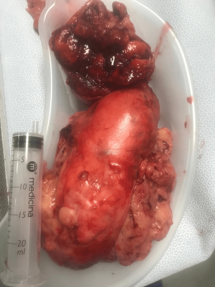
Giant paratesticular liposarcoma (PLS) is an uncommon tumour, often misdiagnosed pre-operatively, which presents as a painless scrotal mass. Early detection and prompt surgical management provide the best outcome. We present an 87-year-old patient with gradually enlarging, painless left scrotal swelling. Ultrasound on initial presentation suggested a benign hernia, resulting in an 11-month treatment delay. Computed tomography (CT) thereafter showed paratesticular scrotal mass measuring 14 x 8 x 7cm. Radical inguinal orchidectomy with high ligation of the spermatic cord was performed. Histopathology and cytogenetics confirmed PLS with both de-differentiated and well-differentiated features involving the spermatic cord margin. The patient had rapid progression to fatal lung metastasis within three months of surgery. Our case highlights that any suspicious fat swelling should be investigated thoroughly and excised promptly if paratesticular liposarcoma is suspected, as delayed management gives poor outcomes.
Keywords: dedifferentiated liposarcoma; giant liposarcoma; paratesticular liposarcoma; paratesticular tumors; testicular mass.
Copyright © 2022, Chan et al.
Conflict of interest statement
The authors have declared that no competing interests exist.
Figures


References
-
- Adult urological sarcoma. Russo P, Brady MS, Conlon K, Hajdu SI, Fair WR, Herr HW, Brennan MF. J Urol. 1992;147:1032–1036. - PubMed
-
- Primary spermatic cord tumors: disease characteristics, prognostic factors, and treatment outcomes. Rodríguez D, Barrisford GW, Sanchez A, Preston MA, Kreydin EI, Olumi AF. Urol Oncol. 2014;32:52–25. - PubMed
-
- Well-differentiated liposarcoma and dedifferentiated liposarcoma: an updated review. Thway K. Semin Diagn Pathol. 2019;36:112–121. - PubMed
Publication types
LinkOut - more resources
Full Text Sources