. 2022 Oct 3:2022:5852786.
doi: 10.1155/2022/5852786. eCollection 2022.

Proteomic Analysis of Exudates from Chronic Ulcer of Diabetic Foot Treated with Scorpion Antimicrobial Peptide

Affiliations

Proteomic Analysis of Exudates from Chronic Ulcer of Diabetic Foot Treated with Scorpion Antimicrobial Peptide

Zhixiang Tan et al. Mediators Inflamm. .

Abstract

Scorpion peptides have good therapeutic effect on chronic ulcer of diabetic foot, but the related pharmacological mechanism has remained unclear. The different proteins and bacteria present in ulcer exudates from chronic diabetic foot patients, treated with scorpion antimicrobial peptide at different stages, were analyzed using isobaric tags for quantification-labeled proteomics and bacteriological methods. According to the mass spectrometry data, a total of 1865 proteins were identified qualitatively, and the number of the different proteins was 130 (mid/early), 401 (late/early), and 310 (mid, late/early). In addition, functional annotation, cluster analysis of effects and the analysis of signal pathway, transcription regulation, and protein-protein interaction network were carried out. The results showed that the biochemical changes of wound microenvironment during the treatment involved activated biological functions such as protein synthesis, cell proliferation, differentiation, migration, movement, and survival. Inhibited biological functions such as cell death, inflammatory response, immune diseases, and bacterial growth were also involved. Bacteriological analysis showed that Burkholderia cepacia was the main bacteria in the early and middle stage of ulcer exudate and Staphylococcus epidermidis in the late stage. This study provides basic data for further elucidation of the molecular mechanism of diabetic foot.


Conflict of interest statement

The authors declare no conflict of interest.

Figures

Figure 1
Figure 1
Changes of ulcer wounds in diabetic foot patients during different treatment periods. (a) is the ulcer wound changes in the lesion site of patients with No. 1 diabetic foot after antimicrobial peptide treatment in the early, middle, and late stage; (b) is the ulcer wound changes in the lesion site of patients with No. 2 diabetic foot after antimicrobial peptide treatment in the early, middle, and late stage; (c) is the ulcer wound changes in the lesion site of patients with No. 3 diabetic foot after antimicrobial peptide treatment in the early, middle, and late stages. The initial ulcer areas of the three patients were about 40 cm2, 10 cm2, and 6 cm2, respectively, and the amount of wound exudate was between 100 μL and 2 mL.
Figure 2
Figure 2
Bacteriological analysis of ulcer wound exudate in diabetic foot patients. (a) The growth morphology of colonies in exudate samples on LB plates, with smooth edges and milky color colonies (about 0.5 mm in diameter). (b) The number of colony-forming units in the exudate of patient No. 1 ranged from 104-7 CFU/mL, and the changes in the early, middle, and late stages were first decreased and then increased. (c) The number of colony-forming units in the exudate of patient No. 2 ranged from 104-5 CFU/mL, and the change trend also decreased first and then increased slightly. (d) The number of colony-forming units in the exudate of patient No. 3 was between 103-8 CFU/mL, with a large range of change and a gradual increase, with a large range in the early and middle stages and a small range in the middle and late stages.
Figure 3
Figure 3
SDS-PAGE separation of serum and exudate protein at different treatment stages.
Figure 4
Figure 4
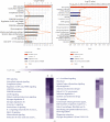
Classification statistics of differential proteins about exudates in different periods. The dark blue column gram showed the classification of differential proteins in C group (medium exudate) compared with B group (early exudate). The light blue column gram showed the classification of differential proteins in D group (late effusion) than that of the B group (early effusion).
Figure 5
Figure 5
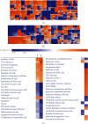
Heatmap of clustering analysis of differential proteins in exudates at different stages. (a) The clustering analysis of differential protein effect in C group (middle exudate) compared with that in B group (early exudate). (b) The clustering analysis of differential protein effect in D group (late exudate) compared with that in B group (early exudate). (c) Analysis of trends in effects of differential proteins in CD/B group.
Figure 6
Figure 6
Statistics of signal pathways involved in differential proteins in exudates at different stages. (a) The signaling pathway analysis of differential proteins in C group (middle exudate) compared with that in B group (early exudate). (b) The signaling pathway analysis of differential proteins in D group (late exudate) compared with that in B group (early exudate). (c) Trend analysis of intergroup signaling pathways.
Figure 7
Figure 7
The interaction network and downstream function of upstream regulators. (a) Regulators derived from differential proteins in C/B group. (b) Regulators derived from differential proteins in D/B group.
Figure 8
Figure 8
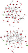
Interaction network of differential proteins. (a) The first interaction network in C/B group. (b) The third interaction network in D/B group.

Similar articles

Cited by

References

    1. Armstrong D. G., Boulton A. J. M., Bus S. A. Diabetic foot ulcers and their recurrence. The New England Journal of Medicine . 2017;376(24):2367–2375. doi: 10.1056/NEJMra1615439. - DOI - PubMed
    1. Skrepnek G. H., Mills J. L., Sr., Lavery L. A., Armstrong D. G. Health care service and outcomes among an estimated 6.7 million ambulatory care diabetic foot cases in the U.S. Diabetes Care . 2017;40(7):936–942. doi: 10.2337/dc16-2189. - DOI - PubMed
    1. Abbott C. A., Carrington A. L., Ashe H., et al. The north-west diabetes foot care study: incidence of, and risk factors for, new diabetic foot ulceration in a community-based patient cohort. Diabetic Medicine . 2002;19(5):377–384. doi: 10.1046/j.1464-5491.2002.00698.x. - DOI - PubMed
    1. Vinik A. I., Nevoret M. L., Casellini C., Parson H. Diabetic neuropathy. Endocrinology and Metabolism Clinics of North America . 2013;42(4):747–787. doi: 10.1016/j.ecl.2013.06.001. - DOI - PubMed
    1. Murray H. J., Young M. J., Hollis S., Boulton A. J. The association between callus formation, high pressures and neuropathy in diabetic foot ulceration. Diabetic Medicine . 1996;13(11):979–982. doi: 10.1002/(SICI)1096-9136(199611)13:11<979::AID-DIA267>3.0.CO;2-A. - DOI - PubMed

Substances