. 2022 Oct 3:2022:3260087.
doi: 10.1155/2022/3260087. eCollection 2022.

The Impact of Different Oxygen Delivery Methods on Corneal Epithelial Repair after Injury

Affiliations

The Impact of Different Oxygen Delivery Methods on Corneal Epithelial Repair after Injury

Shanshan Li et al. J Ophthalmol. .

Abstract

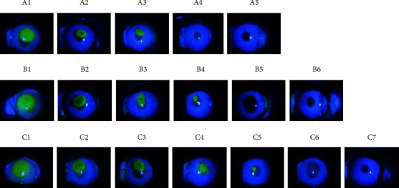
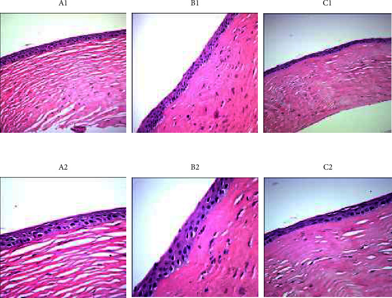
The hyperbaric oxygen therapy is often used in the management of acid and base burns of the eyes. However, oxygen is rarely supplied locally through goggles or face mask in ophthalmology. Therefore, in this study, we aim to investigate how oxygen delivery affects eye recovery after injury. We used a rabbit model with corneal epithelial injury to examine the effects of local oxygen supply via goggles or face mask on the recovery of cornea. A total of 75 healthy New Zealand white rabbits were randomly divided into three groups, A, B, and C, with 25 rabbits in each group. Then, on each rabbit eye (150 eyes in total), a circle of corneal epithelium with 5 mm in diameter was scraped off from the center of the cornea with a corneal epithelial scraper. Group A was given oxygen goggles every day (the oxygen flow rate was 3 L/min, once a day, 2 hours each time); group B was given nasal inhalation of oxygen every day (the oxygen flow rate was 3 L/min, once a day, 2 hours each time); and group C did not receive any treatment and was healed naturally. We found that the group A, which received oxygen supply via goggles, showed the best eye recovery. Transmission electron microscopy showed that the cornea with local oxygen supply via goggles or face mask exhibited intact capillary structure and obvious desmosome/hemidesmosome connections between cells. Moreover, the protein and RNA levels of hypoxia-related genes were lower in group A and B, suggesting that the hypoxia factor is a sensitive and early regulator in the low oxygen environment.


Conflict of interest statement

The authors declare that they have no conflicts of interest.

Figures

Figure 1
Figure 1
Corneal staining analysis in each group at 0 h, 12 h, 36 h, 48 h, 60 h, and 72 h after operation. Group A: 0 h (A1), 12 h (A2), 24 h (A3), 36 h (A4), 48 h (A5); Group B: 0 h (B1), 12 h (B2), 24 h (B3), 36 h (B4), 48 h (B5), 60 h (B6); Group C: 0 h (C1), 12 h (C2), 24 h (C3), 36 h (C4), 48 h (C5), 60 h (C6), 72 h (C7). The corneal fluorescence staining at 12 h, 24 h, 36 h, 48 h, and 72 h after operation: the staining area of group A was significantly smaller than group B at each time point, and the staining area of group B was smaller than group C.
Figure 2
Figure 2
Electron microscope images in each group at 72 h after operation. Electron microscopic images of group A (A1), B (B1), and C (C1) under 200x magnification; electron microscopic images of group A (A2), B (B2), and C (C2) under 400x magnification. Group A: there are 5–6 layers of corneal epithelium; no obvious neovascularization is seen at corneal limbus; the matrix collagen structure is densely and regularly arranged. Group B: there are 4–5 layers of corneal epithelium; new blood vessels can be seen at the corneal limbus; the matrix collagen structure is densely and regularly arranged. Group C: there are 2–3 layers of corneal epithelium; a large number of new blood vessels can be seen at the corneal limbus; the number of fibroblasts in the stromal layer is reduced, and a large number of inflammatory cells are observed; matrix collagen structure is slightly loose but relatively regular.
Figure 3
Figure 3
TEM images in each group at 24 h after operation. TEM images of group A: 5,000x (A1), 15,000x (A2), and 50,000x (A3); TEM images of group B: 5,000x (B1), 15,000x B1 (B2), and 50,000x (B3); TEM images of group C: 5,000x (C1), 15,000x (C2), and 50,000x (C3). (1) The microvilli in A and B groups are closely and regularly arranged. The microvilli in group C are absent, and the surface epithelial cells are necrotic (①). (2) There are a lot of glycocalyx on the cell membrane of microvilli in A and B groups, but no glycocalyx is seen in in group C (①, the enlarged picture is indicated by the arrow). (3) Desmosome junction and hemidesmosome junction can be seen in A and B groups; in group (C), the distribution of desmosome junctions is disordered, the number of desmosomes is reduced, the tension filaments are significantly reduced, and necrotic cells can be seen on one side (②). (4) In group B and C, the mitochondria are swollen, endoplasmic reticulum is expanded, and mitochondrial cristae are damaged (③); myeloid corpuscles are visible. (5) The number of corneal epithelium layers in group C is more than that in group A and B under transmission electron microscope (④). (6) The corneal epithelium layers in group C were more than that in group A and B, but the boundary of each stratification is not obvious (⑤).

References

    1. Eghrari A. O., Riazuddin S. A., Gottsch J. D. Overview of the cornea: structure, function, and development. Progress in Molecular Biology and Translational Science . 2015;134:7–23. - PubMed
    1. Papas E. B., Sweeney D. F. Interpreting the corneal response to oxygen: is there a basis for re-evaluating data from gas-goggle studies? Experimental Eye Research . 2016;151:222–226. - PubMed
    1. Wang L., González S., Dai W., Deng S., Lu L. Effect of hypoxia-regulated polo-like kinase 3 (Plk3) on human limbal stem cell differentiation. Journal of Biological Chemistry . 2016;291(32):16519–16529. doi: 10.1074/jbc.m116.725747. - DOI - PMC - PubMed
    1. Lee A., Karamichos D., Onochie O. E., et al. Hypoxia modulates the development of a corneal stromal matrix model. Experimental Eye Research . 2018;170:127–137. doi: 10.1016/j.exer.2018.02.021. - DOI - PMC - PubMed
    1. Walsh B. K., Smallwood C. D. Pediatric oxygen therapy: a review and update. Respiratory Care . 2017;62(6):645–661. doi: 10.4187/respcare.05245. - DOI - PubMed

LinkOut - more resources