. 2022 Sep 26:9:1012020.
doi: 10.3389/fcvm.2022.1012020. eCollection 2022.

Study of necrotic apoptosis by pulsed electric field ablation in rabbit left ventricular myocardium

Affiliations

Study of necrotic apoptosis by pulsed electric field ablation in rabbit left ventricular myocardium

Zhihong Zhao et al. Front Cardiovasc Med. .

Abstract

Objective: We investigate the characteristics of histological damage to myocardial cells in the ablation region and surrounding areas of the left ventricular epicardium in rabbits using our self-developed cardiac pulsed electric field (PEF) ablation instrument and ablation catheter.

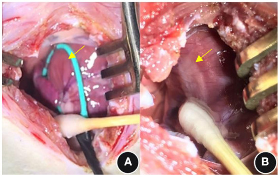
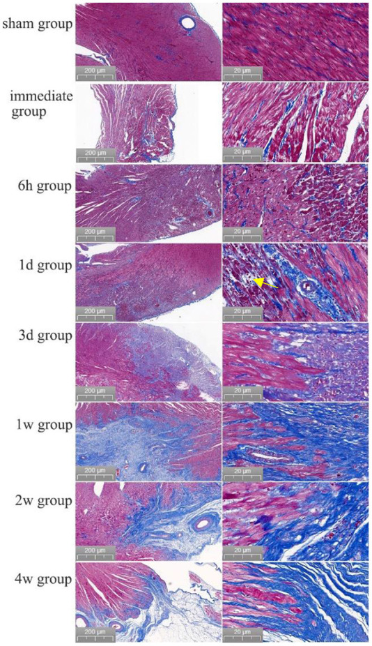
Methods: Forty eight New Zealand rabbits underwent ablation on the left ventricular myocardium after open-heart exposure with a cardiac arrhythmia PEF ablation device and ablation catheter developed by the Medical Translation Laboratory of Pulsed Electric Field Technology in Zhejiang Province. The ablation parameters were set as biphasic electrical pulses; voltage, ±800 V; pulse width, 10 μs; interphase delay, 500 us. Six rabbits were included in the sham group and 42 other rabbits were randomly divided into immediately, 6-h, 1-, 3-day, 1-, 2-, and 4-week post-ablation groups, with six rabbits in each group. Creatine kinase- (CK)-MB isoenzyme (CK-MB), aspartate aminotransferase (AST), and lactate dehydrogenase (LDH) levels were measured before and at different time points after PEF ablation to analyze their dynamic evolution. Masson staining of tissue block sections of left ventricular myocardial ablation and adjacent tissue heart specimens was performed, and the occurrence of TUNEL apoptosis in myocardium tissue was analyzed.

Results: All rabbits completed the PEF ablation procedure and the follow-up process. After PEF ablation, the levels of cardiac enzymes, including CK-MB, CK, and AST, increased significantly, peaking 1-3 days after the procedure. In particular, those of CK and CK-MB increased by 15-20 times but returned to the preoperative level after 2 weeks. Based on general observation, it was found that the myocardium in the ablation area was swollen immediately after PEF ablation. Masson staining analysis revealed that cardiomyocytes were broken and infiltrated by erythrocytes after 6 h. After 1 day, the cells started to experience atrophy and necrosis; after 3 days, fibrotic replacement of the necrotic area became obvious. Then, by 4 weeks, the myocardial cells were completely replaced by hyperplasia. Apoptosis occurred significantly at 6 h and peaked at 24 h post-ablation, demonstrating a 37.7-fold increase; apoptotic cell counts decreased significantly at 3 days post-ablation, and no significant apoptotic cardiomyocytes were seen after 1 week.

Conclusion: After PEF ablation, cardiomyocytes showed apoptotic process and dyed, at least partially, through a secondary necrosis, the ablation boundary was clear, the ablation area was replaced by structurally intact fibroblasts, no island myocardium tissue were seen, and the ablation area vessels and nerves were not affected.

Keywords: ablation; cardiovascular pathology; heart; necrotizing apoptosis; pulsed electric field; rabbit.


Conflict of interest statement

The authors declare that the research was conducted in the absence of any commercial or financial relationships that could be construed as a potential conflict of interest.

Figures

Figure 1
Figure 1
PEF ablation instrument, ablation electrode catheter, and PEF ablation parameters. (A) PEF ablation instrument. (B) PEF ablation electrode under direct epicardial vision. (C) Biphasic PEF ablation parameters. (D) Fifty to one hundred pulse numbers delivered at a frequency of 1 Hz. PEF, Pulsed electric field.
Figure 2
Figure 2
General observations before and immediately after PEF ablation. (A) Pre-ablation and (B) post-ablation; see the yellow arrows. PEF, Pulsed electric field.
Figure 3
Figure 3
Evolution of myocardial necrosis at different time periods before and after pulsed ablation (Masson staining). In the preoperative heart, the myocardium tissue were neatly arranged and tightly packed. Immediately after the operation, it was obvious that the ablation boundary was clear, the myocardium tissue were swollen, and the cell gap became larger. Then, 6 h post-ablation, the boundary of PEF ablation was clearer, the myocardial cells were swollen, and the cell gap became larger; besides, the myocardial bundle was broken into myocardial fragments, and red blood cells appeared in the myocardial cell gap. At 1 day post-ablation, the ablation boundary was still clear, and the cardiomyocytes had begun to atrophy with a large frequency of erythrocyte infiltration (shown by yellow arrows). At 3 days post-ablation, the ablation boundary was clear, and the cardiomyocytes were atrophied and necrotic, with clustered nuclei and aggregated fibrous structures. At 1 week post-ablation, the ablation boundary was clear, most of the myocardial tissue in the ablation area had disappeared, the structure was intact with fibrotic replacement, and the vascular structure was intact without damage. At 2 weeks, the ablation boundary was clear, the myocardial cells in the ablation area had basically disappeared with fibrous connective tissue replacement, and the vascular structure was intact. After 4 weeks, the ablation boundary was clear, the myocardium tissue in the ablation area had almost completely disappeared, the fibrous structure was intact, and the vascular structure was intact.
Figure 4
Figure 4
Morphology of cardiac vessels and nerves in the ablation area (Masson staining). Preoperatively, the endothelial cells of the arterial vessels were neatly and closely arranged with well-defined layers. Then, at 1 day, 1, and 4 weeks after ablation, no abnormal changes had occurred in the arterial vessels or endothelial cells (shown by red arrows). Also, at 1 day after ablation, the nerve structure was intact (shown by black arrows).
Figure 5
Figure 5
Apoptosis of cardiomyocytes, as detected by TUNEL staining, at different time points in the PEF ablation region. Immediately after ablation, no apoptotic cells were generated; at 6 h after ablation, apoptotic cardiomyocytes were seen; at 24 h after ablation, significant apoptotic cells were seen; at 3 days after ablation, a significant decrease in apoptotic cells was observed; and at 1 ~ 2 week after ablation, no apoptotic cells were seen. Positively apoptotic cells are cells with red-stained nuclei.

References

    1. Darby AE. Pulsed field ablation: a novel therapeutic tool for catheter-based treatment of atrial fibrillation. Curr Cardiol Rep. (2022) 24:793–9. 10.1007/s11886-022-01697-2 - DOI - PubMed
    1. Avazzadeh S, O'Brien B, Coffey K, O'Halloran M, Keane D, Quinlan LR. Establishing irreversible electroporation electric field potential threshold in a suspension in vitro model for cardiac and neuronal cells. J Clin Med. (2021) 10:5443. 10.3390/jcm10225443 - DOI - PMC - PubMed
    1. Batista Napotnik T, Polajzer T, Miklavcic D. Cell death due to electroporation - a review. Bioelectrochemistry. (2021) 141:107871. 10.1016/j.bioelechem.2021.107871 - DOI - PubMed
    1. Tang Min NG, Jinrui G, Yu Q, Yulong G, Ke L, Fengyuan Y, et al. . Novel pulsed field ablation system in treatment of paroxysmal atrial fibrillation: 3 cases. Chin J Cardiac Arrhyth. (2022) 26:96–8. 10.3760/cma.j.cn113859-20211203-00243 - DOI
    1. Ye X, Liu S, Yin H, He Q, Xue Z, Lu C, et al. . Study on optimal parameter and target for pulsed-field ablation of atrial fibrillation. Front Cardiovasc Med. (2021) 8:690092. 10.3389/fcvm.2021.690092 - DOI - PMC - PubMed