. 2022 Sep 26:16:987236.
doi: 10.3389/fncel.2022.987236. eCollection 2022.

Pharmacogenetic inhibition of TrkB signaling in adult mice attenuates mechanical hypersensitivity and improves locomotor function after spinal cord injury

Affiliations

Pharmacogenetic inhibition of TrkB signaling in adult mice attenuates mechanical hypersensitivity and improves locomotor function after spinal cord injury

Karmarcha K Martin et al. Front Cell Neurosci. .

Abstract

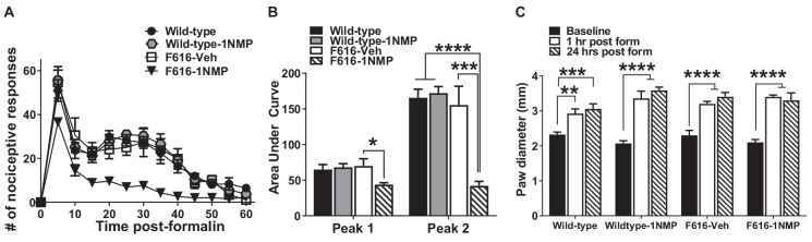
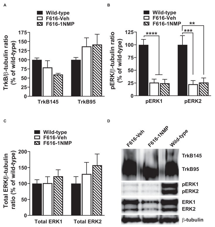
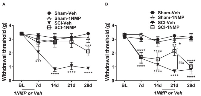
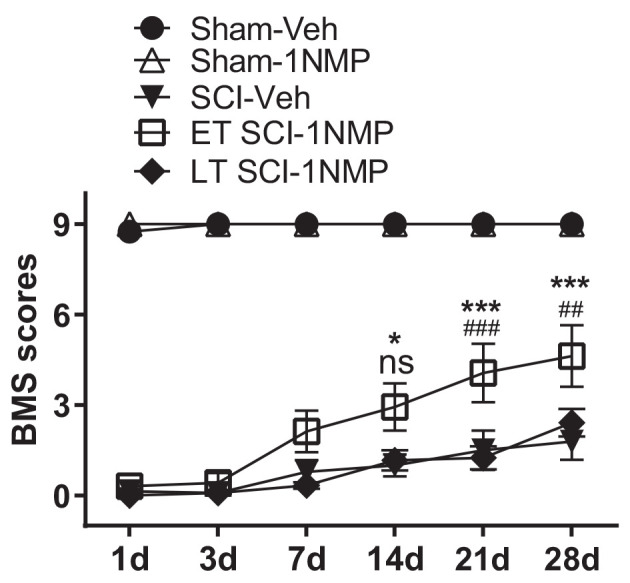
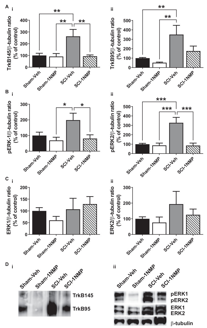
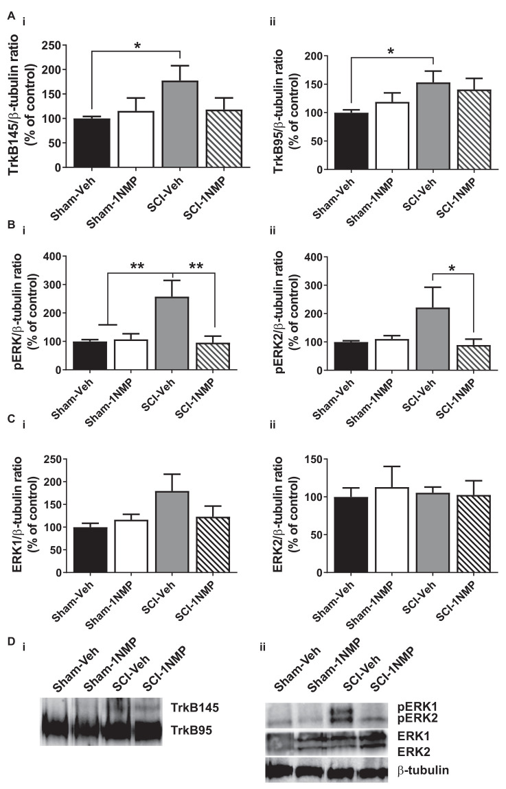
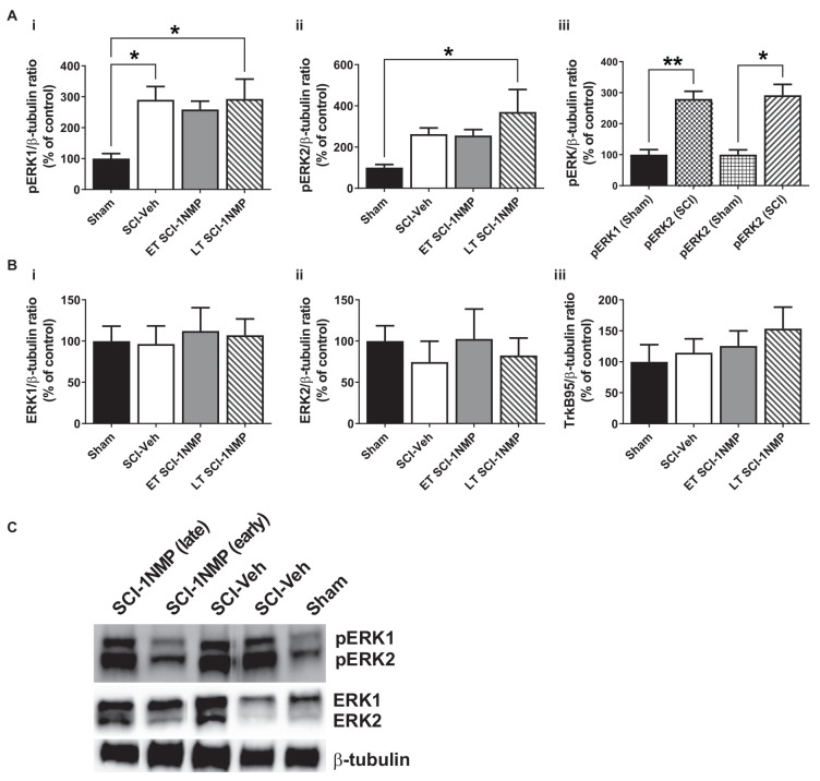
Brain-derived neurotrophic factor (BDNF) signals through tropomyosin receptor kinase B (TrkB), to exert various types of plasticity. The exact involvement of BDNF and TrkB in neuropathic pain states after spinal cord injury (SCI) remains unresolved. This study utilized transgenic TrkBF616 mice to examine the effect of pharmacogenetic inhibition of TrkB signaling, induced by treatment with 1NM-PP1 (1NMP) in drinking water for 5 days, on formalin-induced inflammatory pain, pain hypersensitivity, and locomotor dysfunction after thoracic spinal contusion. We also examined TrkB, ERK1/2, and pERK1/2 expression in the lumbar spinal cord and trunk skin. The results showed that formalin-induced pain responses were robustly attenuated in 1NMP-treated mice. Weekly assessment of tactile sensitivity with the von Frey test showed that treatment with 1NMP immediately after SCI blocked the development of mechanical hypersensitivity up to 4 weeks post-SCI. Contrastingly, when treatment started 2 weeks after SCI, 1NMP reversibly and partially attenuated hind-paw hypersensitivity. Locomotor scores were significantly improved in the early-treated 1NMP mice compared to late-treated or vehicle-treated SCI mice. 1NMP treatment attenuated SCI-induced increases in TrkB and pERK1/2 levels in the lumbar cord but failed to exert similar effects in the trunk skin. These results suggest that early onset TrkB signaling after SCI contributes to maladaptive plasticity that leads to spinal pain hypersensitivity and impaired locomotor function.

Keywords: BDNF; TrkB; mechanical hypersensitivity; pERK; plasticity; spinal cord injury.


Conflict of interest statement

The authors declare that the research was conducted in the absence of any commercial or financial relationships that could be construed as a potential conflict of interest.

Figures

Figure 1
Figure 1
Inhibition of TrkB attenuates formalin-induced inflammatory pain responses. (A) Intraplantar administration of formalin induced a biphasic nociceptive response characterized as phase 1 (0–10 min) and phase 2 (11–60 min) in three (Wild-type, Wild-type-1NMP and F616-Veh) of the four experimental groups of mice. There were no differences in cumulative number of responses between Wild-type, Wild-type-1NMP, and vehicle-treated F616 mice except at 60 min, where Wild-type was different to F616-Veh (p = 0.02). The number of responses in 1NMP-treated F616 mice was significantly reduced compared to wild-type and F616-Veh mice at binned times ranging from 15 to 50 min and from wild-type-1NMP at 5–55 min (p values ranging from p < 0.05 to p < 0.0001; 2-way ANOVA with Tukey’s multiple comparisons tests). (B) Quantitative measurement of the formalin response (area under the curve; AUC) revealed that F616–1NMP mice had a significantly reduced phase 1 response compared to F616-Veh treated mice only (p < 0.05), while their phase 2 response was robustly decreased compared to both wild-type groups (p < 0.0001) and F616-Veh group (p < 0.001). (C) Formalin induced significant paw edema in all four treatment groups, which was evident at both 1 and 24 h post-formalin (*p < 0.05; **p < 0.01; ***p < 0.001; and ****p < 0.0001).
Figure 2
Figure 2
Spinal expression of TrkB, pERK, and ERK in formalin inflammatory pain. (A,C) There were no differences in the spinal protein expression of TrkB and ERK isoforms, respectively, among the three groups of mice 24 h after formalin injection. (B) The levels of both pERK1 and 2 were significantly decreased in the F616 mice compared to Wild-type, although 1NMP treatment had no effect on pERK1/2 expression. (D) Representative Western blot images are shown for TrkB, pERK, and ERK expression in the lumbar spinal cord (**p < 0.01; ***p < 0.001; and ****p < 0.0001).
Figure 3
Figure 3
TrkB inhibition attenuates hind-paw hypersensitivity after SCI. (A) Early administration of 1NMP (days 0–5) after SCI (open squares) blocked the development of mechanical hypersensitivity seen in SCI-Veh mice (filled triangles) which developed significant mechanical hypersensitivity at all timepoints (7–28 dpo) after SCI compared to their pre-surgery baselines (***p < 0.001, and ****p < 0.0001). Only at 28 dpo did SCI-1NMP mice show a decrease in withdrawal threshold compared to baseline (***p < 0.001). However, even then, their average withdrawal threshold was significantly greater than that of the vehicle-treated counterparts at 28 dpo (p < 0.01, not indicated on graph). There were no differences in the withdrawal thresholds of either Sham group across treatment days. (B) Both SCI “late treatment” groups developed mechanical hypersensitivity over time compared to baseline withdrawal thresholds (**p < 0.01 and ****p < 0.0001). Treatment with 1NMP at 16–21 days after SCI (open squares), reversibly attenuated mechanical hypersensitivity at 21 dpo [withdrawal thresholds: significantly higher than SCI-Veh (###p < 0.001); although significantly lower than the Sham-Veh group (*p = 0.05)]. At 28 dpo, mechanical thresholds of SCI-1NMP mice had returned to those seen at 7 and 14 dpo, and were indistinguishable from SCI-Veh treated mice (filled triangles). SCI-Veh mice displayed hind-paw hypersensitivity at all time points, compared to their pre-surgery baselines. Sham mice did not show differences in withdrawal threshold across timepoints.
Figure 4
Figure 4
TrkB inhibition does not block conditioned place aversion (CPA). (A) The dual chamber (“light and dark”) CPA apparatus with a mouse shown inside, visually depicting how: (i) side-to-side transitions are quantified, and (ii) animal preferences are determined by monitoring time spent in each chamber during periods of free exploration, before and after brush stimulation. (B) (i) SCI mice had fewer transitions before and after stimulation thereby revealing continued locomotor impairment several weeks after injury (****p < 0.0001 SCI vs. Sham). (ii, left) SCI mice receiving mechanical brush stimulation while partially restrained in the dark chamber developed a preference for the light chamber post-stimulation (**p < 0.005 pre vs. post). (ii, right) Results broken down by treatment (vehicle or 1NMP) for all SCI and Sham mice, illustrating that 1NMP treatment did not alleviate affective pain behavior. PreC, pre-conditioning; PostC, post-conditioning. The dashed horizontal line in (B) indicates 50% time spent in each chamber (no preference).
Figure 5
Figure 5
Early inhibition of TrkB signaling with 1NMP improves locomotor recovery. SCI mice treated with 1NMP at 0–5 dpo had significantly improved BMS scores at 7, 14, 21 and 28 days compared to day 1 score (p < 0.0001; not indicated). Importantly, early 1NMP treated mice (ET SCI-1NMP) had higher BMS scores than late treated mice (LT SCI-1NMP, 16–21 dpo) at 21 dpo (###p < 0.001) and 28 dpo (##p < 0.01), and SCI-Veh mice at 14 dpo (*p < 0.05), 21 and 28 dpo (***p < 0.001 in both cases), ns indicates no significant difference between early and late treated SCI mice. SCI mice in all three groups had impaired locomotor function compared to Sham groups.
Figure 6
Figure 6
Early inhibition of TrkB signaling reduces spinal TrkB and pERK expression. (A) (i,ii) SCI increased both full length and truncated TrkB expression and, (B) (i,ii) pERK1 and pERK2 levels in the lumbar spinal cord at 34 dpo. Early treatment with 1NMP (day 0–5) blocked these increases. For both TrkB and pERK proteins, the SCI-1NMP group did not differ in expression levels from the Sham-Veh or Sham-1NMP groups. (C) (i,ii) Total ERK expression levels were unchanged among the four experimental groups (*p < 0.05, **p < 0.01, and ***p < 0.001; 1-way ANOVA, n = 6, each group). (D) Representative images of (i) TrkB, and (ii) pERK1/2, ERK1/2, and β-tubulin (loading control) Western blot are shown.
Figure 7
Figure 7
Late inhibition of TrkB signaling modestly reduces spinal TrkB and pERK expression. (A) (i,ii) TrkB expression was significantly increased in the lumbar spinal cord of SCI-Veh mice compared to Sham-Veh subjects only at 34 dpo (*p < 0.05, unpaired t test). (B) (i,ii) Both pERK1 and pERK2 levels were reduced following 1NMP treatment compared to vehicle treatment 16–21 days after SCI (*p < 0.05, and **p < 0.01, 1-way ANOVA). However, only pERK1 levels were significantly increased in SCI-Veh mice in comparison to Sham subjects (**p < 0.01, 1-way ANOVA). (C) (i,ii) Total ERK expression levels were unchanged among the four experimental groups (n = 7, each group). (D) Representative images of (i) TrkB, and (ii) pERK1/2, ERK1/2, and β-tubulin (loading control) Western blot are shown.
Figure 8
Figure 8
SCI increases pERK expression in the adjacent trunk skin. (A) (i,ii) SCI increased the expression of pERK1 and 2 in the adjacent trunk skin compared to sham procedures, although neither early nor late treatment with 1NMP was able to prevent the increases (*p < 0.05, 1-way ANOVA). (iii) Overall, SCI produced a robust increase in trunk pERK1 and pERK2 levels compared to sham procedure (*p < 0.05, **p < 0.01, unpaired t test). (B) Neither (i,ii) ERK1/2 nor (iii) TrkB95 levels were changed in the trunk skin among the experimental groups. (C) Representative images of pERK1/2, ERK1/2, and β-tubulin (loading control) Western blot are shown.

Similar articles

Cited by

References

    1. Adwanikar H., Karim F., Gereau R. W. (2004). Inflammation persistently enhances nocifensive behaviors mediated by spinal group I mGluRs through sustained ERK activation. Pain 111, 125–135. 10.1016/j.pain.2004.06.009 - DOI - PubMed
    1. Akhter E. T., Griffith R. W., English A. W., Alvarez F. J. (2019). Removal of the potassium chloride co-transporter from the somatodendritic membrane of axotomized motoneurons is independent of BDNF/TrkB signaling but is controlled by neuromuscular innervation. eNeuro 6:ENEURO.0172-19.2019. 10.1523/ENEURO.0172-19.2019 - DOI - PMC - PubMed
    1. Apfel S. C., Wright D. E., Wiideman A. M., Dormia C., Snider W. D., Kessler J. A. (1996). Nerve growth factor regulates the expression of brain-derived neurotrophic factor mRNA in the peripheral nervous system. Mol. Cell Neurosci. 7, 134–142. 10.1006/mcne.1996.0010 - DOI - PubMed
    1. Bagdas D., Muldoon P. P., AlSharari S., Carroll F. I., Negus S. S., Damaj M. I. (2016). Expression and pharmacological modulation of visceral pain-induced conditioned place aversion in mice. Neuropharmacology 102, 236–243. 10.1016/j.neuropharm.2015.11.024 - DOI - PMC - PubMed
    1. Bain J., Plater L., Elliott M., Shpiro N., Hastie C. J., Mclauchlan H., et al. . (2007). The selectivity of protein kinase inhibitors: a further update. Biochem. J. 408, 297–315. 10.1042/BJ20070797 - DOI - PMC - PubMed