. 2023 Feb;531(2):217-237.
doi: 10.1002/cne.25419. Epub 2022 Oct 12.

Projections from the five divisions of the orbital cortex to the thalamus in the rat

Affiliations

Projections from the five divisions of the orbital cortex to the thalamus in the rat

Robert P Vertes et al. J Comp Neurol. 2023 Feb.

Abstract

The orbital cortex (ORB) of the rat consists of five divisions: the medial (MO), ventral (VO), ventrolateral (VLO), lateral (LO), and dorsolateral (DLO) orbital cortices. No previous report has comprehensively examined and compared projections from each division of the ORB to the thalamus. Using the anterograde anatomical tracer, Phaseolus vulgaris leucoagglutinin, we describe the efferent projections from the five divisions of the ORB to the thalamus in the rat. We demonstrated that, with some overlap, each division of the ORB distributed in a distinct (and unique) manner to nuclei of the thalamus. Overall, ORB projected to a relatively restricted number of sites in the thalamus, and strikingly distributed entirely to structures of the medial/midline thalamus, while completely avoiding lateral regions or principal nuclei of the thalamus. The main termination sites in the thalamus were the paratenial nucleus (PT) and nucleus reuniens (RE) of the midline thalamus, the medial (MDm) and central (MDc) divisions of the mediodorsal nucleus, the intermediodorsal nucleus, the central lateral, paracentral, and central medial nuclei of the rostral intralaminar complex and the submedial nucleus (SM). With some exceptions, medial divisions of the ORB (MO, VO) mainly targeted "limbic-associated" nuclei such as PT, RE, and MDm, whereas lateral division (VLO, LO, DLO) primarily distributed to "sensorimotor-associated" nuclei including MDc, SM, and the rostral intralaminar complex. As discussed herein, the medial/midline thalamus may represent an important link (or bridge) between the orbital cortex and the hippocampus and between the ORB and medial prefrontal cortex. In summary, the present results demonstrate that each division of the orbital cortex projects in a distinct manner to nuclei of the thalamus which suggests unique functions for each division of the orbital cortex.

Keywords: behavioral flexibility; cognition; medial prefrontal cortex; mediodorsal nucleus; nucleus reuniens; paratenial nucleus; reversal learning.


Conflict of interest statement

Conflict of interest:

The authors declare no conflict of interest.

Figures

Figure 1.
Figure 1.
a: Nissl-stained section through the anterior forebrain showing the locations of the five divisions of the orbital cortex in the rat: the medial orbital (MO), ventral orbital (VO), ventrolateral orbital (VLO), lateral orbital (LO) and dorsolateral orbital (DLO) cortices. b-f: Bright field micrographs showing the locations of PHA-L injections in MO (b), VO (c), VLO (d), LO (e) and DLO (f). Scale bar for a, d = 600 µm; for b, c, e, f = 500 µm.
Figure 2
Figure 2
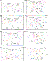
Schematic representation of the pattern of distribution of labeled fibers at select rostral (a) to caudal (j) levels of the thalamus produced by a PHA-L injection in the medial orbital cortex. See list for abbreviations.
Figure 2
Figure 2
Schematic representation of the pattern of distribution of labeled fibers at select rostral (a) to caudal (j) levels of the thalamus produced by a PHA-L injection in the medial orbital cortex. See list for abbreviations.
Figure 3
Figure 3
a-c: Brightfield micrographs through the anterior thalamus showing dense collections of labeled fibers within the paratenial nucleus (PT) of the dorsal midline thalamus, stronger ipsilaterally (left side) than contralaterally. Labeling produced by PHA-L injections in the medial orbital (a), ventral orbital (b) and dorsolateral orbital (c) cortices. Abbreviations. PV, paraventricular nucleus of thalamus. Scale bar for a = 600 µm; for b, c = 500 µm.
Figure 4
Figure 4
Darkfield micrographs at a rostral (a) and caudal level (b) of the anterior thalamus depicting the pattern of distribution of labeled fibers dorsally and ventrally within the midline thalamus produced by a PHA-L injection in the medial orbital cortex. Note the dense collection of labeled fibers in the paratenial nucleus (PT) (a), the medial division of the mediodorsal nucleus (MDm) (a, b), and the nucleus reuniens (RE) (b). Abbreviations: MDc, central division of the mediodorsal nucleus of thalamus; PV, paraventricular nucleus of thalamus; 3V, third ventricle. Scale bar for a, b = 500 µm.
Figure 5
Figure 5
Darkfield micrographs at a rostral (a) and caudal level (b) of the posterior thalamus depicting the pattern of distribution of labeled fibers dorsally and ventrally within the midline thalamus produced by a PHA-L injection in the medial orbital cortex. Note the dense collection of labeled fibers in the medial division of the mediodorsal nucleus (MDm) (a), the intermediodorsal nucleus (IMD) (a) and rostrally (a) and caudally (b) in nucleus reuniens (RE) and the peri-reuniens nucleus (pRE) of thalamus. Abbreviations: RH, rhomboid nucleus of thalamus. Scale bar for a = 650 µm; for b = 500 µm.
Figure 6
Figure 6
Schematic representation of the pattern of distribution of labeled fibers at select rostral (a) to caudal (j) levels of the thalamus produced by a PHA-L injection in the ventral orbital cortex. See list for abbreviations.
Figure 6
Figure 6
Schematic representation of the pattern of distribution of labeled fibers at select rostral (a) to caudal (j) levels of the thalamus produced by a PHA-L injection in the ventral orbital cortex. See list for abbreviations.
Figure 7
Figure 7
Darkfield micrographs at a rostral (a) and caudal level (b) of the anterior thalamus depicting the pattern of distribution of labeled fibers dorsally and ventrally within the midline thalamus produced by a PHA-L injection in the ventral orbital cortex. Note the dense collection of labeled fibers in the paratenial nucleus (PT) (a), the central division of the mediodorsal nucleus (MDc) (b) and the nucleus reuniens (RE) and peri-reuniens nucleus (pRE) at two levels of the anterior thalamus (a, b). Note also the very intense labeling on dorsal/dorsolateral border of the caudal RE (b). Abbreviations: IAM, interanteromedial nucleus of thalamus; MDm, medial division of the mediodorsal nucleus of thalamus; PV, paraventricular nucleus of thalamus. Scale bar for a = 400 µm; for b = 500 µm.
Figure 8
Figure 8
Schematic representation of the pattern of distribution of labeled fibers at select rostral (a) to caudal (h) levels of the thalamus produced by a PHA-L injection in the ventrolateral orbital cortex. See list for abbreviations.
Figure 9
Figure 9
Darkfield micrographs depicting the pattern of distribution of labeled fibers at a rostral (a) and caudal (b) level of the thalamus produced by a PHA-L injection in the ventrolateral orbital cortex. a: Note the presence of labeled fibers coursing laterally into anterior thalamus (left side of a), but a complete lack of terminal labeling at this level in the paraventricular (PV), paratenial (PT) and nucleus reuniens (RE) of the dorsal midline thalamus. b: Note dense collections of labeled fibers in the central lateral (CL), paracentral (PC) and central medial (CM) nuclei of the rostral intralaminar complex of thalamus as well as ventrally in the central division of the mediodorsal nucleus (MDc) and the submedial nucleus (SM) of thalamus. Abbreviations: sm, stria medullaris. Scale bar for a = 650 µm; for b = 500 µm.
Figure 10
Figure 10
Schematic representation of the pattern of distribution of labeled fibers at select rostral (a) to caudal (h) levels of the thalamus produced by a PHA-L injection in the lateral orbital cortex. See list for abbreviations.
Figure 11
Figure 11
Dark field micrographs depicting the pattern of distribution of labeled fibers at a rostral (a) and caudal (b) level of the thalamus produced by a PHA-L injection in the lateral orbital cortex. Note the dense collection of labeled fibers selectively in the central division of the mediodorsal nucleus (MDc) and the submedial nucleus (SM) at the two levels of the thalamus. Abbreviations: PV, paraventricular nucleus of thalamus. Scale bar for a = 550 µm; for b = 500 µm.
Figure 12
Figure 12
Schematic representation of the pattern of distribution of labeled fibers at select rostral (a) to caudal (j) levels of the thalamus produced by a PHA-L injection in the dorsolateral orbital cortex. See list for abbreviations.
Figure 12
Figure 12
Schematic representation of the pattern of distribution of labeled fibers at select rostral (a) to caudal (j) levels of the thalamus produced by a PHA-L injection in the dorsolateral orbital cortex. See list for abbreviations.
Figure 13
Figure 13
Darkfield micrograph depicting dense collections of labeled fibers at a mid-level of the thalamus produced by a PHA-L injection in the dorsolateral orbital cortex. Note dense collection of labeled fibers in the medial division of the mediodorsal nucleus (MDm) and the submedial nucleus (SM) of thalamus as well as moderate labeling of the intermediodorsal nucleus (IMD) of thalamus. Abbreviations: PV, paraventricular nucleus of thalamus, RE, nucleus reuniens of thalamus; RH, rhomboid nucleus of thalamus. Scale bar = 500 µm.
Figure 14.
Figure 14.
Schematic representation of patterns and density of medial orbital (red) and ventral orbital (yellow) projections to the thalamus. Color coded chart depicting strength of projections: - none; +, light; ++, moderate; +++, heavy, ++++, dense projections. See list for abbreviations.
Figure 15.
Figure 15.
Schematic representation of the patterns and density of ventrolateral orbital (green), lateral orbital (blue) and dorsolateral orbital (purple) projections to the thalamus. Color coded chart depicting strength of projections: -, none; +, light; ++, moderate; +++, heavy; ++++, dense projections. See list for abbreviations.

References

    1. Ahmari SE, Spellman T, Douglass NL, Kheirbek MA, Simpson HB, Deisseroth K, Gordon JA, & Hen R. (2013). Repeated cortico-striatal stimulation generates persistent OCD-like behavior. Science. 340(6137), 1234–1239. - PMC - PubMed
    1. Alcaraz F, Marchand AR, Vidal E, Guillou A, Faugère A, Coutureau E, & Wolff M. (2015). Flexible use of predictive cues beyond the orbitofrontal cortex: Role of the submedius thalamic nucleus. Journal of Neuroscience. 35(38), 13183–13193. - PMC - PubMed
    1. Barreiros IV, Panayi MC, & Walton ME. (2021a). Organization of afferents along the anterior-posterior and medial-lateral axes of the rat orbitofrontal cortex. Neuroscience. 460, 53–68. - PMC - PubMed
    1. Barreiros IV, Ishii H, Walton ME, & Panayi MC. (2021b). Defining an orbitofrontal compass: functional and anatomical heterogeneity across anterior-posterior and medial-lateral axes. Behavioral Neuroscience. 135(2), 165–173. - PMC - PubMed
    1. Bedwell SA, Billett EE, Crofts JJ, & Tinsley CJ. (2017). Differences in anatomical connections across distinct areas in the rodent prefrontal cortex. European Journal of Neuroscience. 45(6), 859–873. - PubMed

Publication types

Substances