. 2023 Feb;33(2):136-145.
doi: 10.1111/sms.14246. Epub 2022 Oct 28.

Larger interface area at the human myotendinous junction in type 1 compared with type 2 muscle fibers

Affiliations

Larger interface area at the human myotendinous junction in type 1 compared with type 2 muscle fibers

Jens Rithamer Jakobsen et al. Scand J Med Sci Sports. 2023 Feb.

Abstract

The myotendinous junction (MTJ) is structurally specialized to transmit force. The highly folded muscle membrane at the MTJ increases the contact area between muscle and tendon and potentially the load tolerance of the MTJ. Muscles with a high content of type II fibers are more often subject to strain injury compared with muscles with type I fibers. It is hypothesized that this is explained by a smaller interface area of MTJ in type II compared with type I muscle fibers. The aim was to investigate by confocal microscopy whether there is difference in the surface area at the MTJ between type I and II muscle fibers. Individual muscle fibers with an intact MTJ were isolated by microscopic dissection in samples from human semitendinosus, and they were labeled with antibodies against collagen XXII (indicating MTJ) and type I myosin (MHCI). Using a spinning disc confocal microscope, the MTJ from each fiber was scanned and subsequently reconstructed to a 3D-model. The interface area between muscle and tendon was calculated in type I and II fibers from these reconstructions. The MTJ was analyzed in 314 muscle fibers. Type I muscle fibers had a 22% larger MTJ interface area compared with type II fibers (p < 0.05), also when the area was normalized to fiber diameter. By the new method, it was possible to analyze the structure of the MTJ from a large number of human muscle fibers. The finding that the interface area between muscle and tendon is higher in type I compared with type II fibers suggests that type II fibers are less resistant to strain and therefore more susceptible to injury.

Keywords: confocal microscopy; injury prevention; muscle injury; muscle physiology; myotendinous junction.


Figures

FIGURE 1
FIGURE 1
Schematic illustration showing the myotendinous junction (MTJ) positioned as the interface between the muscle and tendon. The unique structure is highlighted by the folded appearance of the junction. Created with BioRender.com
FIGURE 2
FIGURE 2
Figure shows the workflow of the study, starting with (A) a widefield fluorescent image of all fibers from a subject. Type I MHC is labeled, from which the fiber types can be identified (type I arrow, type II arrowhead) and used for the later analyses. Each fiber is also labeled with collagen XXII antibody and the fiber end is scanned using the confocal part of the microscope, providing images through the whole depth of the fiber, as shown in (B). These confocal images are merged in a z‐stack (image C) which is used to create the 3D surface (D) from which the measurements are made. Scale bars are 10 μm
FIGURE 3
FIGURE 3
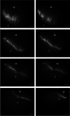
Representative confocal images of the collagen XXII labeled MTJ separating the muscle (M) from the tendon (T). The images are randomly picked from the z‐stack containing the 300 images that are used to make the 3D reconstruction. Scale bars are 10 μm
FIGURE 4
FIGURE 4
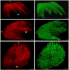
Left column shows z‐stacks of the termination at the myotendinous junction of one whole muscle fiber with the muscle (M) and tendon sides (T) marked. The right column is the software constructed surface based on the z‐stack from left column. Based on these constructed surfaces the interface area was calculated. Scale bars are 10 μm
FIGURE 5
FIGURE 5
Interface area of MTJ type I and type II fibers. In (A) the interface area is plotted as individual median values as well as the median for all type I and II fibers. There is a significant difference between the fiber types (p = 0.023). In (B) the interface area is expressed relative to the muscle fiber diameter of each fiber type per subject, showing the medians for type I and II fibers. The difference between fiber types is significant (p = 0.008). Each subject is represented by an individual symbol. Females are indicated by open symbols and males by closed symbols
FIGURE 6
FIGURE 6
Representative sections of two type II fibers, illustrating that the size of the fiber is not necessarily determining the interface area of the MTJ. The upper row shows 5 images randomly picked from the z‐stack of the whole fiber with a total surface area of 20 692 μm2 and a fiber diameter of 80 μm. The bottom row shows 5 images from a fiber with a diameter of 42 μm but with an MTJ interface area of 76 759 μm2. Scale bars are 10 μm
FIGURE 7
FIGURE 7
Electron microscope image of human MTJ. The width of the base of the foldings (indicated by white arrow) is 220–300 nm. Scale bar 10 μm. Image acquired by Andreas Billa Knudsen, made at the Centre for Integrated Microscopy (CFIM) at the Panum Institute in Copenhagen, Denmark, used by permission

References

    1. Tidball JG. Myotendinous junction: morphological changes and mechanical failure associated with muscle cell atrophy. Exp Mol Pathol. 1984;40:1‐12. - PubMed
    1. Koch M, Schulze J, Hansen U, et al. A novel marker of tissue junctions, collagen XXII. J Biol Chem. 2004;279:22514‐22521. - PMC - PubMed
    1. Huijing PA. Muscle as a collagen fiber reinforced composite: a review of force transmission in muscle and whole limb. J Biomech. 1999;32:329‐345. - PubMed
    1. Tidball JG. Force transmission across muscle cell membranes. J Biomech. 1991;24(Suppl 1):43‐52. - PubMed
    1. Garrett WE. Muscle strain injuries; 1996. - PubMed