. 2023 Jan 31;191(1):47-60.
doi: 10.1093/toxsci/kfac108.

Predicting human cardiac QT alterations and pro-arrhythmic effects of compounds with a 3D beating heart-on-chip platform

Affiliations

Predicting human cardiac QT alterations and pro-arrhythmic effects of compounds with a 3D beating heart-on-chip platform

Roberta Visone et al. Toxicol Sci. .

Abstract

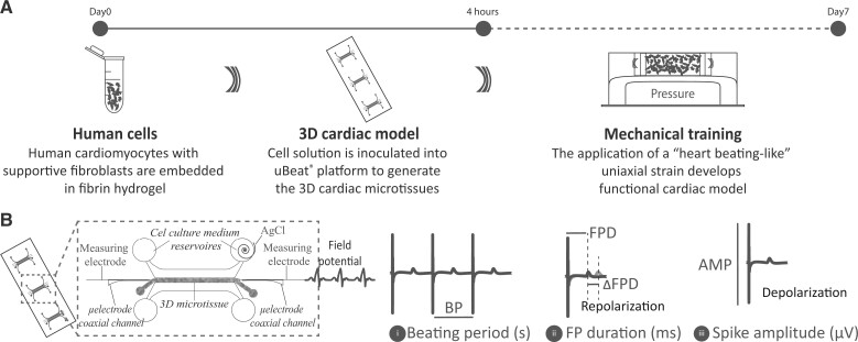
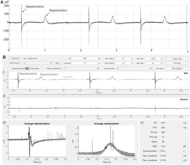
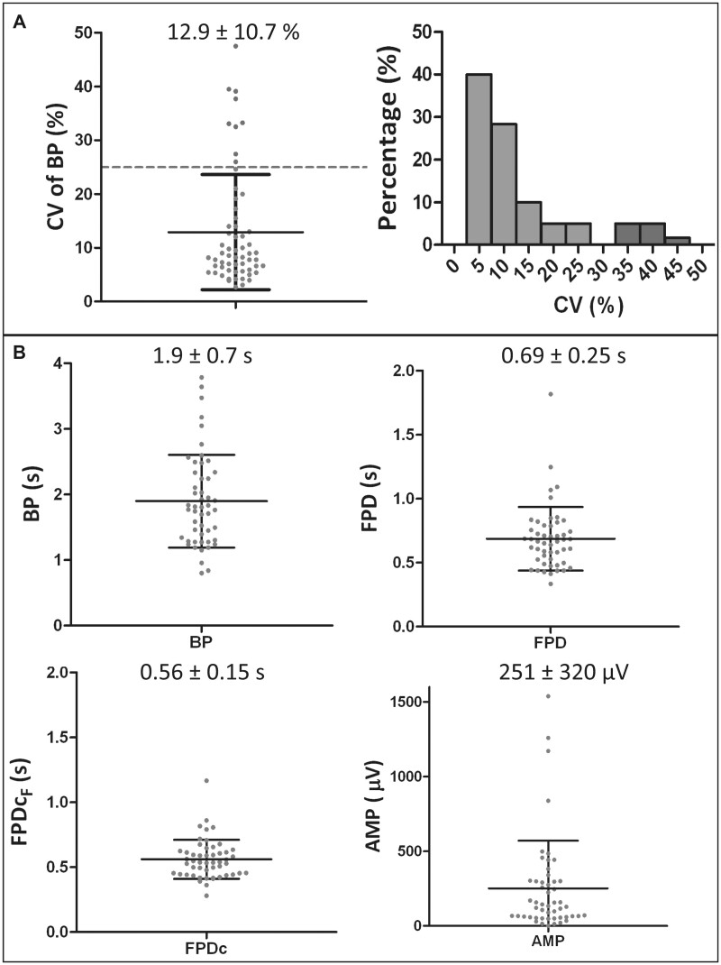
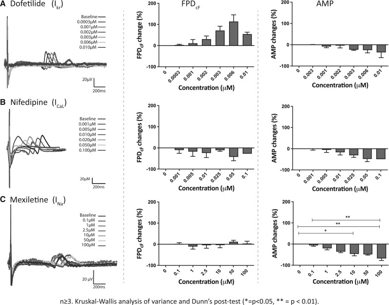
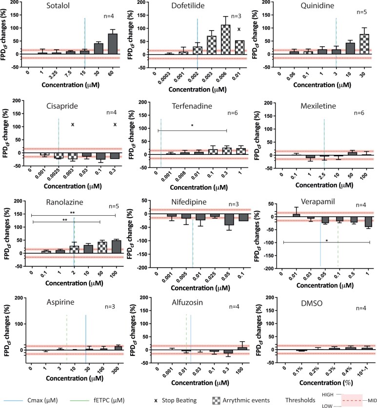
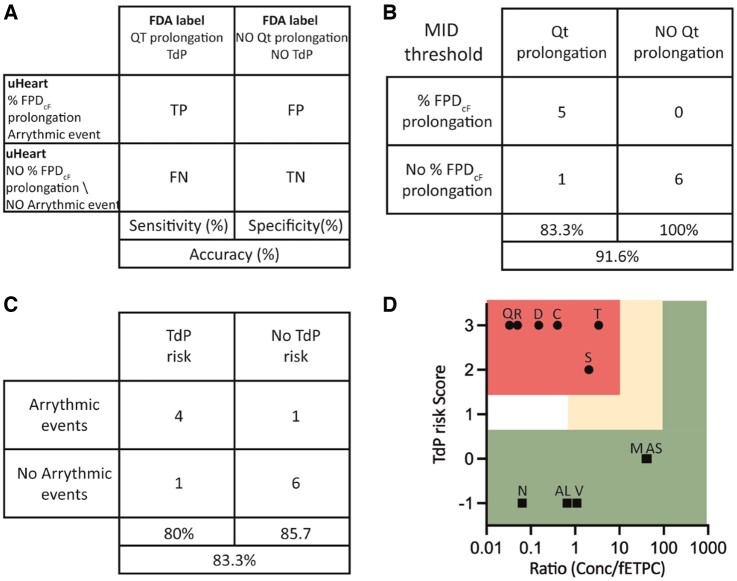
Determining the potential cardiotoxicity and pro-arrhythmic effects of drug candidates remains one of the most relevant issues in the drug development pipeline (DDP). New methods enabling to perform more representative preclinical in vitro studies by exploiting induced pluripotent stem cell-derived cardiomyocytes (iPSC-CM) are under investigation to increase the translational power of the outcomes. Here we present a pharmacological campaign conducted to evaluate the drug-induced QT alterations and arrhythmic events on uHeart, a 3D miniaturized in vitro model of human myocardium encompassing iPSC-CM and dermal fibroblasts embedded in fibrin. uHeart was mechanically trained resulting in synchronously beating cardiac microtissues in 1 week, characterized by a clear field potential (FP) signal that was recorded by means of an integrated electrical system. A drug screening protocol compliant with the new International Council for Harmonisation of Technical Requirements for Pharmaceuticals for Human Use (ICH) guidelines was established and uHeart was employed for testing the effect of 11 compounds acting on single or multiple cardiac ion channels and well-known to elicit QT prolongation or arrhythmic events in clinics. The alterations of uHeart's electrophysiological parameters such as the beating period, the FP duration, the FP amplitude, and the detection of arrhythmic events prior and after drug administration at incremental doses were effectively analyzed through a custom-developed algorithm. Results demonstrated the ability of uHeart to successfully anticipate clinical outcome and to predict the QT prolongation with a sensitivity of 83.3%, a specificity of 100% and an accuracy of 91.6%. Cardiotoxic concentrations of drugs were notably detected in the range of the clinical highest blood drug concentration (Cmax), qualifying uHeart as a fit-to-purpose preclinical tool for cardiotoxicity studies.

Keywords: in vitro cardiac model; QT prolongation; arrhythmias; cardiac toxicity; electrophysiology; organs-on-chip.


Figures

Figure 1.
Figure 1.
Schematic representation of the experimental setup used to (A) develop the uHeart model and (B) to measure and evaluate the electrophysiological parameters of the cardiac microtissues (BP, FPD, FPD changes and AMP).
Figure 2.
Figure 2.
A, Representative FP signal record from uHeart. B, Software developed detect uHeart’s electrophysiological parameters: (1) identification of depolarization spikes, repolarization waves and intervals between the events; (2) residual of the signal after the depolarization and repolarization events are excluded; (3) representation of the averaged depolarizations and repolarizations with reference points indicating the events onset time; (4) means and standard deviations of the computed electrophysiological parameters.
Figure 3.
Figure 3.
A, Frequency distribution and values of the CV characterizing the 60 generated uHeart. B, Beating period (BP), field potential duration (FPD), FPD corrected with the Fridericia methods (FPDcF) and spike amplitude (AMP) characterizing the uHeart models matching inclusion criteria (n = 51). Data are represented as mean ± standard deviation.
Figure 4.
Figure 4.
Representative field potentials (FPs) and electrophysiological parameters (ie, BP and FP duration corrected with Fridericia methods) of uHeart microtissues subjected to drugs acting on single cardiac ion channels: (A) dofetilide, (B) nifedipine, and (C) mexiletine. Data are represented as mean ± standard error.
Figure 5.
Figure 5.
Percentage changes in the FPD corrected with Fridericia formula of uHeart models subjected to drugs acting on single or multiple cardiac ion channels. For each drug, the Cmax (vertical line), the fETPC (vertical dotted line), arrhythmic events (chessboard bars) and the interruption in the spontaneous beating of uHeart (× symbols) are reported in correspondence of the drug concentration. Kruskal-Wallis analysis of variance and Dunn’s post-test (*=p < .05, **=p < .01.). Data are reported as mean ± standard error.
Figure 6.
Figure 6.
A, Schematic representation of the concordance analyses and sensitivity, specificity, and accuracy parameters performed by comparing the results obtained in uHeart with the FDA drug labels. B, uHeart models is considered to show a field potential prolongation when the percentage changes in the FPDcF is higher than the MID threshold. C, uHeart sensitivity, specificity and accuracy for detecting arrhythmic events, correlating with the risk of TdP onset. D, TdP Risk categorization depicted through two-dimensional map indicating high risk drugs (●) in the top left area, intermediate risk drugs (▲) in the empty middle area and low risk drugs (■) in the bottom and right area. The area not enclosed in rectangle indicates insufficient margins to categorize the compound (risk score 1, margin ≤ 1). Letters on the symbol indicate the first letter of each tested dug (for Alfuzosine-AL and Aspirine-AS).

References

    1. Ando H., Yoshinaga T., Yamamoto W., Asakura K., Uda T., Taniguchi T., Ojima A., Shinkyo R., Kikuchi K., Osada T., et al. (2017). A new paradigm for drug-induced torsadogenic risk assessment using human iPS cell-derived cardiomyocytes. J. Pharmacol. Toxicol. Methods 84, 111–127. - PubMed
    1. Blinova K., Dang Q., Millard D., Smith G., Pierson J., Guo L., Brock M., Lu H. R., Kraushaar U., Zeng H., et al. (2018). International multisite study of human-induced pluripotent stem cell-derived cardiomyocytes for drug proarrhythmic potential assessment. Cell Rep. 24, 3582–3592. - PMC - PubMed
    1. Blinova K., Stohlman J., Vicente J., Chan D., Johannesen L., Hortigon-Vinagre M. P., Zamora V., Smith G., Crumb W. J., Pang L., et al. (2017). Comprehensive translational assessment of human- induced pluripotent stem cell derived cardiomyocytes for evaluating drug-induced arrhythmias. Toxicol. Sci. 155, 234–247. - PMC - PubMed
    1. Carlos-Oliveira M., Lozano-Juan F., Occhetta P., Visone R., and Rasponi M. (2021). Current strategies of mechanical stimulation for maturation of cardiac microtissues. Biophys. Rev.13, 717–727. - PMC - PubMed
    1. Clements M., Thomas N. (2014). High-throughput multi-parameter profiling of electrophysiological drug effects in human embryonic stem cell derived cardiomyocytes using multi-electrode arrays. Toxicol. Sci. 140, 445–461. - PubMed

Publication types