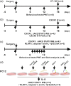
CXCR1 participates in bone cancer pain induced by Walker 256 breast cancer cells in female rats
- PMID: 36227008
- PMCID: PMC9638694
- DOI: 10.1177/17448069221135743
CXCR1 participates in bone cancer pain induced by Walker 256 breast cancer cells in female rats
Abstract
Bone cancer pain (BCP) is a clinically intractable mixed pain, involving inflammation and neuropathic pain, and its mechanisms remain unclear. CXC chemokine receptor 1 (CXCR1, IL-8RA) and 2 (CXCR2, IL-8RB) are high-affinity receptors for interleukin 8 (IL8). According to previous studies, CXCR2 plays a crucial role in BCP between astrocytes and neurons, while the role of CXCR1 remains unclear. The objective of this study was to investigate the role of CXCR1 in BCP. We found that CXCR1 expression increased in the spinal dorsal horn. Intrathecal injection of CXCR1 siRNA effectively attenuated mechanical allodynia and pain-related behaviors in rats. It was found that CXCR1 was predominantly co-localized with neurons. Intrathecal injection of CXCR1-siRNA reduced phosphorylated JAK2/STAT3 protein levels and the NLRP3 inflammasome (NLRP3, caspase1, and IL-1β) levels. Furthermore, in vitro cytological experiments confirmed this conclusion. The study results suggest that the spinal chemokine receptor CXCR1 activation mediates BCP through JAK2/STAT3 signaling pathway and NLRP3 inflammasome (NLRP3, caspase1, and IL-1β).
Keywords: CXCR1; JAK2/STAT3 signaling pathway; NLRP3 inflammasome; bone cancer pain; spinal cord.
Conflict of interest statement
The author(s) declared no potential conflicts of interest with respect to the research, authorship, and/or publication of this article.
Figures


Similar articles
-
P2X7 Receptor-Induced Bone Cancer Pain by Regulating Microglial Activity via NLRP3/IL-1beta Signaling.Pain Physician. 2022 Nov;25(8):E1199-E1210. Pain Physician. 2022. PMID: 36375190
-
Curcumin analogue NL04 inhibits spinal cord central sensitization in rats with bone cancer pain by inhibiting NLRP3 inflammasome activation and reducing IL-1β production.Eur J Pharmacol. 2024 May 5;970:176480. doi: 10.1016/j.ejphar.2024.176480. Epub 2024 Mar 13. Eur J Pharmacol. 2024. PMID: 38490468
-
Anwulignan Alleviates Bone Cancer Pain by Modulating the PPARα/CXCR2 Signaling Pathway in the Rat Spinal Cord.CNS Neurosci Ther. 2025 Mar;31(3):e70302. doi: 10.1111/cns.70302. CNS Neurosci Ther. 2025. PMID: 40079428 Free PMC article.
-
AMPK activation attenuates cancer-induced bone pain by reducing mitochondrial dysfunction-mediated neuroinflammation.Acta Biochim Biophys Sin (Shanghai). 2023 Mar 25;55(3):460-471. doi: 10.3724/abbs.2023039. Acta Biochim Biophys Sin (Shanghai). 2023. PMID: 36971458 Free PMC article.
-
An updated review on the role of the CXCL8-CXCR1/2 axis in the progression and metastasis of breast cancer.Mol Biol Rep. 2021 Sep;48(9):6551-6561. doi: 10.1007/s11033-021-06648-8. Epub 2021 Aug 24. Mol Biol Rep. 2021. PMID: 34426905 Review.
Cited by
-
N4-acetylcytidine acetylation of neurexin 2 in the spinal dorsal horn regulates hypersensitivity in a rat model of cancer-induced bone pain.Mol Ther Nucleic Acids. 2024 Apr 23;35(2):102200. doi: 10.1016/j.omtn.2024.102200. eCollection 2024 Jun 11. Mol Ther Nucleic Acids. 2024. PMID: 38831898 Free PMC article.
-
Fat mass and obesity-related protein contributes to the development and maintenance of bone cancer pain in rats by abrogating m6A methylation of RNA.Mol Pain. 2024 Jan-Dec;20:17448069241295987. doi: 10.1177/17448069241295987. Mol Pain. 2024. PMID: 39415414 Free PMC article.
-
Targeting Members of the Chemokine Family as a Novel Approach to Treating Neuropathic Pain.Molecules. 2023 Jul 30;28(15):5766. doi: 10.3390/molecules28155766. Molecules. 2023. PMID: 37570736 Free PMC article. Review.
-
Targeting triple negative breast cancer stem cells using nanocarriers.Discov Nano. 2024 Mar 7;19(1):41. doi: 10.1186/s11671-024-03985-y. Discov Nano. 2024. PMID: 38453756 Free PMC article. Review.
-
Inhibition of Interleukin-8/C-X-C Chemokine Receptor 2 Signaling Axis Prevents Tumor Growth and Metastasis in Triple-Negative Breast Cancer Cells.Pharmacology. 2025;110(3):178-190. doi: 10.1159/000545659. Epub 2025 Apr 4. Pharmacology. 2025. PMID: 40188812 Free PMC article.
References
Publication types
MeSH terms
Substances
LinkOut - more resources
Full Text Sources
Medical
Molecular Biology Databases
Miscellaneous

