. 2023 Jan 2;35(1):469-487.
doi: 10.1093/plcell/koac301.

Polarly localized WPR proteins interact with PAN receptors and the actin cytoskeleton during maize stomatal development

Affiliations

Polarly localized WPR proteins interact with PAN receptors and the actin cytoskeleton during maize stomatal development

Qiong Nan et al. Plant Cell. .

Abstract

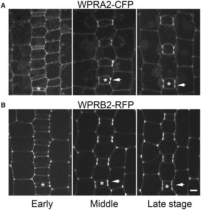
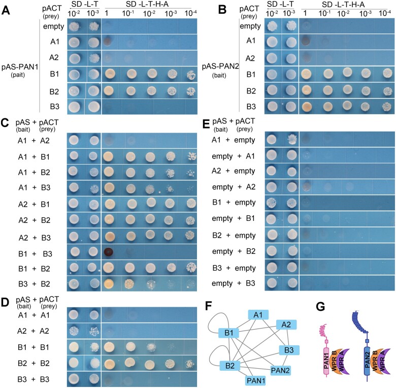
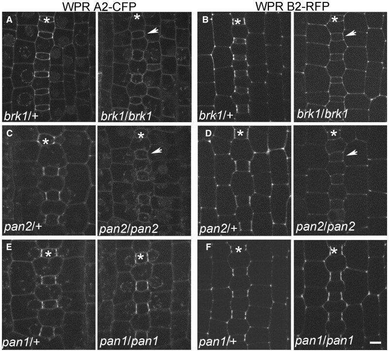
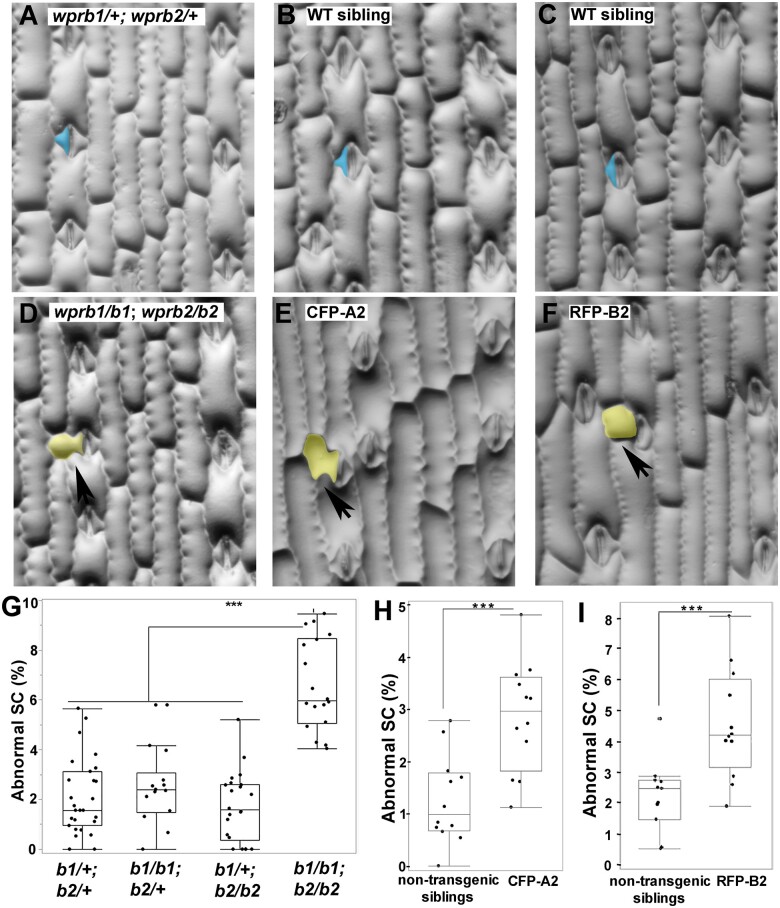
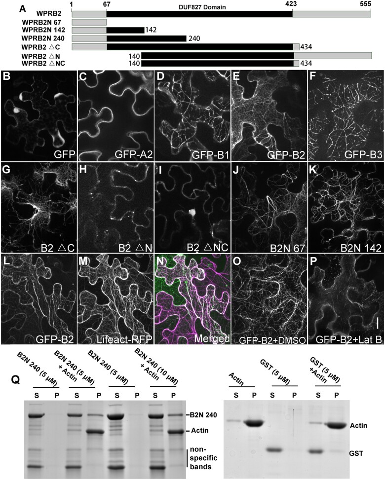
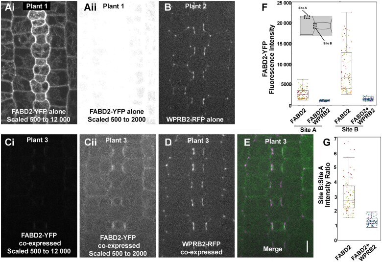
Polarization of cells prior to asymmetric cell division is crucial for correct cell divisions, cell fate, and tissue patterning. In maize (Zea mays) stomatal development, the polarization of subsidiary mother cells (SMCs) prior to asymmetric division is controlled by the BRICK (BRK)-PANGLOSS (PAN)-RHO FAMILY GTPASE (ROP) pathway. Two catalytically inactive receptor-like kinases, PAN2 and PAN1, are required for correct division plane positioning. Proteins in the BRK-PAN-ROP pathway are polarized in SMCs, with the polarization of each protein dependent on the previous one. As most of the known proteins in this pathway do not physically interact, possible interactors that might participate in the pathway are yet to be described. We identified WEAK CHLOROPLAST MOVEMENT UNDER BLUE LIGHT 1 (WEB1)/PLASTID MOVEMENT IMPAIRED 2 (PMI2)-RELATED (WPR) proteins as players during SMC polarization in maize. WPRs physically interact with PAN receptors and polarly accumulate in SMCs. The polarized localization of WPR proteins depends on PAN2 but not PAN1. CRISPR-Cas9-induced mutations result in division plane defects in SMCs, and ectopic expression of WPR-RFP results in stomatal defects and alterations to the actin cytoskeleton. We show that certain WPR proteins directly interact with F-actin through their N-terminus. Our data implicate WPR proteins as potentially regulating actin filaments, providing insight into their molecular function. These results demonstrate that WPR proteins are important for cell polarization.


Figures

Figure 1
Figure 1
CFP-WPRA2 and RFP-WPRB2 polarize in developing subsidiary mother cells. The stomatal division zone of the leaf epidermis of transgenic maize plants expressing fluorescent fusion proteins was dissected and analyzed. Three different developmental stages were selected according to GMC width and SMC division status to observe the localization of CFP-WPRA2 (A) and RFP-WPRB2 (B). Arrows point to sites of CFP-WPRA2 or RFP-WPRB2 accumulation in SMCs where they contact the adjacent GMC. Asterisks mark a GMC within each stomatal row. Scale bar = 5 µm, all images scaled identically.
Figure 2
Figure 2
Y2H analysis of WPR and PAN protein interactions. Interactions were assessed using the GAL4-based Y2H system. Yeast was grown at different dilutions on nonselective (–L–T) and selective (–L–T–A–H) media. Soluble intracellular regions of PAN1 and PAN2 were used. A, B, interactions of PAN1 and PAN2 with WPRA1, WPRA2, WPRB1, WPRB2, and WPRB3. C, Heterodimer formation was assessed between WPRA1, WPRA2, WPRB1, WPRB2, and WPRB3. D, Homodimer formation was assessed between WPRA1, WPRA2, WPRB1, WPRB2, and WPRB3. E, Negative controls using empty bait plasmid pASGW-attR or prey plasmid pACTGW-attR. F, Network diagram showing the observed interactions between WPRs and PAN1/PAN2. G, A model of protein complex formation between PAN and WPR proteins.
Figure 3
Figure 3
WPR proteins polarize in SMCs after BRK1 and before actin. The stomatal division zone of leaf 4 from plants co-expressing (A) CFP-WPRA2 and RFP-WPRB2; (B) BRK1–CFP and RFP-WPRB2; (C) CFP-WPRA2 and PAN1-YFP; (D) CFP-WPRA2 and PAN2-YFP; or (E) CFP-WPRA2 and FABD2-YFP were analyzed for enrichment at the GMC–SMC interface. Fluorescent proteins were scored as polarized when fluorescence was brighter at the SMC–GMC interface than at the cell periphery distal to the GMC. Within each panel, the same GMCs imaged in the different channels (and the subsequent merged panel) are numbered. Arrows point to SMCs where one fluorescent protein is polarized but not the other. Counts of individual SMCs that had only one fluorescent protein, or both, are listed at the right of the images. All images are at the same scale and are Z-projections of seven confocal slices. Scale bar in the merged panel of (E) is 10 µm, all images scaled identically.
Figure 4
Figure 4
Polarization of WPR proteins depends on BRK1 and PAN2 but not PAN1. Localization of CFP-WPRA2 and RFP-WPRB2 in brk1, pan2, and pan1 mutants and nonmutant siblings was analyzed in developing leaf 4. CFP-WPRA2 (A) and RFP-WPRB2 (B) in developing stomata of a brk1 mutant and nonmutant (heterozygous) sibling. CFP-WPRA2 (C) and RFP-WPRB2 (D) in developing stomata of a pan2 mutant and nonmutant (heterozygous) sibling. CFP-WPRA2 (E) and RFP-WPRB2 (F) in developing stomata of a pan1 mutant and nonmutant (heterozygous) sibling. Asterisks mark GMC rows. Arrows point to SMCs without polarized localization of CFP-WPRA2 or RFP-WPRB2. Three to six plants of each genotype were used for data collection. Scale bar, 5 µm, all microscopy images scaled identically.
Figure 5
Figure 5
CRISPR–Cas9-induced wprb1;wprb2 double mutants and CFP-WPRA2- and RFP-WPRB2-expressing lines have subsidiary cell defects. A–F, Representative image of the third leaf epidermis of wprb1/+;wprb2/+ and wprb1/wprb1;wprb2/wprb2 mutants (A and D), CFP-WPRA2 transgenic plants and nontransgenic siblings (B and E), and RFP-WPRB2 transgenic plants and nontransgenic siblings (C and F). Examples of normal subsidiary cells in (A), (B), and (D) are false-colored blue, and abnormal subsidiary cells in (D), (E), and (F) are false-colored yellow and marked with black arrows. G, Quantification of abnormal subsidiary cells in wprb1/+; wprb2/+ (n = 27 plants), wprb1/b1;wprb2/+ (n = 14 plants), wprb1/+;wprb2/wprb2 (n = 22 plants), and wprb1/wprb1;wprb2/wprb2 (n = 18 plants). For each plant, 100–200 subsidiary cells were examined. P-value from Student’s t test comparing each genotype, ***P 0.001. H, I, Quantification of abnormal subsidiary cells in CFP-WPRA2- or RFP-WPRB2-expressing plants and nontransgenic siblings grown in parallel (n = 11–12 plants and 120–200 cells per genotype). Box plots show median values (center line), 25th to 75th interquartile range (box) and 1.5*interquartile range (whiskers). Student’s t tests were performed, ***P 0.001.
Figure 6
Figure 6
WPRB interacts with F-actin. A, Schematic depicting WPRB2 and truncated versions. B–F, Confocal images of transiently expressed full-length GFP fusion proteins in tobacco. Transient expression of GFP only (B), GFP-WPRA2 (C), GFP-WPRB1 (D), GFP-WPRB2 (E), or GFP-WPRB3 (F). G–K, Transient expression of truncated WPRB2 proteins fused to GFP in tobacco leaves. The co-expression of GFP-WPRB2 (L) with Lifeact-RFP labeled actin microfilaments (M); merged image (N) of the same cell. Tobacco leaves expressing GFP-WPRB2 were treated with DMSO (negative control, O) or 40-μM Latrunculin B (P) for 2 h. Scale bar in (P) = 10 µm, all images scaled identically. Q, High-speed co-sedimentation of GST-tagged WPRB2N240 with F-actin. After centrifugation at 100,000g, the proteins in the supernatant (S) and pellet (P) were resolved by SDS–PAGE and visualized with Coomassie Blue staining. GST protein was used as a negative control.
Figure 7
Figure 7
Fluorescence intensity of ABD2-YFP decreases in RFP-WPRB2-expressing plants. Plants expressing RFP-WPRB2 and FABD2-YFP were crossed, and the progeny independently segregated the two markers. A(i) and A(ii) Plant only expressing FABD2-YFP. An identical image is shown in A(i) and A(ii). In Ai, the 16-bit image was scaled from 600 to 12,000 prior to converting to 8 bit. A(ii) was scaled 500 to 2,000. B, Plant only expressing RFP-WPRB2. C–E, Plant co-expressing FABD2-YFP (C, green in E) with RFP-WPRB2 (D, magenta in E). C(i) and C(ii) show an identical image of the ABD2-YFP channel, where C(i) is scaled the same as A(i) and C(ii) is scaled the same as A(ii), for comparative purposes. Image intensity in (B) and (D) are scaled identically. Scale bar in E = 10 microns, all images are scaled identically. F, Quantification of fluorescence intensity of SMC lateral cell side (site A) and SMC–GMC interface (site B) in FABD2-YFP-only plants (n = 5 plants, 292 cells) and RFP-WPRB2- and FABD2-YFP-co-expressing plants (n = 3 plants, 164 cells). One-sided t tests indicate lower values in co-expressing plants (P <0.0001). G, Ratio of intensities measured in (F). A two-sided t test indicates a significantly lower ratio in the co-expressing cells (P <0.0001).

Comment in

Similar articles

Cited by

References

    1. Apostolakos P, Livanos P, Giannoutsou E, Panteris E, Galatis B (2018) The intracellular and intercellular cross-talk during subsidiary cell formation in Zea mays: existing and novel components orchestrating cell polarization and asymmetric division. Ann Bot 122: 679–696 - PMC - PubMed
    1. Ashraf MA, Facette M (2020) Plant biology: BASL gives the plant nucleus a sense of direction. Curr Biol 30: R1375–R1377 - PubMed
    1. Campos R, Goff J, Rodriguez-Furlan C, Van Norman JM (2020) The Arabidopsis receptor kinase IRK is polarized and represses specific cell divisions in roots. Dev Cell 52: 183–195 - PubMed
    1. Cartwright HN, Humphries JA, Smith LG (2009) PAN1: a receptor-like protein that promotes polarization of an asymmetric cell division in maize. Science 323: 649–651 - PubMed
    1. Char SN, Neelakandan AK, Nahampun H, Frame B, Main M, Spalding MH, Becraft PW, Meyers BC, Walbot V, Wang K, et al. (2017) An Agrobacterium-delivered CRISPR/Cas9 system for high-frequency targeted mutagenesis in maize. Plant Biotechnol J 15: 257–268 - PMC - PubMed

Publication types

MeSH terms