

Brain structure in autoimmune Addison's disease
- PMID: 36227196
- PMCID: PMC10110435
- DOI: 10.1093/cercor/bhac389
Brain structure in autoimmune Addison's disease
Abstract
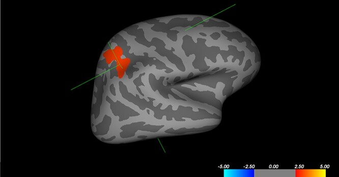
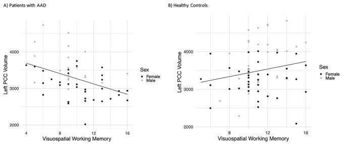
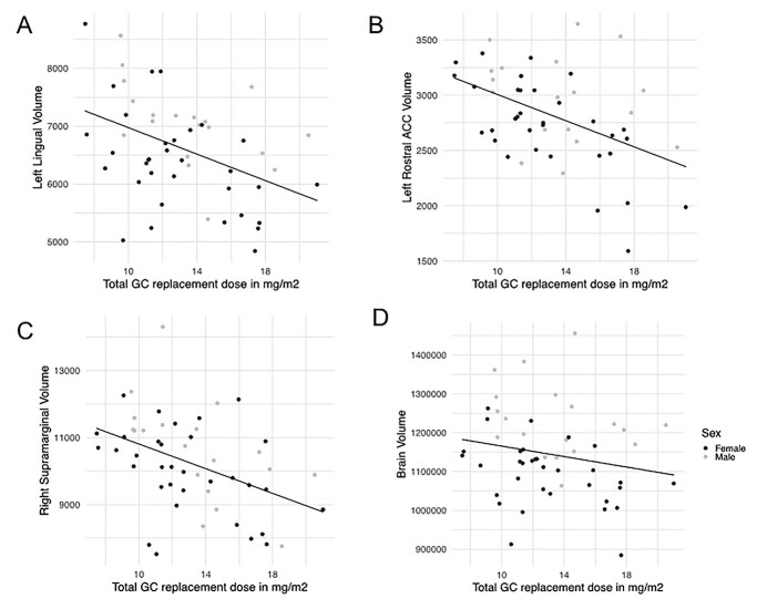
Long-term disturbances in cortisol levels might affect brain structure in individuals with autoimmune Addison's disease (AAD). This study investigated gray and white matter brain structure in a cohort of young adults with AAD. T1- and diffusion-weighted images were acquired for 52 individuals with AAD and 70 healthy controls, aged 19-43 years, using magnetic resonance imaging. Groups were compared on cortical thickness, surface area, cortical gray matter volume, subcortical volume (FreeSurfer), and white matter microstructure (FSL tract-based spatial statistics). Individuals with AAD had 4.3% smaller total brain volume. Correcting for head size, we did not find any regional structural differences, apart from reduced volume of the right superior parietal cortex in males with AAD. Within the patient group, a higher glucocorticoid (GC) replacement dose was associated with smaller total brain volume and smaller volume of the left lingual gyrus, left rostral anterior cingulate cortex, and right supramarginal gyrus. With the exception of smaller total brain volume and potential sensitivity of the parietal cortex to GC disturbances in men, brain structure seems relatively unaffected in young adults with AAD. However, the association between GC replacement dose and reduced brain volume may be reason for concern and requires follow-up study.
Keywords: Addison; brain structure; cortisol; executive function; working memory.
© The Author(s) 2022. Published by Oxford University Press.
Figures
References
-
- Allolio B, Arlt W, Hahner S. DHEA: why, when, and how much--DHEA replacement in adrenal insufficiency. Ann Endocrinol. 2007:68(4):268–273. - PubMed
-
- Andela CD, Haalen FM, Ragnarsson O, Papakokkinou E, Johannsson G, Santos A, Webb SM, Biermasz NR, Wee NJ, Pereira AM. MECHANISMS IN ENDOCRINOLOGY: Cushing’s syndrome causes irreversible effects on the human brain: a systematic review of structural and functional magnetic resonance imaging studies. Eur J Endocrinol. 2015:173(1):R1–R14. - PubMed
-
- Andersson JLR, Graham MS, Zsoldos E, Sotiropoulos SN. Incorporating outlier detection and replacement into a non-parametric framework for movement and distortion correction of diffusion MR images. NeuroImage. 2016:141:556–572. - PubMed
-
- Barkley RA. Barkley deficits in executive functioning scale (BDEFS for adults). New York: Guilford Press; 2011
Publication types
MeSH terms
LinkOut - more resources
Full Text Sources
Medical
Miscellaneous
