. 2023:2563:135-148.
doi: 10.1007/978-1-0716-2663-4_6.

Fluorescence Lifetime Imaging Microscopy of Biomolecular Condensates

Affiliations

Fluorescence Lifetime Imaging Microscopy of Biomolecular Condensates

My Diem Quan et al. Methods Mol Biol. 2023.

Abstract

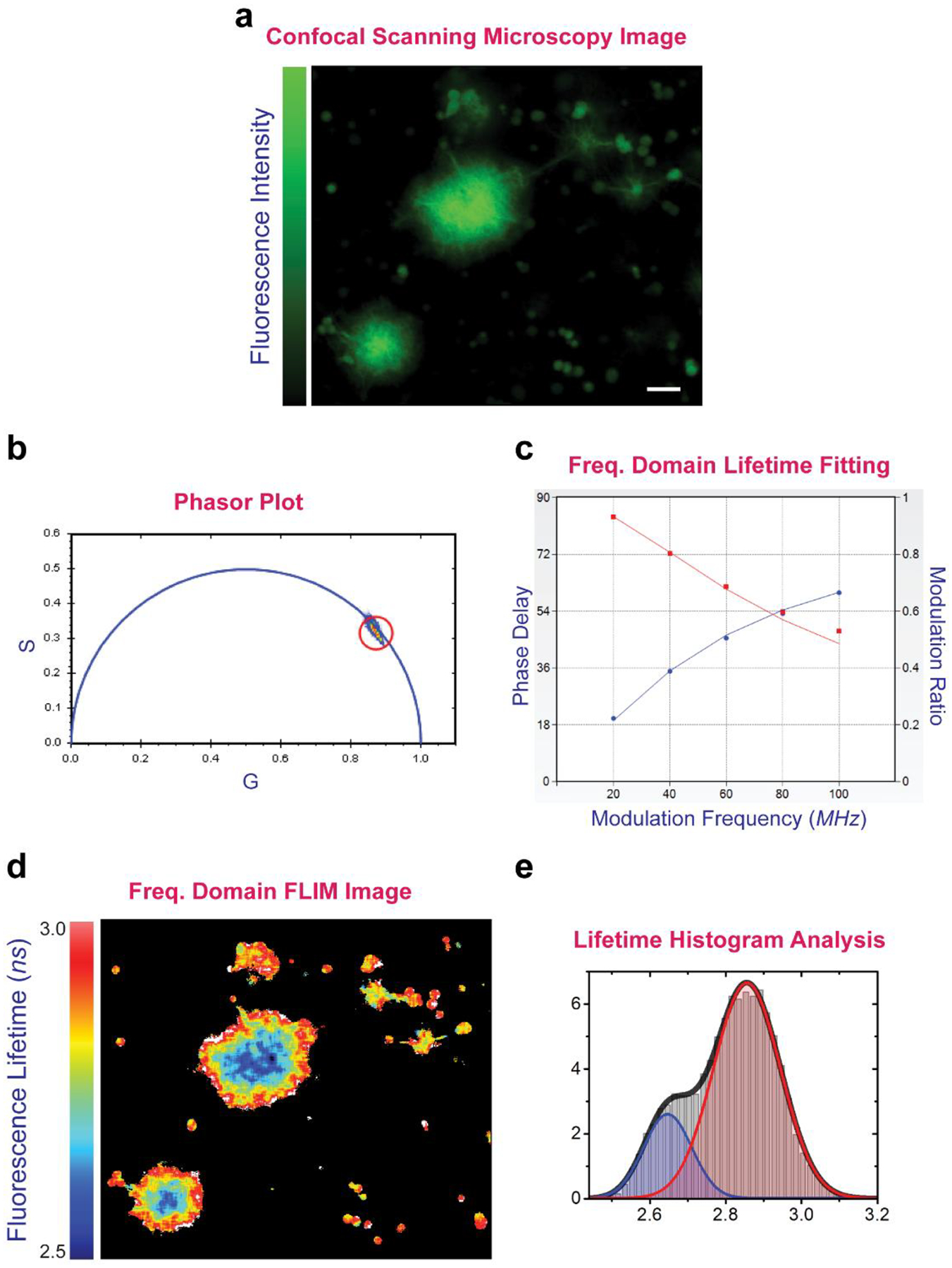
Biomolecular condensates of ribonucleoproteins (RNPs) such as the transactivation response element (TAR) DNA-binding protein 43 (TDP-43) arise from liquid-liquid phase separation (LLPS) and play vital roles in various biological processes including the formation-dissolution of stress granules (SGs). These condensates are thought to be directly linked to neurodegenerative diseases, providing a depot of aggregation-prone proteins and serving as a cauldron of protein aggregation and fibrillation. Despite recent research efforts, biochemical processes and rearrangements within biomolecular condensates that trigger subsequent protein misfolding and aggregation remain to be elucidated. Fluorescence lifetime imaging microscopy (FLIM) provides a minimally intrusive high-sensitivity and high-resolution imaging method to monitor in-droplet spatiotemporal changes that initiate and lead to protein aggregation. In this chapter, we describe a FLIM application for characterizing chemical chaperone-assisted decoupling of TDP-43 liquid-liquid phase separation and aggregation/fibrillation, highlighting potential therapeutic strategies to combat pathological RNP-associated aggregates without compromising cellular stress responses.

Keywords: Droplet maturation; Fluorescence lifetime imaging microscopy; Intrinsically disordered proteins; Liquid-liquid phase separation; Neurodegenerative diseases; Protein aggregation; TDP-43.


Figures

Fig. 1:
Fig. 1:
The chemical chaperone TMAO decouples phase separation and fibrillation of the low-complexity domain of the ALS-associated protein TDP-43. (a) Confocal fluorescence microscopy image of TDP-43LCD in a solution condition that allows both protein droplet formation and droplet “maturation” to fibrillar aggregated state/s (condition: 0.5 M TMAO, 200 mM NaCl, 10 mM sodium acetate, 10 mM NaH2PO4, 10 mM glycine, pH 7.5), presented using an intensity map that highlights morphological contrast. (b) Increasing TMAO concentration enhances LLPS and disfavors fibrillation of TDP-43LCD. (c) Differential destabilization of the disordered denatured states (D) of TDP-43LCD in the protein-depleted/low-density phase (L1) and protein-enriched/high-density phase (L2) resulting from TMAO-induced LLPS. Increasing TMAO concentration favors compaction of D ensembles and LLPS; increasing protein concentration enhances protein fibrillation and LLPS (see Note 1). Panels b and c were adapted from Choi et al. (2018) Biochemistry 57 (50): 6822–6826 [6].
Fig. 2:
Fig. 2:
Schematic of a fluorescence lifetime confocal laser microscopy system applicable for single or dual color detection.
Fig. 3:
Fig. 3:
Sample data analyses performed on fluorescence lifetime measurements of TDP-43LCD condensates. (a) Confocal scanning microscopy image shows phase separated TDP-43LCD droplets and aggregates in 0.5M TMAO. (b) Pixels of the scanning microscopy image lie as a cluster on the Universal Circle, indicating a single average lifetime [20] (see Note 9). (c) FD lifetime fitting extract information about the phase delay and modulation ratio at digital modulation frequencies of 20 to 100 MHz. (d) FD FLIM image maps non-uniform lifetime distributions within populations of “young” and “aged” TDP-43LCD droplets. (e) Fluorescence lifetimes are estimated by fitting to Gaussian functions with the distribution showing two distinct species characterized by different lifetimes.

References

    1. Boeynaems S, Alberti S, Fawzi NL, Mittag T, Polymenidou M, Rousseau F, Schymkowitz J, Shorter J, Wolozin B, Van Den Bosch L, Tompa P, Fuxreiter M (2018) Protein Phase Separation: A New Phase in Cell Biology. Trends Cell Biol 28 (6):420–435. doi:10.1016/j.tcb.2018.02.004 - DOI - PMC - PubMed
    1. Hyman AA, Weber CA, Julicher F (2014) Liquid-liquid phase separation in biology. Annu Rev Cell Dev Biol 30:39–58. doi:10.1146/annurev-cellbio-100913-013325 - DOI - PubMed
    1. Li YR, King OD, Shorter J, Gitler AD (2013) Stress granules as crucibles of ALS pathogenesis. J Cell Biol 201 (3):361–372. doi:10.1083/jcb.201302044 - DOI - PMC - PubMed
    1. Sharma R, Choi KJ, Quan MD, Sharma S, Sankaran B, Park H, LaGrone A, Kim JJ, MacKenzie KR, Ferreon ACM, Kim C, Ferreon JC (2021) Liquid condensation of reprogramming factor KLF4 with DNA provides a mechanism for chromatin organization. Nat Commun 12 (1):5579. doi:10.1038/s41467-021-25761-7 - DOI - PMC - PubMed
    1. Purice MD, Taylor JP (2018) Linking hnRNP Function to ALS and FTD Pathology. Front Neurosci 12:326. doi:10.3389/fnins.2018.00326 - DOI - PMC - PubMed

Publication types

MeSH terms

LinkOut - more resources