

Numerical and experimental investigation of a lighthouse tip drainage cannula used in extracorporeal membrane oxygenation
- PMID: 36227654
- PMCID: PMC10092507
- DOI: 10.1111/aor.14421
Numerical and experimental investigation of a lighthouse tip drainage cannula used in extracorporeal membrane oxygenation
Abstract
Background: Extracorporeal membrane oxygenation is a life-saving therapy used in case of acute respiratory/circulatory failure. Exposure of blood to non-physiological surfaces and high shear stresses is related to hemolytic damage and platelet activation. A detailed knowledge of the fluid dynamics of the components under different scenarios is thus paramount to assess the thrombogenicity of the circuit.
Methods: An investigation of the flow structures developing in a conventional lighthouse tip (single-staged) drainage cannula was performed with cross-validated computational fluid dynamics and particle image velocimetry. The aim was to quantify the variation in drainage performance and stress levels induced by different fluid models, hematocrit and vessel-to-cannula flow rate ratios.
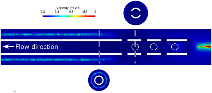
Results: The results showed that the 90° bends of the flow through the side holes created a recirculation zone inside the cannula which increased residence time. Flow structures resembling a jet in a crossflow were also observed. The use of different hematocrits did not significantly affect drainage performances. The most proximal set of holes drained the largest fraction of fluid. However, different flow rate ratios altered the flow rate drained through the tip. The use of 2D data led to a 50% underestimation of shear rate levels. In the drainage zone the non-Newtonian behavior of blood was less relevant.
Conclusions: The most proximal holes drained the largest amount of fluid. The flow features and distribution of flow rates among the holes showed little dependence on the hematocrit. The non-Newtonian behavior of blood had a small influence on the dynamics of the flow.
Keywords: CFD; ECMO; PIV; drainage; flow structures; jet in crossflow; non-Newtonian.
© 2022 The Authors. Artificial Organs published by International Center for Artificial Organ and Transplantation (ICAOT) and Wiley Periodicals LLC.
Conflict of interest statement
The authors declare that they have no conflicts of interest with the contents of this article.
Figures


Similar articles
-
Cannulation configuration and recirculation in venovenous extracorporeal membrane oxygenation.Sci Rep. 2022 Sep 30;12(1):16379. doi: 10.1038/s41598-022-20690-x. Sci Rep. 2022. PMID: 36180496 Free PMC article.
-
Effect of flow rate ratio and positioning on a lighthouse tip ECMO return cannula.Biomech Model Mechanobiol. 2023 Dec;22(6):1891-1899. doi: 10.1007/s10237-023-01741-2. Epub 2023 Jul 16. Biomech Model Mechanobiol. 2023. PMID: 37454305 Free PMC article.
-
Performance Comparison of Centered and Tilted Blunt and Lighthouse Tip Cannulae for Drainage in Extracorporeal Life Support.Cardiovasc Eng Technol. 2025 Apr;16(2):238-250. doi: 10.1007/s13239-024-00770-x. Epub 2025 Feb 10. Cardiovasc Eng Technol. 2025. PMID: 39930265 Free PMC article.
-
Recirculation in venovenous extracorporeal membrane oxygenation.ASAIO J. 2015 Mar-Apr;61(2):115-21. doi: 10.1097/MAT.0000000000000179. ASAIO J. 2015. PMID: 25423117 Review.
-
Recirculation in venovenous extracorporeal membrane oxygenation.J Crit Care. 2016 Dec;36:107-110. doi: 10.1016/j.jcrc.2016.05.027. Epub 2016 Jun 6. J Crit Care. 2016. PMID: 27546757 Review.
Cited by
-
Cannulation configuration and recirculation in venovenous extracorporeal membrane oxygenation.Sci Rep. 2022 Sep 30;12(1):16379. doi: 10.1038/s41598-022-20690-x. Sci Rep. 2022. PMID: 36180496 Free PMC article.
-
Clinical assessment of cannula performance during adult minimally invasive valve surgery using predictive mathematical models.Interdiscip Cardiovasc Thorac Surg. 2025 Jun 4;40(6):ivaf127. doi: 10.1093/icvts/ivaf127. Interdiscip Cardiovasc Thorac Surg. 2025. PMID: 40442945 Free PMC article.
-
Hemodynamic and recirculation performance of dual lumen cannulas for venovenous extracorporeal membrane oxygenation.Sci Rep. 2023 May 8;13(1):7472. doi: 10.1038/s41598-023-34655-1. Sci Rep. 2023. PMID: 37156961 Free PMC article.
-
Investigating optimal drainage cannula for venovenous extracorporeal membrane oxygenation: impact of side holes on blood oxygenation - an in vitro study.J Artif Organs. 2025 Aug 20. doi: 10.1007/s10047-025-01525-8. Online ahead of print. J Artif Organs. 2025. PMID: 40836181
-
Effect of flow rate ratio and positioning on a lighthouse tip ECMO return cannula.Biomech Model Mechanobiol. 2023 Dec;22(6):1891-1899. doi: 10.1007/s10237-023-01741-2. Epub 2023 Jul 16. Biomech Model Mechanobiol. 2023. PMID: 37454305 Free PMC article.
References
MeSH terms
Grants and funding
LinkOut - more resources
Full Text Sources
Miscellaneous
