. 2022 Oct 13;6(10):e22.00097.
doi: 10.5435/JAAOSGlobal-D-22-00097. eCollection 2022 Oct 1.

Premalignant Conditions of Bone

Affiliations

Premalignant Conditions of Bone

Michael D Eckhoff et al. J Am Acad Orthop Surg Glob Res Rev. .

Abstract

Development of malignancy is a multifactorial process, and there are multitude of conditions of bone that may predispose patients to malignancy. Etiologies of malignancy include benign osseous conditions, genetic predisposition, and extrinsic conditions. New-onset pain or growth in a previously stable lesion is that should concern for malignant change and should prompt a diagnostic workup for malignancy.


Figures

Figure 1
Figure 1
Diagrams showing multiple hereditary exostoses: (A and B) demonstrate AP and lateral radiographs with characteristic exostoses with secondary deformity of the knee joint. C, Axial T2 MRI cut with signal intense cartilaginous cap. D, Complete resection of this lesion shows a cartilaginous cap with underlying trabecular bone and marrow contents.
Figure 2
Figure 2
Diagrams showing enchondroma. A, AP and (B) lateral radiographs of a small finger middle phalanx base enchondroma. Often these lesions are purely lytic and expansile when present in the small bones of the hand. C, Hyaline cartilage is seen with variable atypia. In this case, the atypia is minimal, but sometimes the atypia may resemble low-grade chondrosarcoma due to hypercellularity, nuclear atypia, and myxoid changes.
Figure 3
Figure 3
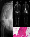
Diagrams showing polyostotic Paget disease of bone. A, Proximal humerus and (B) proximal femur and pelvic involvement with classic moth-eaten appearance. B, Varus deformity can develop secondary to proximal femoral involvement. Technicium-99 bone scan (C) anterior and (D) posterior showing increased metabolic activity in bilateral proximal humeri, the right clavicle, and right hip. E, This microscopic image shows trabecular bone with a mosaic (or jigsaw puzzle) pattern, along prominent cement lines.
Figure 4
Figure 4
Diagrams showing polyostotic fibrous dysplasia. A, AP pelvis x-ray demonstrating bilateral expansile and ground-glass appearing lesions with secondary varus deformity of the proximal femur. B, CT of the patient's left femur after hardware removal with Sheppard crook deformity. C, These lesions typically show irregular shaped, thin bone trabeculae in a background of fibroblast-like spindle cells, which lack conspicuous osteoblastic rimming.
Figure 5
Figure 5
Diagram showing synovial chondromatosis. A and B, Soft-tissue ossifications around fifth toe. C and D, Sequential coronal T1 postcontrast MRI with multiple bodies with peripheral enhancement. E, Nodules of mature hyaline cartilage are seen with variable degrees of cellularity and nuclear atypia.

References

    1. Vasquez L, Silva J, Chavez S, et al. : Prognostic impact of diagnostic and treatment delays in children with osteosarcoma. Pediatr Blood Cancer 2020;67:e28180. - PubMed
    1. Levine AJ, Momand J, Finlay CA: The p53 tumour suppressor gene. Nature 1991;351:453-456. - PubMed
    1. Rodriguez-Galindo C, Orbach DB, VanderVeen D: Retinoblastoma. Pediatr Clin North Am 2015;62:201-223. - PubMed
    1. Knudson AG, Jr: Mutation and cancer: Statistical study of retinoblastoma. Proc Natl Acad Sci U S A 1971;68:820-823. - PMC - PubMed
    1. Jhiang SM: The RET proto-oncogene in human cancers. Oncogene 2000;19:5590-5597. - PubMed

MeSH terms

LinkOut - more resources