Review
. 2022 Oct 13;18(10):e1010849.
doi: 10.1371/journal.ppat.1010849. eCollection 2022 Oct.

Not your Mother's MAPKs: Apicomplexan MAPK function in daughter cell budding

Affiliations
Review

Not your Mother's MAPKs: Apicomplexan MAPK function in daughter cell budding

William J O'Shaughnessy et al. PLoS Pathog. .

Abstract

Reversible phosphorylation by protein kinases is one of the core mechanisms by which biological signals are propagated and processed. Mitogen-activated protein kinases, or MAPKs, are conserved throughout eukaryotes where they regulate cell cycle, development, and stress response. Here, we review advances in our understanding of the function and biochemistry of MAPK signaling in apicomplexan parasites. As expected for well-conserved signaling modules, MAPKs have been found to have multiple essential roles regulating both Toxoplasma tachyzoite replication and sexual differentiation in Plasmodium. However, apicomplexan MAPK signaling is notable for the lack of the canonical kinase cascade that normally regulates the networks, and therefore must be regulated by a distinct mechanism. We highlight what few regulatory relationships have been established to date, and discuss the challenges to the field in elucidating the complete MAPK signaling networks in these parasites.


Conflict of interest statement

The authors have declared that no competing interests exist.

Figures

Fig 1
Fig 1. Overview of MAPK signaling.
(A) In a canonical MAPK signaling cascade, a signal results in activation of the upstream MAP3K (the “MAPK kinase kinase”), which phosphorylates and activates the MAP2K (the “MAPK kinase”), which in turn phosphorylates its target MAPK on both Thr and Tyr. Apicomplexan parasites lack the STE kinase family, to which all MAP2Ks belong. They therefore encode no MAP2Ks or MAP3Ks. (B) Domain architecture of the apicomplexan MAPKs (not to scale). ERK7 and MAPKL1 both have long CTEs that are predicted to be intrinsically disordered by IUPRED [75]. The ERK7 CTE contains sequence repeats (green; Sarcocystidae only). In Toxoplasma, both phosphorylation (yellow diamonds) and fucosylation (orange hexagons) sites have been identified. The Plasmodium ERK7 CTE also has one or more predicted nuclear localization signals (purple rectangles), though no post-translational modifications of the CTE have been identified as of yet. The Toxoplasma MAPKL1 CTE is also phosphorylated. Many MAPKL1 family members have an extended activation loop that reaches a maximum of approximately 100 residues in Sarcocystidae. Most MAPK2 proteins have a disordered N-terminal extension. The activation motifs for each of the apicomplexan MAPK subfamilies are indicated above the kinase domain. CTE, C-terminal extension; MAPK, mitogen-activated protein kinase.
Fig 2
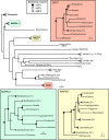
Fig 2. Phylogenetic analysis of the apicomplexan MAPKs.
Phylogenetic trees of multiple sequence alignments of the MAPKs from the indicated organisms were estimated using IQTREE2 [76]. Subtrees were collapsed for space considerations, indicated by triangles. When present, numbers to the right of the triangles indicate number of kinases in the subtree. Expanded subtrees for the apicomplexan MAPK clades are shown in inset boxes. MAPK activation loop motifs (e.g., TDY, TxH) have been indicated, where appropriate, to demonstrate that the TxH motif is not confined to the MAPK2 clade. Organisms: Metazoan (human, mouse, fruit fly), Apicomplexa (T. gondii, E. falciformis, E. maxima, C. parvum, G. niphandrodes, P. berghei, P. falciparum, P. vivax), Chromerid (C. velia, V. brassicaformis), Dinoflagellate (S. microadriaticum), Ciliate (T. thermophila), green plant (A. thaliana, C. reinhardtii), red algae (C. crispus, P. purpureum, P. yezoensis). Note: The estimated number of MAPKs in V. brassicaformis is approximately 40 and approximately 75 in C. velia; note that analysis is complicated by current low quality of genome build and lack of verification of gene models by transcript sequencing. MAPK, mitogen-activated protein kinase.
Fig 3
Fig 3. MAPKs use docking sites to recognize substrates and regulators.
(A) Canonical MAPKs use the conserved D-site (orange) to recognize kinase-interaction-motifs such as that of MKP3 bound to ERK2 (blue; PDB: 2FYS). This site lies distal to the active site and substrate recognition region. Some MAPKs use a second docking site, the F-site (yellow) to recognize F-x-F-P motifs such as that found in ORP45 (purple; PDB: 7OPM). (B) The inhibitory scaffold AC9 wraps around the Toxoplasma ERK7 kinase domain (blue; PDB: 6V6A) and occupies both the D-site (orange), active site, and substrate-recognition region. The site where the F-site would be localized on the TgERK7 structure is indicated in yellow, though no F-site binding partners have yet been identified for an apicomplexan MAPK. It is possible that apicomplexan MAPKs do not use F-site recognition. (C) Alignment of the sequences comprising the D-site of the indicated MAPKs. Polar (mostly acidic) sites that are thought to provide specificity to the KIMs recognized are indicated by arrowheads and red text. Sites that typically make backbone or hydrophobic interactions with KIMs are boxed. (Hs–human; Tg–T. gondii; Cp–C. parvum; Pf–P. falciparum) KIM, kinase-interacting motif; MAPK, mitogen-activated protein kinase.
Fig 4
Fig 4. MAPK function in Toxoplasma tachyzoite replication.
ERK7 (red kinase) localizes to the maternal and daughter bud apical caps, just below the conoid (orange rings). MAPKL1 (green kinase) localizes to the pericentrosomal material surrounding the centrosome (green circle). MAPK2 (yellow kinase) is cytosolic. Right panels show phenotypes resulting from knockdown of each of MAPKs. Loss of ERK7 results in destruction of the conoid. Loss of MAPKL1 and MAPK2 has opposing effects on centrosome duplication. MAPK, mitogen-activated protein kinase.

References

    1. Cobb MH, Goldsmith EJ. How MAP kinases are regulated. J Biol Chem. 1995;270:14843–14846. doi: 10.1074/jbc.270.25.14843 - DOI - PubMed
    1. Lewis TS, Shapiro PS, Ahn NG. Signal transduction through MAP kinase cascades. Adv Cancer Res. 1998;74:49–139. doi: 10.1016/s0065-230x(08)60765-4 - DOI - PubMed
    1. Peti W, Page R. Molecular basis of MAP kinase regulation. Protein Sci Publ Protein Soc. 2013;22:1698–1710. doi: 10.1002/pro.2374 - DOI - PMC - PubMed
    1. Raman M, Chen W, Cobb MH. Differential regulation and properties of MAPKs. Oncogene. 2007;26:3100–3112. doi: 10.1038/sj.onc.1210392 - DOI - PubMed
    1. Talevich E, Mirza A, Kannan N. Structural and evolutionary divergence of eukaryotic protein kinases in Apicomplexa. BMC Evol Biol. 2011;11:321. doi: 10.1186/1471-2148-11-321 - DOI - PMC - PubMed

Publication types

Substances