. 2022 Oct 13;17(10):e0275972.
doi: 10.1371/journal.pone.0275972. eCollection 2022.

Loss of Ecrg4 improves calcium oxalate nephropathy

Affiliations

Loss of Ecrg4 improves calcium oxalate nephropathy

Daniela Cabuzu et al. PLoS One. .

Abstract

Kidney stone is one of the most frequent urinary tract diseases, affecting 10% of the population and displaying a high recurrence rate. Kidney stones are the result of salt supersaturation, including calcium and oxalate. We have previously identified Esophageal cancer-related gene 4 (Ecrg4) as being modulated by hypercalciuria. Ecrg4 was initially described as a tumor suppressor gene in the esophagus. Lately, it was shown to be involved as well in apoptosis, cell senescence, cell migration, inflammation and cell responsiveness to chemotherapy. To the best of our knowledge, nothing is known about ECRG4's function in the renal tissue and its relationship with calciuria. We hypothesized that the increased expression of Ecrg4 mRNA is triggered by hypercalciuria and might modulate intratubular calcium-oxalate precipitation. In this study, we have first (i) validated the increased Ecrg4 mRNA in several types of hypercalciuric mouse models, then (ii) described the Ecrg4 mRNA expression along the nephron and (iii) assessed ECRG4's putative role in calcium oxalate nephropathy. For this, Ecrg4 KO mice were challenged with a kidney stone-inducing diet, rich in calcium and oxalate precursor. Taken together, our study demonstrates that Ecrg4's expression is restricted mainly to the distal part of the nephron and that the Ecrg4 KO mice develop less signs of tubular obstruction and less calcium-oxalate deposits. This promotes Ecrg4 as a modulator of renal crystallization and may open the way to new therapeutic possibilities against calcium oxalate nephropathy.


Conflict of interest statement

The authors have declared that no competing interests exist.

Figures

Fig 1
Fig 1. Ecrg4 is expressed in the kidney and along the distal part of the nephron.
(A) Ecrg4 mRNA expression in the indicated organs relative to Actb and to the expression in the esophagus (dotted line) in control male mice (n = 3). (B) Ecrg4 mRNA expression in microdissected renal tubular segments related to Actb and to the expression in the DCT-CNT segment (dotted line) in control male mice (n = 3). Data are shown as individual dots and mean ± SD. Stars indicate *p<0.05, calculated using t-test. PROX: proximal tubule; TAL: thick ascending limb; DCT-CNT: distal convoluted tubule and connecting tubule; CD: collecting duct.
Fig 2
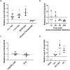
Fig 2. Ecrg4 transcript is upregulated in several hypercalciuric mouse models.
(A) Ecrg4 mRNA expression (related to Actb and appropriate control mice for each condition, dotted line) was found increased in the kidneys of mice issued from several established hypercalciuric models (n = 4 males/condition), but not in mice exposed to chronic furosemide (injected with 20mg furosemide/kg body weight for 6 consecutive days). Ncx1 KO: Ncx1 KO mice. Cldn2 KO: Claudin-2 KO mice. DHTS: Dihydrotachysterol-treated mice (1,5mg/kg food) for 7 days. (B) Renal Ecrg4 expression after acute furosemide injection relative to renal expression of vehicle-injected male mice (dotted line). The mice were injected with 20mg furosemide/kg body weight and kidneys were harvested after 15, 30, 60 or 240min (n = 3–5 animals/group). (C) Renal Ecrg4 mRNA expression is not changed by the calcitropic hormones PTH or 1,25(OH)2-vitamin D. The kidneys were harvested 6h after calcitriol injection (n = 5), and 2h after PTH injection (n = 6). The dotted line at 1 represents renal Ecrg4 mRNA expression of vehicle-treated male mice. (D) Ecrg4’s regulation along the nephron of the hypercalciuric Ncx1 KO mouse model compared to control littermates (dotted line) (n = 3 males/condition). Data are shown as individual dots and mean ± SD. Stars indicate *p<0.05, calculated using t test and Dunnett’s multiple correction.
Fig 3
Fig 3. Ecrg4 KO male mice are resistant to induced hypercalciuria.
(A) Ecrg4 mRNA expression in the kidney of Ecrg4 KO mice. P value was calculated using t-test. (B) Schematic representation of the experimental plan. Male mice were habituated in metabolic cages for 2 days. Then, control and Ecrg4 KO mice were exposed to either the regular chow diet or to a diet enriched in calcium (1.5%) and the oxalate precursor hydroxyproline (2%) (CaOx diet) for 8 days. The mice were housed individually in metabolic cages for urine collection at baseline, day 1 and day 8. (C) Increase in the urinary calcium-creatinine ratio in both control and Ecrg4 KO mice under CaOx diet. The ratios were calculated from the urine collected at baseline, day 1 and 8 under chow diet or CaOx diet. At 8 days of the CaOx diet, the Ecrg4 KO male mice had a significantly lower calcium-creatinine excretion compared to control mice. (D) Urinary sodium-creatinine ratio in both control and Ecrg4 KO male mice. (E) Urinary calcium/sodium ratio after 8 days of CaOx diet. Data are shown as individual points and mean ± SD (n = 4). For data B-D, p value was calculated using two-way ANOVA with Tukey correction for multiple comparisons. Stars indicate * p<0.05 between control and Ecrg4 KO mice; § indicates p<0.05 for comparison of control mice in the two different diets (chow vs. CaOx); # indicates p<0.05 of Ecrg4 KO mice in the two different diets (chow vs. CaOx).
Fig 4
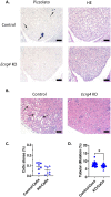
Fig 4. Ecrg4 KO male mice have less renal tubular obstruction after 8 days of CaOx diet.
(A) Representative pictures of renal papilla stained by Pizzolato (left panels) or Hematoxylin-eosin (HE, right panels) at 20x magnification. Intraluminal crystals and tubular dilation are visible in control mice but not in Ecrg4 KO mice. Bars represent 100um. (B) HE staining of sections of the cortex of kidneys from control and Ecrg4 KO male mice kidneys, after 8 days of CaOx diet, at 20x magnification. Bars represent 100um. The black arrows indicate the intraluminal crystals (A), as well as the tubular dilatation (B) in the control mice. (C) Quantification of the CaOx crystals in kidney tissue expressed as percentage of crystal surface over the kidney section (n = 7). (D) Quantification of tubular dilation of the cortex expressed as percentage of surface dilated over the whole surface (5 sections/mouse; n = 7). Data are shown individually and as mean ± SD. P values are calculated using t-test.

Similar articles

Cited by

References

    1. Chen Z, Prosperi M, Bird VY. Prevalence of kidney stones in the USA: The National Health and Nutrition Evaluation Survey. Journal of Clinical Urology. 2019;12(4):296–302.
    1. Moor MB, Bonny O. Ways of calcium reabsorption in the kidney. Am J Physiol Renal Physiol. 2016;310(11):F1337–50. doi: 10.1152/ajprenal.00273.2015 - DOI - PubMed
    1. Qin X, Zhang P. ECRG4: a new potential target in precision medicine. Frontiers of medicine. 2019;13(5):540–6. doi: 10.1007/s11684-018-0637-9 - DOI - PubMed
    1. Schmidt T, Samaras P, Frejno M, Gessulat S, Barnert M, Kienegger H, et al.. ProteomicsDB. Nucleic Acids Research. 2017;46(D1):D1271–D81. - PMC - PubMed
    1. Yue C-M, Deng D-J, Bi M-X, Guo L-P, Lu S-H. Expression of ECRG4, a novel esophageal cancer-related gene, downregulated by CpG island hypermethylation in human esophageal squamous cell carcinoma. World J Gastroenterol. 2003;9(6):1174–8. doi: 10.3748/wjg.v9.i6.1174 - DOI - PMC - PubMed

Publication types