. 2022 Oct 13;18(10):e1010912.
doi: 10.1371/journal.ppat.1010912. eCollection 2022 Oct.

Glaesserella parasuis serotype 5 breaches the porcine respiratory epithelial barrier by inducing autophagy and blocking the cell membrane Claudin-1 replenishment

Affiliations

Glaesserella parasuis serotype 5 breaches the porcine respiratory epithelial barrier by inducing autophagy and blocking the cell membrane Claudin-1 replenishment

Mingxing Liu et al. PLoS Pathog. .

Abstract

Glaesserella parasuis (G. parasuis), the primary pathogen of Glässer's disease, colonizes the upper respiratory tract and can break through the epithelial barrier of the respiratory tract, leading to lung infection. However, the underlying mechanisms for this adverse effect remain unclear. The G. parasuis serotype 5 SQ strain (HPS5-SQ) infection decreased the integrity of piglets' lung Occludin and Claudin-1. Autophagy regulates the function of the epithelial barrier and tight junction proteins (TJs) expression. We tested the hypothesis that HPS5-SQ breaking through the porcine respiratory epithelial barrier was linked to autophagy and Claudin-1 degradation. When HPS5-SQ infected swine tracheal epithelial cells (STEC), autophagosomes encapsulated, and autolysosomes degraded oxidatively stressed mitochondria covered with Claudin-1. Furthermore, we found that autophagosomes encapsulating mitochondria resulted in cell membrane Claudin-1 being unable to be replenished after degradation and damaged the respiratory tract epithelial barrier. In conclusion, G. parasuis serotype 5 breaks through the porcine respiratory epithelial barrier by inducing autophagy and interrupting cell membrane Claudin-1 replenishment, clarifying the mechanism of the G. parasuis infection and providing a new potential target for drug design and vaccine development.


Conflict of interest statement

The authors have declared that no competing interests exist.

Figures

Fig 1
Fig 1. HPS5-SQ infection causes damage to the porcine respiratory epithelial barrier.
(A) H&E staining of healthy piglets and piglets infected with HPS5-SQ. Scale bar: 50 μm. (B) Piglets’ lung Occludin and Claudin-1 levels were determined by Western blot. (C) Quantification of Occludin and Claudin-1. Data shown are means ± SEM; ** P < 0.01; *** P < 0.001. (two-tailed Student’s t-tests). (D) CFU of HPS5-SQ across the porcine respiratory epithelial barrier model were counted. Data shown are means ± SEM. (E) The porcine respiratory epithelial barrier was uninfected or infected with HPS5-SQ (MOI of 100). TER of the epithelial barrier was measured. Data shown are means ± SEM; * P < 0.05; ** P < 0.01. (two-way ANOVA). (F) STEC were either control-treated or infected by HPS5-SQ at an MOI of 100, and whole-cell extracts were prepared from control-infected and HPS5-SQ-infected cells at 6, 12, 18, and 24 hpi. Occludin and Claudin-1 levels were determined by Western blot. (G) Quantification of Occludin and Claudin-1. Data shown are means ± SEM; ** P < 0.01; *** P < 0.001. (one-way ANOVA).
Fig 2
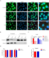
Fig 2. HPS5-SQ infection degrades cytoplasm Claudin-1 in STEC.
(A) STEC were infected with control or HPS5-SQ (MOI 100) for 12 h and 24 h in media. STEC were fixed and subjected to immunofluorescence analysis to detect Claudin-1 (green). Scale bar: 10 μm. (B) STEC were control-treated or infected by HPS5-SQ at an MOI of 100, and whole-cell extracts were prepared from control-infected and HPS5-SQ-infected cells at 12 and 24 hpi. (C) Quantification of cytoplasm Claudin-1 and cytomembrane Claudin-1. Data shown are means ± SEM; ** P < 0.01; *** P < 0.001. (one-way ANOVA). (D) STEC were uninfected or infected with HPS5-SQ (MOI of 100). LDH of the STEC was measured. Data shown are means ± SEM; ** P < 0.01. (two-tailed Student’s t-tests). (E) STEC were either control treated or infected by HPS5-SQ at an MOI of 100. mRNA levels of Claudin-1 were detected using qRT-PCR.
Fig 3
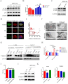
Fig 3. Autophagy cuts off the replenishment of Claudin-1 on the cell membrane.
(A) STEC were either controlled or infected by HPS5-SQ at an MOI of 100; whole-cell extracts were prepared from control-infected and HPS5-SQ-infected cells at 3, 6, 9, and 12 hpi. LC3BII and p62 levels were determined by Western blot. (B) Quantification of LC3BII and p62. Data shown are means ± SEM; ** P < 0.01; *** P < 0.001. (one-way ANOVA). (C) STEC were treated with or without Baf-A1 for the final 4 h and then infected with or without HPS5-SQ (MOI of 100); whole-cell extracts were prepared from control-infected and HPS5-SQ-infected cells at 12 hpi. LC3B levels were determined by Western blot. (D) Immunofluorescence microscopy analysis of autophagosome-like vesicles in STEC infected by HPS5-SQ for 6 and 12 h. Scale bar: 5 μm. (E) Quantification of STEC autolysosomes. Data shown are means ± SEM; * P < 0.05; *** P < 0.001. (two-tailed Student’s t-tests). (F) STEC were either control-treated or infected with HPS5-SQ at an MOI of 100. Electron microscopy images revealed the STEC ultrastructure. In the images, membrane-like vesicles in HPS5-SQ-infected cells were observed. Scale bar: 5 μm, 500 nm. (G) STEC were treated with 3-MA for the final 3 h or Baf-A1 for the final 4 h, then infected with or without HPS5-SQ (MOI of 100); cytoplasm Claudin-1 and cytomembrane Claudin-1 were determined by Western blot. (H) Quantification of cytoplasm Claudin-1 and cytomembrane Claudin-1. Data shown are means ± SEM; * P < 0.05; ** P < 0.01. (one-way ANOVA). (I) The porcine respiratory epithelial barrier was treated with 3-MA for the final 3 h or untreated with 3-MA, then infected with or without HPS5-SQ (MOI of 100). TER of the epithelial barrier was measured. Data shown are means ± SEM; * P < 0.05. (one-way ANOVA). (J) STEC were transfected with ATG5 siRNA or siNC. STEC were infected at MOI = 100. ATG5 and Claudin-1 were determined via Western blot. (K) Quantification of ATG5 and Claudin-1. Data shown are means ± SEM; ** P < 0.01; *** P < 0.001. (one-way ANOVA).
Fig 4
Fig 4. HPS5-SQ-mediated autophagy degrades Claudin-1 covering mitochondria in STEC.
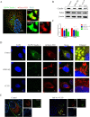
(A) The expression of the mCherry-FIS1 and EGFP-Claudin-1 in STEC. Immunofluorescence microscopy images detected the subcellular localization of mitochondria and Claudin-1. Scale bars: 5 μm. (B) Western blot showing Claudin-1 and Parkin expression in HPS5-SQ-infected cells or treated with mitophagy activator: CCCP for 6 and 12 h. (C) Quantification of Claudin-1 and Parkin. Data shown are means ± SEM; * P < 0.05; *** P < 0.001. (one-way ANOVA). (D) STEC expressing mCherry-FIS1 and EGFP-Claudin-1 were control-treated or infected with HPS5-SQ (MOI of 100) and treated with CCCP for 12 h as a positive control. Immunofluorescence microscopy images detected the Claudin-1 and mitochondria. Scale bar: 5 μm. (E) The expression of the mCherry-FIS1 and EGFP-Claudin-1 in STEC were treated with Baf-A1 for the final 4 h or untreated with Baf-A1, then CCCP was added to STEC pretreated with Baf-A1 for 12 h. Immunofluorescence microscopy images detected the subcellular localization of mitochondria and Claudin-1 in STEC pretreated with Triton-100 for more than 12 h. Scale bars: 5 μm.
Fig 5
Fig 5. HPS5-SQ-mediated autophagy envelops mitochondria under oxidative stress and kills invading bacteria.
(A) STEC were fixed and subjected to immunofluorescence analysis to detect LC3 (pink) and mitochondrial fluorescence (red and green). Scale bar: 10 μm. (B) Immunofluorescence of control, HPS5-SQ, and HPS5-SQ+NAC (5 mM for the final 1 h) infected STEC stained for mROS and LC3B. The white triangle refers to HPS5-SQ (blue). Scale bar: 10 μm. (C) STEC were treated with 3-MA for the final 3 h or Baf-A1 for the final 4 h, quantifying the invading HPS5-SQ in the infection of HPS5-SQ for 6 and 12 h. Data shown are means ± SEM; ** P < 0.01. (two-way ANOVA). (D) STEC were treated with ATG5 siRNA for 24 h and infected with HPS5-SQ for 12 h. Quantification of the invading HPS5-SQ. Data shown are means ± SEM; ** P < 0.01. (one-way ANOVA).
Fig 6
Fig 6. HPS5-SQ-mediated autophagy involves the decline of porcine respiratory epithelial Claudin-1.
(A) The mROS staining of piglets’ lung tissue section of the autopsy showed the mROS (red) and the nucleus (blue). Scale bars: 1000 μm, 100 μm. (B) Pictures were marked by @ and & in Fig 6A, and immunofluorescence microscopy detected the Claudin-1 (pink) and DAPI (blue) in piglets lung tissue. The picture was marked by ii and iv in Fig 6A, and immunofluorescence microscopy detected the localization of mROS(red), LC3(green), and DAPI (blue) in healthy piglets lung tissue infected with HPS5-SQ. Scale bars: 200 μm, 50 μm, 20 μm.
Fig 7
Fig 7. Diagram of the destruction of the respiratory epithelial barrier in the infection of HPS5-SQ.
Autophagy induced by mROS facilitates the damage of the respiratory epithelial barrier in the infection of HPS5-SQ. In brief, autolysosomes can degrade mitochondrion and Claudin-1. In addition, cytotoxicity induced by HPS5-SQ infection accelerates the escape of Claudin-1 from the cytoplasm. Consequently, the Claudin-1 degradation and escape in the STEC make cell membrane Claudin-1 unable to replenish in time, and the respiratory tract epithelial barrier is damaged.

References

    1. Luppi A, Bonilauri P, Dottori M, Iodice G, Gherpelli Y, Merialdi G, et al.. Haemophilus parasuisSerovars Isolated from Pathological Samples in Northern Italy. Transboundary and Emerging Diseases. 2013;60(2):140–2. 10.1111/j.1865-1682.2012.01326.x. - DOI - PubMed
    1. Luo X, Chang X, Zhou H, Lin H, Fan H. Glaesserella parasuis induces inflammatory response in 3D4/21 cells through activation of NLRP3 inflammasome signaling pathway via ROS. Vet Microbiol. 2021;256:109057. Epub 2021/04/03. doi: 10.1016/j.vetmic.2021.109057 - DOI - PubMed
    1. Cao Q, Wei W, Wang H, Wang Z, Lv Y, Dai M, et al.. Cleavage of E-cadherin by porcine respiratory bacterial pathogens facilitates airway epithelial barrier disruption and bacterial paracellular transmigration. Virulence. 2021;12(1):2296–313. Epub 2021/09/07. doi: 10.1080/21505594.2021.1966996 - DOI - PMC - PubMed
    1. Schlingmann B, Molina SA, Koval M. Claudins: Gatekeepers of lung epithelial function. Semin Cell Dev Biol. 2015;42:47–57. Epub 2015/05/09. doi: 10.1016/j.semcdb.2015.04.009 - DOI - PMC - PubMed
    1. Buckley A, Turner JR. Cell Biology of Tight Junction Barrier Regulation and Mucosal Disease. Cold Spring Harb Perspect Biol. 2018;10(1). Epub 2017/05/17. doi: 10.1101/cshperspect.a029314 - DOI - PMC - PubMed

Publication types