. 2023 Apr;23(7-8):e2200021.
doi: 10.1002/pmic.202200021. Epub 2022 Oct 13.

Data-independent acquisition and quantification of extracellular matrix from human lung in chronic inflammation-associated carcinomas

Affiliations

Data-independent acquisition and quantification of extracellular matrix from human lung in chronic inflammation-associated carcinomas

Joanna Bons et al. Proteomics. 2023 Apr.

Abstract

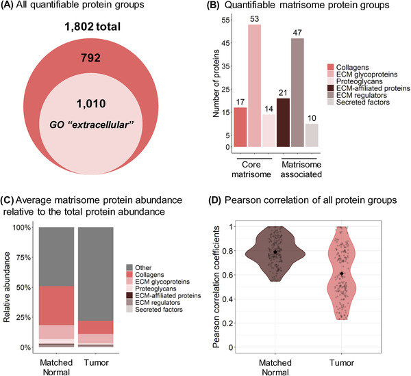
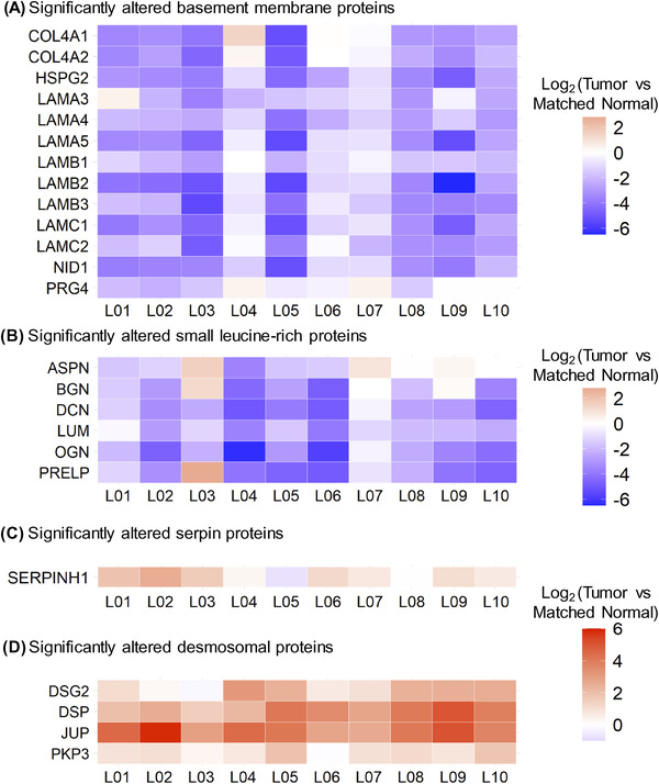
Early events associated with chronic inflammation and cancer involve significant remodeling of the extracellular matrix (ECM), which greatly affects its composition and functional properties. Using lung squamous cell carcinoma (LSCC), a chronic inflammation-associated cancer (CIAC), we optimized a robust proteomic pipeline to discover potential biomarker signatures and protein changes specifically in the stroma. We combined ECM enrichment from fresh human tissues, data-independent acquisition (DIA) strategies, and stringent statistical processing to analyze "Tumor" and matched adjacent histologically normal ("Matched Normal") tissues from patients with LSCC. Overall, 1802 protein groups were quantified with at least two unique peptides, and 56% of those proteins were annotated as "extracellular." Confirming dramatic ECM remodeling during CIAC progression, 529 proteins were significantly altered in the "Tumor" compared to "Matched Normal" tissues. The signature was typified by a coordinated loss of basement membrane proteins and small leucine-rich proteins. The dramatic increase in the stromal levels of SERPINH1/heat shock protein 47, that was discovered using our ECM proteomic pipeline, was validated by immunohistochemistry (IHC) of "Tumor" and "Matched Normal" tissues, obtained from an independent cohort of LSCC patients. This integrated workflow provided novel insights into ECM remodeling during CIAC progression, and identified potential biomarker signatures and future therapeutic targets.

Keywords: data-independent acquisition; extracellular matrix; lung squamous cell carcinoma; quantification; serpins.


Conflict of interest statement

The authors declare no conflict of interest.

Figures

FIGURE 1
FIGURE 1
Study Design and Proteomic Pipeline for ECM Analysis of Lung Squamous Cell Carcinoma. “Tumor” and “Matched Normal” fresh tissue specimens were collected from 10 patients with lung squamous cell carcinoma by the Ferri team at McGill University Health Centre (QC, Canada) or through the Cooperative Human Tissue Network Western Division (TN, USA). Fresh samples in UW Cold Storage Solution were sent to the Tlsty team at UCSF (CA, USA) for enrichment for insoluble extracellular matrix (ECM) proteins. ECM‐enriched samples were further processed by the Schilling team at the Buck Institute (Novato, CA); proteins were solubilized, in‐gel digested with Lys‐C and trypsin, and extracted proteolytic peptides were de‐glycosylated with PNGase F. All resulting samples were analyzed in duplicate on a nanoLC‐TripleTOF 6600 system (QqTOF) operated in data‐independent acquisition (DIA) mode, and data were processed with Spectronaut (Biognosys). Finally, candidates were validated on independent cohorts by immunofluorescence‐based immunohistochemistry by the Tlsty team
FIGURE 2
FIGURE 2
Proteomic Analysis of ECM and Matrisome Components from Human Lung Squamous Cell Carcinoma. (A) 1802 protein groups with at least two unique peptides were identified, including 1,010 protein groups matching the Gene Ontology (GO) Cellular Component “extracellular” term. (B) 162 of these quantified protein groups are reported in the human MatrisomeDB [50]. (C) Average protein abundance relative to the total protein abundance of the matrisomal (colored) and non‐matrisomal (grey) protein groups. Abundance is based on the MS/MS peak area of the 3–6 best fragment ions per precursor ion. Protein abundances were obtained with summing peptide/precursor abundances as described in the Methods section. (D) Violin plots of the Pearson coefficients of correlation between the “Matched Normal” or “Tumor” replicates. The Pearson correlation compares all MS acquisitions within one condition to each other (one by one). The filled diamonds represent the average value of the coefficients: 0.76 for the “Matched Normal” group and 0.61 for the “Tumor” group. High heterogeneity of the “Tumor” ECM enrichments across cancer patients (right plot) contrasts with a more homogeneous profile for “Matched Normal” ECM enrichments (left plot)
FIGURE 3
FIGURE 3
“Complete” Remodeling of the ECM in Lung Squamous Cell Cancer. (A) Supervised clustering analysis using partial least squares‐discriminant analysis (PLS‐DA) performed on protein groups quantified in the “Matched Normal” (brown) and “Tumor” (pink) samples. (B) Volcano plot of the 1802 quantified protein groups showing 202 down‐regulated and 327 up‐regulated protein groups for “Tumor” versus “Matched Normal” comparison. (C) 66 significantly altered protein groups are reported in the human MatrisomeDB [50] and are listed in Figure S4. Specific proteins from these significantly altered protein groups are listed in (D). (E‐F) Dot plots showing the ConsensusPathDB [47, 48] Gene Ontology (GO) biological processes enriched for protein groups significantly down‐regulated (E) and up‐regulated (F) in “Tumor” vs. “Matched Normal”
FIGURE 4
FIGURE 4
Alteration of Core Protein Signatures in Lung Squamous Cell Carcinoma across Patients. Heatmap showing log2‐fold protein changes (“Tumor” vs. ’Matched Normal’) for each individual LSCC patient referred to as L01, L02, L03 …, and L10 assessing Core Protein Signatures. (A) Thirteen basement membrane proteins and (B) six small leucine‐rich proteins were significantly down‐regulated in “Tumor” vs. “Matched Normal”, while (C) SERPINH1 (Hsp47) and (D) desmosomal proteins were significantly up‐regulated across patients. For all displayed proteins, all Q‐values (not displayed) were smaller than 9.38e‐8, when comparing “Tumor” to “Matched Normal” group (Table S3)
FIGURE 5
FIGURE 5
Dramatical upregulation of SERPINH1 (Hsp47) in Lung Squamous Cell Carcinoma (LSCC) Compared to Histologically Normal Lung (NL) Tissues. (A) Six cases of lung squamous cell carcinomas and matched histologically normal lung tissues from two of these cases and four additional histologically normal lung tissue specimens adjacent to lung cancers were probed for SERPINH1 level by immunofluorescence‐based immunohistochemistry (IHC). (B) Left: SERPINH1 level was quantified based on the percentage of area with positive (FITC; green) staining in five independent images per specimen (pixels with positive staining above baseline threshold/total number of pixels per image). An example of pseudo‐colorized positive area (pink) is shown for a matched set of specimens. Right: Plot corresponding to averaged values of positive staining of five images for each of 12 human specimens (six “Matched Normal” and six “Tumor”). Statistical analysis was carried out as described in the Methods section. Magnification: 20×

References

    1. Tlsty, T. D. , & Gascard, P. (2019). Stromal directives can control cancer. Science, 365(6449), 122–123. - PMC - PubMed
    1. Zhao, H. , Wu, L. , Yan, G. , Chen, Y. , Zhou, M. , Wu, Y. , & Li, Y. (2021). Inflammation and tumor progression: signaling pathways and targeted intervention. Signal Transduction and Targeted Therapy, 6(1), 263. - PMC - PubMed
    1. Qian, S. , Golubnitschaja, O. , & Zhan, X. (2019). Chronic inflammation: key player and biomarker‐set to predict and prevent cancer development and progression based on individualized patient profiles. EPMA Journal, 10(4), 365–381. - PMC - PubMed
    1. Hecht, S. S. (2011). Tobacco smoke carcinogens and lung cancer. In Penning T. M. (Ed.), Chemical carcinogenesis (pp. 53–74). Humana Press.
    1. Tsao, A. S. , Liu, D. , Lee, J. J. , Spitz, M. , & Hong, W. K. (2006). Smoking affects treatment outcome in patients with advanced nonsmall cell lung cancer. Cancer, 106(11), 2428–2436. - PubMed

Publication types

Substances