. 2023 Feb 1:253:123992.
doi: 10.1016/j.talanta.2022.123992. Epub 2022 Oct 8.

Label free electrochemical DNA biosensor for COVID-19 diagnosis

Affiliations

Label free electrochemical DNA biosensor for COVID-19 diagnosis

Atchara Lomae et al. Talanta. .

Abstract

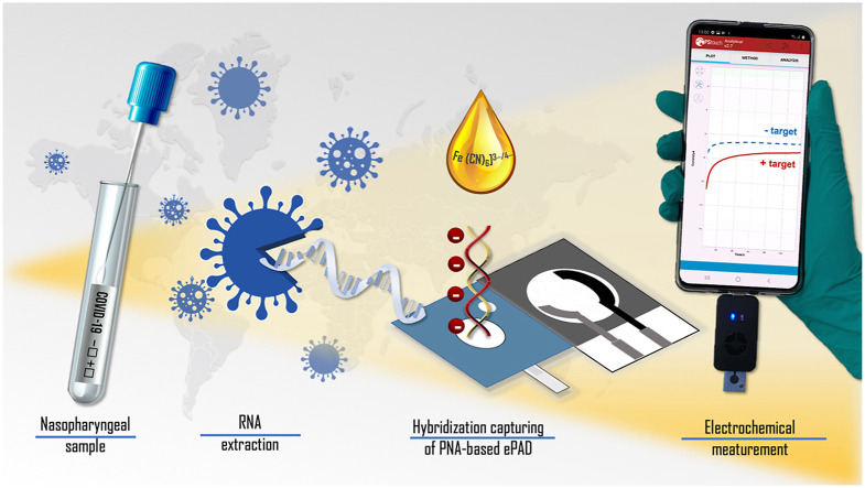
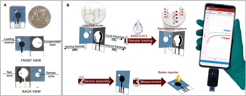
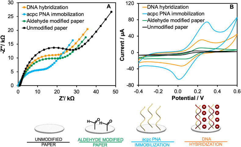
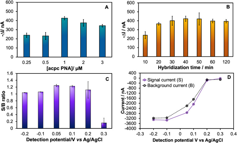
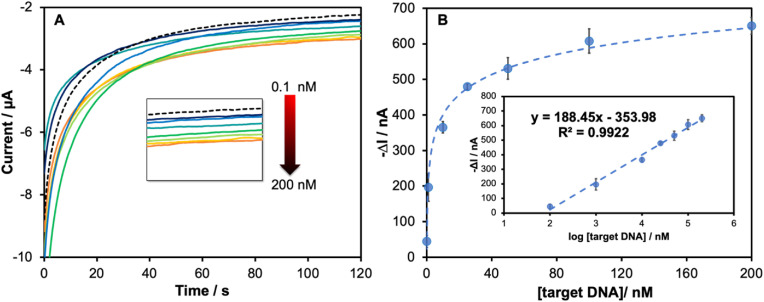
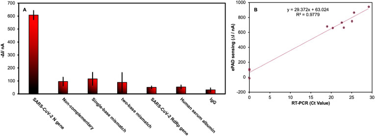
The COVID-19 pandemic has significantly increased the development of the development of point-of-care (POC) diagnostic tools because they can serve as useful tools for detecting and controlling spread of the disease. Most current methods require sophisticated laboratory instruments and specialists to provide reliable, cost-effective, specific, and sensitive POC testing for COVID-19 diagnosis. Here, a smartphone-assisted Sensit Smart potentiostat (PalmSens) was integrated with a paper-based electrochemical sensor to detect severe acute respiratory syndrome coronavirus 2 (SARS-CoV-2). A disposable paper-based device was fabricated, and the working electrode directly modified with a pyrrolidinyl peptide nucleic acid (acpcPNA) as the biological recognition element to capture the target complementary DNA (cDNA). In the presence of the target cDNA, hybridization with acpcPNA probe blocks the redox conversion of a redox reporter, leading to a decrease in electrochemical response correlating to SARS-CoV-2 concentration. Under optimal conditions, a linear range from 0.1 to 200 nM and a detection limit of 1.0 pM were obtained. The PNA-based electrochemical paper-based analytical device (PNA-based ePAD) offers high specificity toward SARS-CoV-2 N gene because of the highly selective PNA-DNA binding. The developed sensor was used for amplification-free SARS-CoV-2 detection in 10 nasopharyngeal swab samples (7 SARS-CoV-2 positive and 3 SARS-CoV-2 negative), giving a 100% agreement result with RT-PCR.

Keywords: COVID-19; PNA; Paper-based device; Point-of-care testing; Portable potentiostat; SARS-CoV-2.


Conflict of interest statement

Declaration of competing interest The authors declare that they have no known competing financial interests or personal relationships that could have appeared to influence the work reported in this paper.

Figures

Image 1
Graphical abstract
Scheme 1
Scheme 1
(A) Schematic illustration of the proposed PNA-based ePAD with front and back views; (B) the detection principle and procedure for SARS-CoV-2 detection using the PNA-based ePAD sensor connected with a smartphone-based potentiostat.
Fig. 1
Fig. 1
Electrochemical characterization of each modification step including unmodified paper substrate (black line), aldehyde-modified paper substrate (green line), acpcPNA immobilization (blue line) and DNA hybridization (orange line) via (A) EIS and (B) CV techniques. (All EIS Nyquist plots were fitted with the Randles equivalent circuit). (For interpretation of the references to color in this figure legend, the reader is referred to the Web version of this article.)
Fig. 2
Fig. 2
Assay optimization of the important parameters for SARS-CoV-2 DNA based detection including: (A) acpcPNA concentrations; (B) hybridization times; (C) detection potentials for measuring 50 nM SARS-CoV-2 DNA (N gene) via amperometric technique at 120 s sampling times; (D) the amperometric current response for 5 mM [Fe(CN)6]3-/4- in presence (S) and in absence (B) of 50 nM SARS-CoV-2 target. The error bar of all experiment represented the standard deviation of three repetitive measurements using three independent devices (n = 3).
Fig. 3
Fig. 3
(A) Amperograms and (B) calibration plot of the current response at 120 s in a function of SARS-CoV-2 DNA concentrations in the range from 0.1 nM to 200 nM showing the enlarged amperogram in each different DNA concentration in the inset (A); linear calibration plot as a function of logarithmic concentration of SARS-CoV-2 DNA in the inset (B) (n = 3).
Fig. 4
Fig. 4
(A) ΔI value in the presence of SARS-CoV-2 (N gene), non-complementary, single-base mismatch, two-base mismatch, SARS-CoV-2 (RdRp gene) at 100 nM and 15.6 mg/mL of HSA and IgG (n = 3); (B) correlation of SARS-CoV-2 (N gene) detection in nasopharyngeal swab samples using ePAD sensing (y-axis) vs RT-PCR assay (x-axis).

References

    1. Li X., Zai J., Wang X., Li Y. Potential of large “first generation” human‐to‐human transmission of 2019‐nCoV. J. Med. Virol. 2020;92 doi: 10.1002/jmv.25693. - DOI - PMC - PubMed
    1. World Health Organization (WHO) 2022. Weekly Epidemiological Update on COVID-19 - 25 January 2022.https://www.who.int/publications/m/item/weekly-epidemiological-update-on... (accessed January 28, 2022)
    1. Sun X., Andoh E.A., Yu H. A simulation-based analysis for effective distribution of COVID-19 vaccines: a case study in Norway. Transp. Res. Interdiscip. Perspect. 2021;11 doi: 10.1016/j.trip.2021.100453. - DOI - PMC - PubMed
    1. Binagwaho A., Mathewos K., Davis S. Equitable and effective distribution of the COVID-19 vaccines – a scientific and moral obligation. Int. J. Health Pol. Manag. 2021 doi: 10.34172/ijhpm.2021.49. - DOI - PMC - PubMed
    1. Burgos R.M., Badowski M.E., Drwiega E., Ghassemi S., Griffith N., Herald F., Johnson M., Smith R.O., Michienzi S.M. The race to a COVID-19 vaccine: opportunities and challenges in development and distribution. Drugs Context. 2021;10:1–10. doi: 10.7573/dic.2020-12-2. - DOI - PMC - PubMed