. 2022 Oct 13;5(12):e202201491.
doi: 10.26508/lsa.202201491.

High-resolution structure of a fish aquaporin reveals a novel extracellular fold

Affiliations

High-resolution structure of a fish aquaporin reveals a novel extracellular fold

Jiao Zeng et al. Life Sci Alliance. .

Abstract

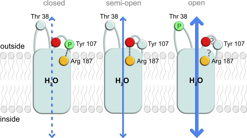
Aquaporins are protein channels embedded in the lipid bilayer in cells from all organisms on earth that are crucial for water homeostasis. In fish, aquaporins are believed to be important for osmoregulation; however, the molecular mechanism behind this is poorly understood. Here, we present the first structural and functional characterization of a fish aquaporin; cpAQP1aa from the fresh water fish climbing perch (<i>Anabas testudineus</i>), a species that is of high osmoregulatory interest because of its ability to spend time in seawater and on land. These studies show that cpAQP1aa is a water-specific aquaporin with a unique fold on the extracellular side that results in a constriction region. Functional analysis combined with molecular dynamic simulations suggests that phosphorylation at two sites causes structural perturbations in this region that may have implications for channel gating from the extracellular side.


Conflict of interest statement

The authors declare that they have no conflict of interest.

Figures

Figure 1.
Figure 1.. Water passage in climbing perch and water flow in the fish intestine.
(A) Schematic illustration of the assumed main water flow for climbing perch in fresh water, seawater (SW), and air, respectively, where the size of the arrow corresponds to the water flow and the tissue barrier is indicated by a dashed red line. In fresh water, Anabas testudineus is a hyperosmotic osmoregulator, aiming to minimize the diffusional inflow of water across the gills and producing dilute urine to get rid of excess water. In SW, on the other hand, A. testudineus will acclimate and become a hypoosmotic osmoregulator with the reverse problem with water diffusing out across all epithelia. This is counteracted by an ion-coupled water uptake across the intestine and a decreased glomerular filtration rate and urine production. During terrestrial exposure, A. testudineus must reduce water loss from the large surface areas constituted especially by the gills but also by the skin. The intestinal water absorption is also likely to be reduced as that is dependent on drinking, which ceases in air. (B) During SW acclimation, a total water flow is directed from the mucosal to the serosal side of the intestine driven by an ionic gradient of mainly K+, Na+, and Cl, involving ion transport via NKCC, Na,K-ATPase, and CFTR. In the SW trout, the increased intestinal water absorption is facilitated by aquaporin isoforms in the apical (AQP1a and AQP8ab) and basolateral (AQP8ab) membranes, respectively (reviewed in Madsen, 2015). The figure shows two enterocytes separated by tight junctions (TJ).
Figure 2.
Figure 2.. Crystal structure of cpAQP1aa.
(A, B) The cpAQP1aa tetramer viewed (A) parallel to the membrane and (B) from the extracellular side. The two half-helices forming the seventh pseudo-transmembrane segment are colored yellow in the frontmost monomer. Water molecules inside the channel are showed as red spheres. (C) HOLE analysis of the water-conducting channel with the channel dimensions visualized as dots. Residues in the aromatic-arginine (ar/R) and NPA regions as well as the extracellular vestibule are shown in stick representation. The graph shows a comparison between the channel profiles of cpAQP1aa (teal), human AQP4 (orange, PDB code 3GD8), human AQP7 (gray, PDB code 6QZI), and Arabidopsis TIP2;1 (green, PDB code 5I32). The zero channel position corresponds to the midpoint between the two NPA motifs. For cpAQP1aa, the channel is significantly narrower on the extracellular side of the ar/R region with a channel radius of 1.1 Å around Leu 117 (∼16 Å from the NPA region). (D) Close-up view of the hydrogen-bonding network involving Tyr 107, Arg 187, and residues 114–119 that stabilizes the unique conformation of loop C in cpAQP1aa. Hydrogen bonds are depicted as blue dashed lines. Electron density shown as gray mesh represents the 2FoFc-map contoured at 1.0 σ. (E) Structural overlay of cpAQP1aa (teal), human AQP4 (orange), and human AQP7 (gray), viewed from the extracellular side, showing that the main structural differences are found in loops A and C. Residues in the cpAQP1aa extracellular constriction region and putative phosphorylation site at Thr 38 in loop A are shown in stick representation. Water molecules in the channel are shown as red spheres. (E, F) Zoom-in on boxed region in (E), viewed from the side of the membrane (90 degree rotation), showing significant structural differences in loop C between the three overlaid structures. In cpAQP1aa, the side chain of Tyr 107 has flipped compared with AQP4 and AQP7, pushing loop C further into the extracellular vestibule closer to the channel opening.
Figure S1.
Figure S1.. Sequence alignment of selected aquaporin homologs from various species.
Strictly conserved residues are rendered with white characters on a red background in the column. Well-conserved residues (>70% similarity) are rendered as red characters in the column. Specific amino acids of importance for cpAQP1aa regulation or modification are marked by black boxes and highlighted by arrows; namely, T38 and Y107 involved in channel gating and the three confirmed C-terminal phosphorylation sites. Sequence alignment was performed using Clustal Omega, EMBL-EBI (https://www.ebi.ac.uk/Tools/msa/clustalo/), and the figure was made with ESPript 3.0 (http://espript.ibcp.fr/ESPript/ESPript/).
Figure S2.
Figure S2.. Structural comparison of eukaryotic aquaporins.
(A, B) Overlay of crystal structures of cpAQP1aa (teal), bovine AQP1 (blue), human AQP4 (orange), human AQP7 (gray), and Arabidopsis TIP2;1 (green) viewed (A) parallel to the membrane and (B) from the extracellular side. Water molecules are shown as red spheres.
Figure S3.
Figure S3.. Crystal packing for cpAQP1aa at different pH.
Cartoon representation of crystal packing in cpAQP1aa crystals. (A, B) Crystal packing in cpAQP1aa grown at pH 7.8. The views differ by a 90° rotation toward the viewer. (C, D) same as in (A) and (B) but for cpAQP1aa crystals grown at pH 6.5. The protein molecules corresponding to the asymmetric unit are colored teal (pH 7.8) and purple (pH 6.5).
Figure S4.
Figure S4.. Structure of cpAQP1aa at pH 6.5.
(A, B) Overlay of the four monomers in the cpAQP1aa tetramer viewed (A) parallel to the membrane and (B) from the extracellular side, displaying only minor structural differences between the monomers. In one monomer only, monomer C (purple) and loops A and C could be built in full. (C) Structural overlay of the cpAQP1aa structures at pH 7.8 (teal) and 6.5 (monomer C, purple). The two structures are highly similar but differ in loops A and C. Although these loops could be fully built in monomer C of the low pH structure, they were mostly disordered in all monomers in the high pH structure. (D) Zoom-in on the extracellular part of the overlay in (C), showing that despite the differences in loop C, the distal part of the loop occupies a similar position in both structures, including the residues that participate in the hydrogen-bonding network that stabilizes its unique conformation. Electron density (gray mesh) for loop C and Arg 187 in the low pH structure is a composite omit map displayed at 1.0 σ. (E) Graph comparing the channel profiles of cpAQP1aa structures at pH 6.5 (monomer C, purple) and 7.8 (teal), as calculated by HOLE. In both structures, a similar constriction can be seen on the extracellular side of the ar/R region. The zero channel position corresponds to the midpoint between the two NPA motifs.
Figure S5.
Figure S5.. Functional analysis of the cpAQP1aa.
(A) Representative curves for the water flow of hAQP4 (positive control), cpAQP1aa-243, cpAQP1aa-FL, and empty liposome (negative control), showing that purified protein of both cpAQP1aa-FL and cpAQP1aa-243 are functional. (B) Initial rate (i.r.) of the water flow of hAQP4 (positive control), cpAQP1aa-243, cpAQP1aa-FL, and empty liposome (negative control), showing that purified protein of both cpAQP1aa-FL and cpAQP1aa-243 are functional (n = 9–25 ± SD). (C) Representative curves for glycerol flow of cpAQP1aa-FL, cpAQP1aa-243, hAQP4, and empty liposome (negative control) (n = 3).
Figure S6.
Figure S6.. Immunoblots showing liposome reconstitution.
(A) Immunoblot using the 6× His monoclonal antibody (Takara Clontech) showing typical proteoliposome preps for the empty liposome (negative control), cpAQP1aa-FL, cpAQP1-243, and hAQP4 (positive control) for the functional analysis of the water flow. (B) Immunoblot using the 6× His monoclonal antibody (Takara Clontech) showing typical proteoliposome preps for the empty liposome (negative control), hAQP4 (positive control), cpAQP1aa-FL, and cpAQP1-243 for the functional analysis of the glycerol flow. (C) Immunoblot using the 6× His monoclonal antibody (Takara Clontech) showing the liposome preps for the empty liposome (negative control), cpAQP1aa-FL, cpAQP1aa-Y107S, cpAQP1aa-Y107A, cpAQP1aa-T38E, cpAQP1aa-T38A, and cpAQP1aa-L117A. The relative protein reconstitution efficiencies are averages from two immunoblots. (D) Immunoblot using the 6× His monoclonal antibody (Takara Clontech) showing the liposome preps for the empty liposome (negative control), cpAQP1aa-FL, and cpAQP1aa-Y107E. The relative protein reconstitution efficiencies are averages from four immunoblots.
Figure 3.
Figure 3.. Structural comparison between cpAQP1aa and the ammonia-facilitating aquaporin AtTIP2;1.
(A) Overlay of cpAQP1aa (teal) and AtTIP2;1 (green) viewed from the extracellular side. The arginine in the ar/R region is shown in stick representation. Water molecules in the channel are shown as spheres of the respective protein color. (A, B) Zoom-in on the boxed area in (A), showing the different side-chain orientation of the arginine in the ar/R region and the hydrogen-bonding pattern between water molecules and residues within the channel. Hydrogen bonds are shown as dotted lines in the respective protein color. In cpAQP1aa, the carbonyl position of Cys 181 and its hydrogen bonds to Asn 120 and two water molecules correspond to what is typically seen in water-specific AQPs. In contrast, the carbonyl of Gly 194 in AtTIP2;1 occupies a different position, observed mainly in non–water-specific AQPs, and shows a different hydrogen-bonding pattern.
Figure 4.
Figure 4.. Mutational and in silico analysis of the extracellular constriction region.
(A) The initial water permeability rates for wild-type and mutant cpAQP1aa with the rate of hAQP4 shown for comparison. All values are normalized taking the protein amount into account (n = 6–31 ± SD). The normalized initial water flow rates show that the Y107A, T38A, and T38E mutants are more efficient than the wild-type protein, in the range of hAQP4 (no significant differences), whereas the L117A and Y107E are similar to wild-type. (B) Zoom-in the pore opening in an energy-minimized final snapshot from the 100-ns molecular dynamic simulation of the wild-type system with Tyr 107, Leu 117, Gly 118, Leu 119, Gly 180, and Arg 187 highlighted. Distances are given in Ångström. (B, C) Same as (B) but for the pY107 system. The extracellular loop C has shifted to the left, narrowing the pore opening, and is held in place by the interactions between Arg 187 and phosphorylated Tyr 107. (D, E) Overlay of the final snapshots from the simulations for wild-type (cyan) and pY107 (blue). In pY107, Arg 187 is pulled away from the pore center because of its interaction with the phosphorylated tyrosine side chain, with its new position causing the distal end of transmembrane helix 1 and loop A to be displaced. (E) Analysis of the pore dimensions using HOLE (E) shows that the constriction region (channel position ∼15 Å) is narrower in pY107 than in the wild-type system. In addition, the new position of Arg 187 results in a widening of the ar/R region in pY107.
Figure S7.
Figure S7.. Confirmation of phosphorylation of cpAQP1aa expressed in Pichia pastoris.
SDS–PAGE and phosphostain for (1) positive control (lysozyme), (2) negative control (SPY), and (3) cpAQP1aa-243.
Figure S8.
Figure S8.. Snapshots from molecular dynamic simulations of wild-type and mutant cpAQP1aa.
Overlay of the energy-minimized structures of the final snapshots after 100-ns simulation of wild-type cpAQP1aa (cyan), pY107 (blue), pT38 (magenta), Y107E (green), Y107A (white), and L118A (gray). (A, B) Side view and (B) view from the extracellular side. The structures are highly similar except for structural variability in loops A and C.
Figure S9.
Figure S9.. Comparison of PfAQP and cpAQP1aa.
(A) Crystal structure of Plasmodium falciparum AQP (PfAQP, PDB code 3C02) showing the interaction between Arg 196 of the ar/R.-region and the main chain carbonyl of Trp 124 in loop C. (B) Similar interaction between Arg 187 and the main chain of loop C is seen in cpAQP1aa. The resulting saturation of the arginine guanidino group hydrogen-bonding sites has been suggested to play a role in water selectivity. Water and glycerol molecules in the PfAQP and cpAQP1aa pores are shown in sphere and stick representation, respectively.
Figure 5.
Figure 5.. Proposed regulatory mechanism for cpAQP1aa.
Schematic of a possible extracellular gating mechanism where phosphorylation of Thr 38 and Tyr 107 controls the water permeability. The crystal structure represents the non-phosphorylated state (middle) in which a hydrogen-bonding network between Tyr 107, backbone residues in the distal part of loop C (represented by a red sphere), and the ar/R region arginine (Arg 187, orange sphere) results in an extracellular constriction region that is consistent with a gate caught in a semi-open state. Upon phosphorylation of Tyr 107 (left), this hydrogen-bonding network is rearranged, and the phosphorylated tyrosine side chain instead interacts with Arg 187. This causes structural changes in this region, including further narrowing of the constriction region that is indicative of pore closure and a widened ar/R region. Phosphorylation of Thr 38 (right) results in an open pore; however, because of the structural flexibility of this region, a possible structural mechanism for this remains elusive.
Figure S10.
Figure S10.. Structural comparison with other gated aquaporins.
(A, B) Overlay of cpAQP1aa (teal) and mammalian AQP0 in its junctional (magenta, PDB code 2B6O) and non-junctional (white, PDB code 1YMG) form viewed (A) parallel to the membrane and (B) from the extracellular side. (A) Pore profiles of junctional AQP0 and cpAQP1aa as calculated by HOLE are indicated by small spheres in (A). In junctional (closed, magenta) AQP0, the side chain of Met 176 points into the channel, causing restriction, whereas in non-junctional AQP0 (open, white), it points away from the channel. In cpAQP1aa, this position corresponds to Ile 176 which together with Leu 117 and Gly 180 constitutes an extracellular constriction site. The cpAQP1aa constriction site is a result of a unique conformation of loop C, whereby the side chain of Tyr 107 pushes it toward the center of the channel. In AQP0, the corresponding residue Tyr 105 points in the opposite direction, as seen for other AQPs. (C) Comparison of the pore profiles of cpAQP1aa (teal), junctional AQP0 (magenta), and spinach PIP2;1 (SoPIP2;1, brown) as calculated by HOLE. Constriction regions at the extracellular side of the pore are observed in cpAQP1aa and AQP0, whereas cytoplasmic constriction regions are present in AQP0 and SoPIP2;1. (D, E) Crystal structures of SoPIP2;1 in closed (brown, PDB code 1Z98) and open (beige, PDB code 2B5F) conformations viewed (D) parallel to the membrane and (E) from the cytoplasmic side. Channel closure is achieved through a conformational change of loop D, resulting in the insertion of Leu 197 into the channel opening. A Ca2+-binding site at the N-terminus, occupied by Cd2+ in the crystal structure that is involved in stabilizing loop D in the closed conformation is indicated by a gray sphere. Residues involved in gating by pH (His 193), phosphorylation (Ser 115 and Ser 274), and the Ca2+-ligands (Asp 28 and Glu 31) are highlighted in stick representation.
Figure S11.
Figure S11.. Putative phosphorylation site in cpAQP1.
Extracellular tyrosine kinase PKDCC, also known as vertebrate lonesome kinase, has been shown to phosphorylate the protein osteopontin that has a very similar sequence as cpAQP1aa at Tyr 107. This supports the possibility that cpAQP1aa is phosphorylated in vivo, even though this kinase is suggested to not exist in yeast.

Similar articles

Cited by

References

    1. Afonine PV, Grosse-Kunstleve RW, Echols N, Headd JJ, Moriarty NW, Mustyakimov M, Terwilliger TC, Urzhumtsev A, Zwart PH, Adams PD (2012) Towards automated crystallographic structure refinement with phenix.refine. Acta Crystallogr D Biol Crystallogr 68: 352–367. 10.1107/s0907444912001308 - DOI - PMC - PubMed
    1. Ampah-Korsah H, Anderberg HI, Engfors A, Kirscht A, Norden K, Kjellstrom S, Kjellbom P, Johanson U (2016) The aquaporin splice variant NbXIP1;1α is permeable to boric acid and is phosphorylated in the N-terminal domain. Front Plant Sci 7: 862. 10.3389/fpls.2016.00862 - DOI - PMC - PubMed
    1. Anthis NJ, Haling JR, Oxley CL, Memo M, Wegener KL, Lim CJ, Ginsberg MH, Campbell ID (2009) β integrin tyrosine phosphorylation is a conserved mechanism for regulating talin-induced integrin activation. J Biol Chem 284: 36700–36710. 10.1074/jbc.m109.061275 - DOI - PMC - PubMed
    1. Beitz E, Pavlovic-Djuranovic S, Yasui M, Agre P, Schultz JE (2004) Molecular dissection of water and glycerol permeability of the aquaglyceroporin from Plasmodium falciparum by mutational analysis. Proc Natl Acad Sci U S A 101: 1153–1158. 10.1073/pnas.0307295101 - DOI - PMC - PubMed
    1. Biasini M, Bienert S, Waterhouse A, Arnold K, Studer G, Schmidt T, Kiefer F, Cassarino TG, Bertoni M, Bordoli L, et al. (2014) SWISS-MODEL: Modelling protein tertiary and quaternary structure using evolutionary information. Nucleic Acids Res 42: W252–W258. 10.1093/nar/gku340 - DOI - PMC - PubMed

Publication types

LinkOut - more resources