. 2023 Mar;44(3):499-512.
doi: 10.1038/s41401-022-01004-3. Epub 2022 Oct 13.

Mitofusin-2 mediates cannabidiol-induced neuroprotection against cerebral ischemia in rats

Affiliations

Mitofusin-2 mediates cannabidiol-induced neuroprotection against cerebral ischemia in rats

Bing-Tian Xu et al. Acta Pharmacol Sin. 2023 Mar.

Abstract

Cannabidiol (CBD) reportedly exerts protective effects against many psychiatric disorders and neurodegenerative diseases, but the mechanisms are poorly understood. In this study, we explored the molecular mechanism of CBD against cerebral ischemia. HT-22 cells or primary cortical neurons were subjected to oxygen-glucose deprivation insult followed by reoxygenation (OGD/R). In both HT-22 cells and primary cortical neurons, CBD pretreatment (0.1, 0.3, 1 μM) dose-dependently attenuated OGD/R-induced cell death and mitochondrial dysfunction, ameliorated OGD/R-induced endoplasmic reticulum (ER) stress, and increased the mitofusin-2 (MFN2) protein level in HT-22 cells and primary cortical neurons. Knockdown of MFN2 abolished the protective effects of CBD. CBD pretreatment also suppressed OGD/R-induced binding of Parkin to MFN2 and subsequent ubiquitination of MFN2. Overexpression of Parkin blocked the effects of CBD in reducing MFN2 ubiquitination and reduced cell viability, whereas overexpressing MFN2 abolished Parkin's detrimental effects. In vivo experiments were conducted on male rats subjected to middle cerebral artery occlusion (MCAO) insult, and administration of CBD (2.5, 5 mg · kg-1, i.p.) dose-dependently reduced the infarct volume and ER stress in the brains. Moreover, the level of MFN2 within the ischemic penumbra of rats was increased by CBD treatment, while the binding of Parkin to MFN2 and the ubiquitination of MFN2 was decreased. Finally, short hairpin RNA against MFN2 reversed CBD's protective effects. Together, these results demonstrate that CBD protects brain neurons against cerebral ischemia by reducing MFN2 degradation via disrupting Parkin's binding to MFN2, indicating that MFN2 is a potential target for the treatment of cerebral ischemia.

Keywords: MFN2; Parkin; cannabidiol; cerebral ischemia; oxidative stress.


Conflict of interest statement

The authors declare no competing interests.

Figures
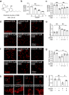
Fig. 1
Fig. 1. CBD attenuated OGD/R-induced HT-22 cell death and mitochondrial dysfunction.
a The chemical structure of CBD. b HT-22 cells were subjected to OGD insult at various times (0, 3, 6, and 9 h) followed by 24 h of reoxygenation. The cell viability was detected by the CCK-8 assay (n = 4). c HT-22 cells pretreated with CBD (0.1, 0.3, 1 μM) were subjected to 6 h of OGD followed by 24 h of reoxygenation, and then cell viability was detected by the CCK-8 assay (n = 4). d, e HT-22 cells pretreated with CBD (0.1, 0.3, 1 μM) were subjected to 6 h of OGD followed by 1 h of reoxygenation. The variance of intracellular ROS was detected by CellROX staining (n = 3). f, g HT-22 cells pretreated with CBD (0.1, 0.3, 1 μM) were subjected to 6 h of OGD followed by 1 h of reoxygenation. The variance of MMP was detected by TMRE staining (n = 3). h, i HT-22 cells pretreated with CBD (1 μM) were subjected to 6 h of OGD followed by 1 h of reoxygenation. The variance of mitochondrial morphology was visualized with Mito-tracker staining. The mitochondrial area was calculated with ImageJ (n = 3). *P < 0.05, **P < 0.01, versus the indicated group. n represents the number of independent cultures.
Fig. 2
Fig. 2. CBD ameliorated OGD/R-induced ER stress in HT-22 cells.
HT-22 cells pretreated with CBD (1 μM) were subjected to 6 h of OGD followed by 1 h of reoxygenation. af Then the variances of GRP78, p-IRE1α, IRE1α, p-eIF2α, and eIF2α were detected by immunoblotting. The changes in GRP78/β-tubulin (n = 3), p-IRE1α/IRE1α (n = 3), and p-eIF2α/eIF2α (n = 4) were determined by densitometry of the blots. g, h HT-22 cells pretreated with DTT (100 μM) were treated with CBD (1 μM) and then subjected to 6 h of OGD followed by 1 h of reoxygenation. The variance of p-IRE1α and IRE1α was detected by immunoblotting, and the change in p-IRE1α/IRE1α was determined by densitometry of the blots (n = 3). i HT-22 cells pretreated with DTT (100 μM) were treated with CBD (1 μM) and then subjected to 6 h of OGD followed by 24 h of reoxygenation. The change in cell viability was determined by the CCK-8 assay (n = 4). *P < 0.05, **P < 0.01, versus the indicated group. n represents the number of independent cultures.
Fig. 3
Fig. 3. MFN2 conferred protective effects of CBD against ER and mitochondrial dysfunction.
a, b HT-22 cells pretreated with CBD (0.1, 0.3, 1 μM) were subjected to 6 h of OGD followed by 1 h of reoxygenation. Then, the variance of MFN2 was detected by immunoblotting, and the change in MFN2/β-tubulin was determined by densitometry of the blots (n = 5). HT-22 cells pre-transfected with siMFN2 were treated with CBD (1 μM) and subjected to 6 h of OGD. c, d At 1 h after reoxygenation, the change of MMP was measured with TMRE staining (n = 3). e At 24 h after reoxygenation, cell viability was measured with the CCK-8 assay (n = 3). HT-22 cells pre-transfected with siMFN2 or MFN2 plasmid were treated with CBD (1 μM) and then subjected to 6 h of OGD. f At 24 h after reoxygenation, cell viability was measured with the CCK-8 assay (n = 3). g, h At 1 h after reoxygenation, the variance of intracellular ROS was detected with CellROX staining and quantified with ImageJ software (n = 3). HT-22 cells pre-transfected with siMFN2 were treated with CBD (1 μM) and subjected to 6 h of OGD. il At 1 h after reoxygenation, the variances of p-IRE1α, IRE1α, p-eIF2α, and eIF2α were detected by immunoblotting. Then, the changes of p-IRE1α/IRE1α (n = 3) and p-eIF2α/eIF2α (n = 4) were determined by densitometry of the blots. *P < 0.05, **P < 0.01, versus the indicated group. n represents the number of independent cultures.
Fig. 4
Fig. 4. CBD reduced OGD/R-induced cellular death and ER stress in primary cortical neurons.
a Primary cortical neurons pretreated with CBD (0.1, 0.3, 1 μM) were subjected to 1 h of OGD followed by 24 h of reoxygenation, and then cell viability was detected by the CCK-8 assay (n = 3). Primary cortical neurons pretreated with CBD (1 μM) were subjected to 1 h of OGD. b, c At 1 h after reoxygenation, the levels of caspase 3 and cleaved caspase 3 were detected by immunoblotting, and the change in cleaved caspase 3/caspase 3 was determined by densitometry of the blots (n = 3). d, e At 6 h after reoxygenation, the morphological change of dendrite was visualized with MAP2 staining (n = 3). fh At 1 h after reoxygenation, the levels of p-IRE1α, IRE1α, p-eIF2α, and eIF2α were detected by immunoblotting. The changes of p-IRE1α/IRE1α (n = 3) and p-eIF2α/eIF2α (n = 4) were determined by densitometry of the blots. *P < 0.05, **P < 0.01, versus the indicated group. n represents the number of independent cultures.
Fig. 5
Fig. 5. MFN2 mediated the protective effects of CBD against OGD/R-induced injury in primary cultured neurons.
a, b Primary cortical neurons pretreated with CBD (1 μM) were subjected to 1 h of OGD followed by 6 h of reoxygenation. The levels of MFN2 were detected by immunoblotting, and the change in MFN2/β-tubulin was determined by densitometry of the blots (n = 3). c Primary cortical neurons were transfected with siMFN2, and then the knockdown efficacy was verified by immunoblotting. Primary cortical neurons pre-transfected with siMFN2 were treated with CBD (1 μM) and then subjected to 1 h of OGD. d, e At 6 h after reoxygenation, the dendrite morphological variance was detected with MAP2 staining (n = 3). f, g The levels of caspase 3 and cleaved caspase 3 were detected by immunoblotting, and the change in cleaved caspase 3/caspase 3 was determined by densitometry of the blots (n = 5). *P < 0.05, **P < 0.01, versus the indicated group.
Fig. 6
Fig. 6. CBD ameliorated Parkin-mediated ubiquitination of MFN2 induced by OGD/R.
HT-22 cells pretreated with CBD (1 μM) were subjected to 6 h of OGD followed by 1 h of reoxygenation. a, b The change in Parkin binding to MFN2 was detected by co-immunoprecipitation, and the variance of Parkin/MFN2 was determined by densitometry of the blots (n = 3). c The input level of Parkin and MFN2 was detected by immunoblotting. d, e The ubiquitination of MFN2 was detected by immunoprecipitation, and the variance of ubiquitin/MFN2 was determined by densitometry of the blots (n = 3). f The change in MFN2 mRNA level was detected with qPCR (n = 5). HT-22 cells pretreated with CBD (1 μM) or MG132 (20 μM) were subjected to 6 h of OGD followed by 1 h of reoxygenation. g, h, i The changes in MFN2 and cleaved caspase 3 were detected by immunoblotting, and the variance of MFN2/β-tubulin (n = 4) and cleaved caspase 3/caspase 3 (n = 3) were determined by densitometry of the blots. HT-22 cells pre-transfected with Parkin plasmid were treated with CBD (1 μM) and then subjected to 6 h of OGD followed by 1 h of reoxygenation. j, k The variance of MFN2 was detected by immunoblotting, and the variance of MFN2/β-tubulin was determined by densitometry of the blots (n = 4). l, m The ubiquitination of MFN2 was detected by immunoprecipitation, and the variance of Ubiquitin/MFN2 was determined by densitometry of the blots (n = 3). n HT-22 cells pre-transfected with Parkin plasmid were treated with CBD (1 μM) and then subjected to 6 h of OGD followed by 24 h of reoxygenation. Cell viability was measured with the CCK-8 assay (n = 3). o HT-22 cells pre-transfected with Parkin plasmid or MFN2 plasmid were treated with CBD (1 μM) and then subjected to 6 h of OGD followed by 24 h of reoxygenation. Cell viability was measured with the CCK-8 assay (n = 3). *P < 0.05, **P < 0.01, versus the indicated group. n represents the number of independent cultures.
Fig. 7
Fig. 7. CBD reduced ER and ubiquitination of MFN2 in the cortex penumbra of SD rats subjected to MCAO.
SD rats were subjected to 2 h of MCAO. At the time of reperfusion, rats were administered CBD (2.5 mg/kg or 5 mg/kg) or the vehicle. a, b At 24 h after reperfusion, the infarct size was detected by TTC staining (n = 10 each group). c Before TTC staining, the neurological deficit scores were recorded (n = 10, each group). SD rats were subjected to MCAO for 2 h. At the time of reperfusion, CBD (5 mg/kg) or the vehicle was administered. At 24 h after reperfusion, the cortex penumbra was collected. dg The levels of MFN2, p-IRE1α, p-eIF2α, and eIF2α were detected by immunoblotting, and the variances of MFN2/β-tubulin, p-IRE1α/β-tubulin, and p-eIF2α/eIF2α were determined by densitometry of the blots (n = 4). h, i The change in Parkin binding to MFN2 was detected by co-immunoprecipitation, and the variance of Parkin/MFN2 was determined by densitometry of the blots (n = 4). j, k The ubiquitination of MFN2 was detected by immunoprecipitation, and the variance of ubiquitin/MFN2 was determined by densitometry of the blots (n = 4). *P < 0.05, **P < 0.01, versus the indicated group.
Fig. 8
Fig. 8. MFN2 conferred protective effects of CBD against MCAO-induced SD rat injury.
a, b SD rats were transfected with AAV2/9 loading shMFN2 or NC intracerebroventricularly at least for 2 weeks. Then, knockdown efficacy was verified by immunoblotting, and the level of MFN2/GAPDH was determined by densitometry of the blots (n = 4). SD rats were subjected to MCAO for 2 h. At the time of reperfusion, CBD (5 mg/kg) or the vehicle was administered. ce At 24 h after reperfusion, the infarct size was measured with TTC staining (n = 8, 9, 9 for each group), and the neurological deficit scores were recorded (n = 8, 9, 9 for each group). At 24 h after reperfusion, the cortex penumbra was collected. fi The variance of MFN2, p-IRE1α, p-eIF2α, and eIF2α was detected by immunoblotting, and the levels of MFN2/β-tubulin, p-IRE1α/β-tubulin and p-eIF2α/eIF2α were determined by densitometry of the blots (n = 4). *P < 0.05, **P < 0.01, versus the indicated group.

Similar articles

Cited by

References

    1. Hankey GJ. Stroke. Lancet. 2017;389:641–54. doi: 10.1016/S0140-6736(16)30962-X. - DOI - PubMed
    1. Amar AP, Sagare AP, Zhao Z, Wang Y, Nelson AR, Griffin JH, et al. Can adjunctive therapies augment the efficacy of endovascular thrombolysis? A potential role for activated protein C. Neuropharmacology. 2018;134:293–301. doi: 10.1016/j.neuropharm.2017.09.021. - DOI - PMC - PubMed
    1. Campbell BCV, De Silva DA, Macleod MR, Coutts SB, Schwamm LH, Davis SM, et al. Ischaemic stroke. Nat Rev Dis Prim. 2019;5:70. doi: 10.1038/s41572-019-0118-8. - DOI - PubMed
    1. Larrea D, Pera M, Gonnelli A, Quintana-Cabrera R, Akman HO, Guardia-Laguarta C, et al. MFN2 mutations in Charcot-Marie-Tooth disease alter mitochondria-associated ER membrane function but do not impair bioenergetics. Hum Mol Genet. 2019;28:1782–800. doi: 10.1093/hmg/ddz008. - DOI - PMC - PubMed
    1. Martorell-Riera A, Segarra-Mondejar M, Munoz JP, Ginet V, Olloquequi J, Perez-Clausell J, et al. Mfn2 downregulation in excitotoxicity causes mitochondrial dysfunction and delayed neuronal death. EMBO J. 2014;33:2388–407. doi: 10.15252/embj.201488327. - DOI - PMC - PubMed

MeSH terms